
Rationale and Objectives
The aim of this study was to assess the ability of a semiautomated process to produce three-dimensional reconstructions of the ventricles and calculate ventricular volumes from magnetic resonance (MR) imaging data in children with structural brain abnormalities.
Materials and Methods
Fourteen children referred for MR imaging of the brain for neurologic symptoms were selected. Seven participants had structural brain abnormalities on MR imaging; seven further participants were age-matched controls with normal brain morphology. MR imaging included T1-weighted volumetric images in all cases. Semiautomated postprocessing techniques were performed on the MR imaging data to generate three-dimensional reconstructions of the ventricles. These were analyzed for morphologic changes, and volumes were calculated. Inter- and intrarater agreement of ventricular volumes were calculated.
Results
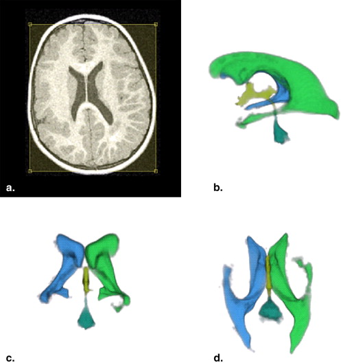
This technique produced detailed three-dimensional reconstructions of the ventricles, even in children with grossly abnormal ventricular morphology. All MR imaging data were successfully postprocessed in <5 minutes. Inter- and intrarater reliability was excellent, with correlation coefficients of 0.99 and 0.92, respectively.
Conclusion
This methodology can create detailed three-dimensional visualizations and volumetric measurements of morphologically abnormal ventricles. This technique could help physicians and parents comprehend abnormal ventricular anatomy better and may have future clinical uses in monitoring disease progression or neurosurgical planning.
There are many congenital abnormalities affecting the structure of children’s brains. Acquired brain injuries are also seen in childhood, including those associated with premature birth, hypoxic ischemic encephalopathy, or neurodegenerative disorders. Many of these conditions are associated with abnormal ventricular anatomy.
Outside the neonatal period, magnetic resonance (MR) imaging has become the first-line method of investigating developmental brain abnormalities . One valuable sequence in that assessment is the T1-weighted volume acquisition of the whole brain. This can be acquired relatively quickly, with high morphologic and contrast resolution. These data can be manipulated in many different ways. Reformations can be made in orthogonal, nonorthogonal, or nonlinear planes at its simplest. At its most complex, cortical surface reconstructions and segmentation can be produced, generating gray-matter and white-matter maps for volumetric assessments of both structures .
Get Radiology Tree app to read full this article<
Get Radiology Tree app to read full this article<
Get Radiology Tree app to read full this article<
Material and methods
Subjects
Get Radiology Tree app to read full this article<
Get Radiology Tree app to read full this article<
MR Imaging
Get Radiology Tree app to read full this article<
Postprocessing and Three-dimensional Ventricular Visualization
Get Radiology Tree app to read full this article<
Get Radiology Tree app to read full this article<
Get Radiology Tree app to read full this article<
Get Radiology Tree app to read full this article<
Get Radiology Tree app to read full this article<
Get Radiology Tree app to read full this article<
Get Radiology Tree app to read full this article<
Results
Get Radiology Tree app to read full this article<
Table 1
Measured Ventricular Volumes of the 14 Children Studied (seven with ventricular abnormalities and seven age-matched controls)
Brain Malformation Type Ventricular Volume of Abnormal Brain (mm 3 ) Ventricular Volume of Age-Matched Patient (mm 3 ) Age (y) Lissencephaly 54.3 6.5 2 Semilobar holoprocephaly 224.5 10.4 5 Unilateral polymicrogyria 25.1 11.2 5 Closed-lip schizencephaly 74.7 12.5 9 Agenesis of corpus callosum 66.3 13.7 9 Hemimegalencephaly 45.5 13.8 10 Subependymal heterotopia 50.3 19.4 14
Get Radiology Tree app to read full this article<
Get Radiology Tree app to read full this article<

Get Radiology Tree app to read full this article<
Discussion
Get Radiology Tree app to read full this article<
Get Radiology Tree app to read full this article<
Get Radiology Tree app to read full this article<
Get Radiology Tree app to read full this article<
Get Radiology Tree app to read full this article<
Get Radiology Tree app to read full this article<
Get Radiology Tree app to read full this article<
Get Radiology Tree app to read full this article<
Get Radiology Tree app to read full this article<
References
1. Brunel H., Girard N., Confort-Gouny S., et. al.: Fetal brain injury. J Neuroradiol 2004; 31: pp. 123-137.
2. Levine D., Trop I., Mehta T.S., Barnes P.D.: MR imaging appearance of fetal cerebral ventricular morphology. Radiology 2002; 223: pp. 652-660.
3. Jain H., Natarajan K., Sgouros S.: Influence of the shunt type in the difference in reduction of volume between the two lateral ventricles in shunted hydrocephalic children. Childs Nerv Syst 2005; 21: pp. 552-558.
4. Steen R.G., Emudianughe T., Hankins G.M., et. al.: Brain imaging findings in pediatric patients with sickle cell disease. Radiology 2003; 228: pp. 216-225.
5. Poe L.B., Coleman L.L., Mahmud F.: Congenital central nervous system anomalies. RadioGraphics 1989; 9: pp. 801-826.
6. Giesel F.L., Hahn H.K., Thomann P.A., et. al.: Temporal horn index and volume using a new semi-automated method for rapid and precise assessment of medial temporal lobe atrophy. AJNR Am J Neuroradiol 2006; 27: pp. 1454-1458.
7. Melhem E.R., Hoon A.H., Ferrucci J.T., et. al.: Periventricular leukomalacia: relationship between lateral ventricular volume on brain MR images and severity of cognitive and motor impairment. Radiology 2000; 214: pp. 199-204.
8. Condon B., Patterson J., Wyper D., et. al.: Use of magnetic resonance imaging to measure intracranial cerebrospinal fluid volume. Lancet 1986; 1: pp. 1355-1357.
9. Tsunoda A., Mitsuoka H., Sato K., Kanayama S.: A quantitative index of intracranial cerebrospinal fluid distribution in normal pressure hydrocephalus using an MRI-based processing technique. Neuroradiology 2000; 42: pp. 424-429.
10. Schierlitz L., Dumanli H., Robinson J.N., et. al.: Three-dimensional magnetic resonance imaging of fetal brains. Lancet 2001; 357: pp. 1177-1178.
11. Thompson P.M., Hayashi K.M., De Zubicaray G.I., et. al.: Mapping hippocampal and ventricular change in Alzheimer disease. Neuroimage 2004; 22: pp. 1754-1766.
12. Hahn H.K., Peitgen H.O.: IWT—interactive watershed transform: a hierarchical method for efficient interactive and automated segmentation of multidimensional gray-scale images. Med Imaging Image Process 2003; 5032: pp. 643-653.
13. Hildebrandt H., Hahn H.K., Kraus J.A., Schulte-Herbrüggen A., Schwarze B., Schwendemann G.: Memory performance in multiple sclerosis patients correlates with central brain atrophy. Mult Scler 2006; 12: pp. 428-436.
14. Hahn H.K., Millar W.S., Klinghammer O., Durkin M.S., Tulipano P.K., Peitgen H.O.: A reliable and efficient method for cerebral ventricular volumetry in pediatric neuroimaging. Methods Inf Med 2004; 43: pp. 376-382.
15. Hahn H.K., Lentschig M.G., Deimling M., Terwey B., Peitgen H.O.: MRI-based volumetry of intra- and extracerebral CSF spaces.CARS—computer aided radiology and surgery, ICS 1230.2001.ElsevierAmsterdam, the Netherlands:pp. 384-389.
16. Hahn H.K., Jolly B., Lee M., et. al.: How accurate is brain volumetry? A methodological evaluation.MICCAI—medical image computing and computer-assisted intervention, LNCS 3216.2004.SpringerBerlin, Germany:pp. 335-342.
17. Barkovich A.J.: Magnetic resonance imaging: role in the understanding of cerebral malformations. Brain Dev 2002; 24: pp. 2-12.
18. Barkovich A.J.: Congenital malformations of the brain and spine.Pediatric neuroimaging.2005.Raven PressNew York:pp. 177-276.
19. Barkovich A.J., Kuzniecky R.I., Dobyns W.B., Jackson G.D., Becker L.E., Evrard P.: A classification scheme for malformations of cortical development. Neuropediatrics 1996; 27: pp. 59-63.
20. Hahn HK Morphological volumetry—theory, concepts, and application to quantitative medical imaging. PhD dissertation, Department of Mathematics and Computer Science, University of Bremen, Bremen, Germany.
21. March K.: Intracranial pressure monitoring: why monitor?. AACN Clin Issues 2005; 16: pp. 456-475.
22. Fritsch M.J., Kienke S., Manwaring K.H., Mehdorn H.M.: Endoscopic aqueductoplasty and interventriculostomy for the treatment of isolated fourth ventricle in children. Neurosurgery 2004; 55: pp. 372-377.