
Purpose
To intraindividually compare the delineation of intracranial arterial vasculature in nonenhanced versus contrast-enhanced magnetization prepared rapid gradient echo (MPRAGE) imaging at 7 Tesla (T).
Materials and Methods
Sixteen subjects were examined on a 7 T whole-body magnetic resonance system (Magnetom 7T) equipped with a 32-channel transmit/receive head coil. MPRAGE imaging was performed pre- and postcontrast after the application of 0.1 mmol/kg bodyweight gadobutrol. For qualitative analysis, the delineation of the intracranial arteries, overall image quality, and image impairment were assessed in the nonenhanced and contrast-enhanced datasets using a 5-point scale (5 = excellent to 1 = nondiagnostic). Additionally, contrast ratios (CR) of the middle cerebral artery in correlation to surrounding gray matter in nonenhanced and postcontrast images were obtained. For statistical analysis a Wilcoxon signed-rank test was applied.
Results
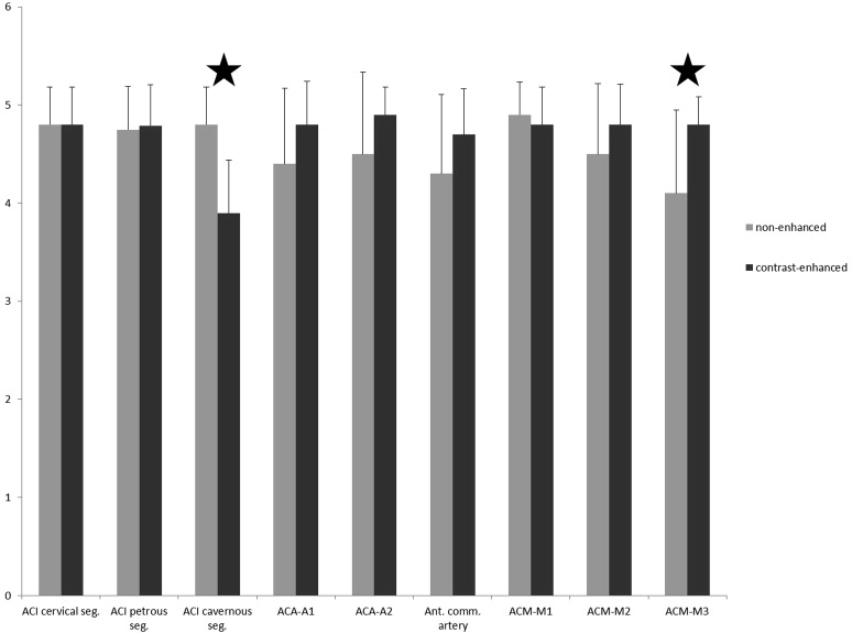
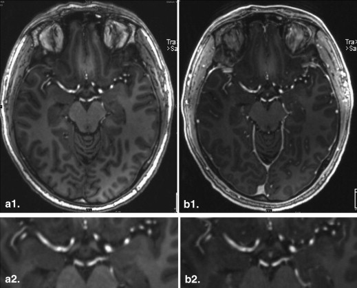
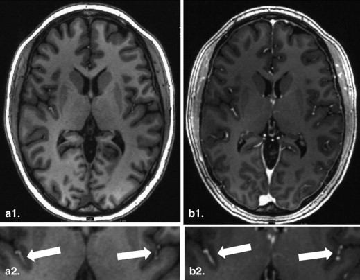
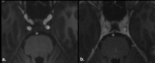
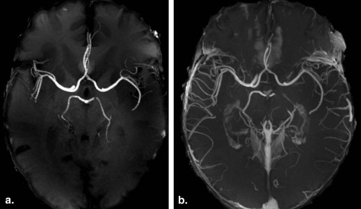
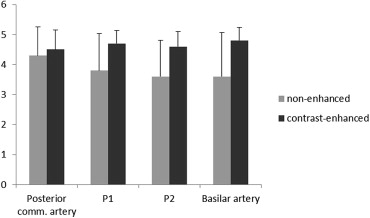
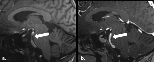
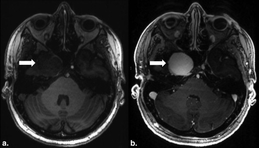
Nonenhanced MPRAGE imaging offered an excellent delineation of the central vessel segments of the anterior circulation (mean anterior circulation 4.6) and a moderate- to high-quality assessment of the vessels of the posterior circulation (mean posterior circulation 3.9). Vessel delineation was improved in all assessed segments in the contrast-enhanced datasets, except for the cavernous segment of the internal carotid artery. Quantitative analysis revealed a mild, nonsignificant increase in CR mean values of the M1 segment (CR nonenhanced 0.67; CR contrast-enhanced 0.69).
Conclusion
Our results demonstrate the high diagnostic value of nonenhanced 7 T MPRAGE imaging for the assessment of the intracranial arterial vasculature, with improved assessment of the peripheral segments because of the application of a contrast agent.
Magnetization prepared rapid acquisition gradient echo sequence (MPRAGE) imaging is a commonly applied pulse sequence for T1-weighted anatomical imaging of the brain. The increase of the magnetic field strength >1.5 T has been proven beneficial in neuro imaging , allowing for a successful transformation of the associated increase in signal-to-noise ratio into imaging at higher spatiotemporal resolution.
Furthermore, initial 7T neuro and abdominal magnetic resonance imaging (MRI) studies have revealed an additional incidental finding by means of a homogeneous hyperintense delineation of nonenhanced arterial vasculature in T1-weighted MRI, offering the diagnostic potential for nonenhanced MR angiographic applications. Maderwald et al and Zwanenburg et al demonstrated the strong diagnostic ability of nonenhanced 7T MPRAGE MRI in the assessment of intracranial arterial vasculature and their related anatomical structures . Although the feasibility of first-pass contrast-enhanced MR angiography of the renal arteries at 7 T has been successfully demonstrated, the administration of gadolinium for 7 T brain imaging has mainly focused on the assessment of parenchymatous features up to current status .
Get Radiology Tree app to read full this article<
Material and methods
Get Radiology Tree app to read full this article<
Get Radiology Tree app to read full this article<
Get Radiology Tree app to read full this article<
Get Radiology Tree app to read full this article<
Get Radiology Tree app to read full this article<
Get Radiology Tree app to read full this article<
Get Radiology Tree app to read full this article<
Get Radiology Tree app to read full this article<
Get Radiology Tree app to read full this article<
Results
Get Radiology Tree app to read full this article<
Get Radiology Tree app to read full this article<
Qualitative Analysis: Anterior Circulation
Get Radiology Tree app to read full this article<
Get Radiology Tree app to read full this article<
Qualitative Analysis: Posterior Circulation
Get Radiology Tree app to read full this article<
Get Radiology Tree app to read full this article<
Overall Image Quality and Overall Image Impairment
Get Radiology Tree app to read full this article<
Quantitative Analysis
Get Radiology Tree app to read full this article<
Patient Analysis
Get Radiology Tree app to read full this article<
Get Radiology Tree app to read full this article<
Discussion
Get Radiology Tree app to read full this article<
Get Radiology Tree app to read full this article<
Get Radiology Tree app to read full this article<
Get Radiology Tree app to read full this article<
Get Radiology Tree app to read full this article<
Get Radiology Tree app to read full this article<
Get Radiology Tree app to read full this article<
Conclusion
Get Radiology Tree app to read full this article<
References
1. Kollia K., Maderwald S., Putzki N., et. al.: First clinical study on ultra-high-field MR imaging in patients with multiple sclerosis: comparison of 1.5T and 7T. AJNR Am J Neuroradiol 2009; 30: pp. 699-702.
2. Kuhl C.K., Träber F., Gieseke J., et. al.: Whole-body high-field-strength (3.0-T) MR imaging in clinical practice part II. Technical considerations and clinical applications. Radiology 2008; 247: pp. 16-35.
3. Kuhl C.K., Träber F., Schild H.H.: Whole-body high-field-strength (3.0-T) MR imaging in clinical practice part I. Technical considerations and clinical applications. Radiology 2008; 246: pp. 675-696.
4. Moenninghoff C., Maderwald S., Theysohn J.M., et. al.: Evaluation of intracranial aneurysms with 7 T versus 1.5 t time-of-flight MR angiography: initial experience. Rofo 2009; 181: pp. 16-23.
5. Moenninghoff C., Maderwald S., Theysohn J., et. al.: Imaging of adult astrocytic brain tumours with 7 T MRI: preliminary results. Eur Radiol 2010; 20: pp. 704-713.
6. Moenninghoff C., Maderwald S., Theysohn J.M., et. al.: Imaging of brain metastases of bronchial carcinomas with 7 T MRI: initial results. Rofo 2010; 182: pp. 764-772.
7. Mönninghoff C., Maderwald S., Wanke I.: Pre-interventional assessment of a vertebrobasilar aneurysm with 7 Tesla time-of-flight MR angiography. Rofo 2009; 181: pp. 266-268.
8. Theysohn J.M., Kraff O., Maderwald S., et. al.: 7 tesla MRI of microbleeds and white matter lesions as seen in vascular dementia. J Magn Reson Imaging 2011; 33: pp. 782-791.
9. Maderwald S., Ladd S., Gizewski E., et. al.: To TOF or not to TOF: strategies for non-contrast-enhanced intracranial MRA at 7 T. Magn Reson Materials Phys Biol Med 2008; 21: pp. 159-167.
10. Zwanenburg J.J., Hendrikse J., Takahara T., et. al.: MR angiography of the cerebral perforating arteries with magnetization prepared anatomical reference at 7T: comparison with time-of-flight. J Magn Reson Imaging 2008; 28: pp. 1519-1526.
11. Umutlu L., Maderwald S., Kinner S., et. al.: First-pass contrast-enhanced renal MRA at 7 Tesla: initial results. Eur Radiol 2012; pp. 1-8.
12. Wrede K.H., Johst S., Dammann P., et. al.: Caudal image contrast inversion in MPRAGE at 7 Tesla: problem and solution. Acad Radiol 2012; 19: pp. 172-178.
13. Grinstead J.W., Rooney W., G L.: The origins of bright blood MPRAGE at 7 Tesla and a simultaneous method for T1 imaging and non-contrast MRA. Proc Intl Soc Mag Reson Med 2010; 18: pp. 1429.
14. Metzger G.J., van de Moortele P.F.: Non-contrast enhanced renal MRA at 7T. Proc Intl Soc Mag Reson Med 2011;
15. Umutlu L., Kraff O., Orzada S., et. al.: Dynamic contrast-enhanced renal MRI at 7 Tesla: preliminary results. Invest Radiol 2011; 46: pp. 425-433.
16. Umutlu L., Orzada S., Kinner S., et. al.: Renal imaging at 7 Tesla: preliminary results. Eur Radiol 2010; 21: pp. 841-849.
17. Umutlu L, Bitz AK, Maderwald S, et al. Contrast-enhanced ultra-high-field liver MRI: a feasibility trial. Eur J Radiol Epub ahead of print.
18. Umutlu L., Maderwald S., Kraff O., et. al.: New look at renal vasculature: 7 tesla nonenhanced T1-weighted FLASH imaging. J Magn Reson Imaging 2012; 36: pp. 714-721.
19. Michaely H.J., Kramer H., Dietrich O., et. al.: Intraindividual comparison of high-spatial-resolution abdominal MR angiography at 1.5 T and 3.0 T: initial experience. Radiology 2007; 244: pp. 907-913.
20. Noebauer-Huhmann I.M., Szomolanyi P., Juras V., et. al.: Gadolinium-based magnetic resonance contrast agents at 7 tesla: in vitro t1 relaxivities in human blood plasma. Invest Radiol 2010; 45: pp. 554-558.
21. Rohrer M., Bauer H., Mintorovitch J., et. al.: Comparison of magnetic properties of MRI contrast media solutions at different magnetic field strengths. Invest Radiol 2005; 40: pp. 715-724.
22. Lauenstein T.C., S K., Morreira R., Tata S., et. al.: Nephrogenic systemic fibrosis: center case review. J Magn Reson Imaging 2007; 26: pp. 1198-1203.