
Rationale and Objectives
Diffusion tensor imaging can depict rarefaction of the optical fibers. Manual segmentation is time consuming. The purposes of the study were (1) to present a new semiquantitative segmentation approach for analyzing 3-T diffusion tensor imaging of optical fibers and (2) to clinically test the new approach by comparing optic fiber rarefaction in patients with glaucoma to that in age-matched, healthy controls.
Materials and Methods
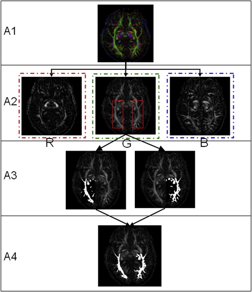
To perform semiautomated and quantitative segmentation of the optical radiation, a Mathcad-based software program was developed. The results were compared to the manual evaluation of the images performed by two experienced neuroradiologists. The eyes of 42 subjects (22 patients with glaucoma and 20 controls) aged 37 to 86 years were assessed in full ophthalmologic examinations. Magnetic resonance imaging was performed using a 3-T high-field scanner.
Results
The evaluation using the new approach matched 94% with manually acquired rarefaction of the optical radiation; Cronbach’s α was >0.81 for calculation of the manually and semiautomatically derived volumes.
Conclusion
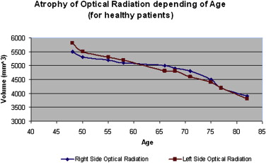
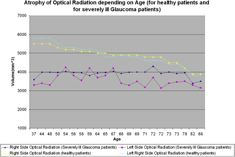
The new approach seems to be robust and is clearly faster compared to the more tedious manual segmentation. Using diffusion tensor imaging at 3 T, it could be shown that there was increasing atrophy of the optical radiation (fourth neuron) with increasing age in patients with glaucoma. Compared to age-matched, healthy patients, more pronounced atrophy of the fourth neuron was found in patients with glaucoma.
Whether glaucoma-related rarefaction of the optical radiation exists is still dubious, because there are no experimental or clinical studies dealing with this topic. Loss of ganglion cells in the retina and their axons representing the third neuron and atrophy of the fourth neuron have been described in diseases such as traumatic injury and glaucoma . The axons of various retinal ganglion cell subtypes, differing in specific morphology and function, exit the eyeball and finally converge to anatomically distinguishable layers of the lateral geniculate nucleus (LGN) , where a loss of neural cells has also been described for glaucoma .
Diffusion tensor imaging (DTI) can depict the optic nerve (third neuron) and optical radiation (fourth neuron) . However, manual analysis is time consuming. We used DTI with a newly developed semiautomatic segmentation approach to assess rarefaction of the fourth neuron in 20 control subjects aged 45 to 83 years and in 22 severely ill patients with glaucoma aged 37 to 86 years.
Materials and methods
Get Radiology Tree app to read full this article<
Get Radiology Tree app to read full this article<
Get Radiology Tree app to read full this article<
Get Radiology Tree app to read full this article<
Get Radiology Tree app to read full this article<
Results
Get Radiology Tree app to read full this article<
Get Radiology Tree app to read full this article<
Get Radiology Tree app to read full this article<
Discussion
Get Radiology Tree app to read full this article<
Get Radiology Tree app to read full this article<
Get Radiology Tree app to read full this article<
Get Radiology Tree app to read full this article<
Get Radiology Tree app to read full this article<
Conclusion
Get Radiology Tree app to read full this article<
Get Radiology Tree app to read full this article<
References
1. Conti A.C., Raghupathi R., Trojanowski J.Q., et. al.: Experimental brain injury induces regionally distinct apoptosis during the acute and delayed post-traumatic period. J Neurosci 1998; 18: pp. 5663-5672.
2. Dacey D.M.: Circuitry for color coding in the primate retina. Proc Natl Acad Sci U S A 1996; 93: pp. 582-588.
3. Yücel Y.H., Zhang Q., Weinreb R.N., et. al.: Atrophy of relay neurons in magno- and parvocellular layers in the lateral geniculate nucleus in experimental glaucoma. Invest Ophthalmol Vis Sci 2001; 42: pp. 3216-3222.
4. Weber A.J., Chen H., Hubbard W.C., et. al.: Experimental glaucoma and cell size, density, and number in the primate lateral geniculate nucleus. Invest Ophthalmol Vis Sci 2000; 41: pp. 1370-1379.
5. Staempfli P., Rienmueller A., Reischauer C., et. al.: Reconstruction of the human visual system based on DTI fiber tracking. J Magn Reson Imaging 2007; 26: pp. 886-893.
6. Taoka T., Sakamoto M., Iwasaki S., et. al.: Diffusion tensor imaging in cases with visual field defect after anterior temporal lobectomy. AJNR Am J Neuroradiol 2005; 26: pp. 797-803.
7. Shimony J.S., Burton H., Epstein A.A., et. al.: Diffusion tensor imaging reveals white matter reorganization in early blind humans. Cereb Cortex 2006; 16: pp. 1653-1661.
8. Meyer A.: The connections of the occipital lobes and the presents statues of the cerebral visual affections. Trans Assoc Phys 1907; 22: pp. 7-23.