
Rationale and Objectives
To compare the performance of two shortened breast magnetic resonance imaging (MRI) protocols to a standard MRI protocol for breast cancer screening.
Materials and Methods
In this Health Insurance Portability and Accountability Act compliant, institutional review board–approved pilot study, three fellowship-trained breast imagers evaluated 48 breast MRIs (24 normal, 12 benign, and 12 malignant) selected from a high-risk screening population. MRIs were presented in three viewing protocols, and a final Breast Imaging-Reporting and Data System assessment was recorded for each case. The first shortened protocol (abbreviated 1) included only fat-saturated precontrast T2-weighted, precontrast T1-weighted, and first pass T1-weighted postcontrast sequences. The second shortened protocol (abbreviated 2) included the abbreviated 1 protocol plus the second pass T1-weighted postcontrast sequence. The third protocol (full), reviewed after a 1-month waiting period, included a nonfat-saturated T1-weighted sequence, fat-saturated T2-weighted, precontrast T1-weighted, and three or four dynamic postcontrast sequences. Interpretation times were recorded for the abbreviated 1 and full protocols. Sensitivity and specificity were compared via a chi-squared analysis. This pilot study was designed to detect a 10% difference in sensitivity with a power of 0.8.
Results
There was no significant difference in sensitivity between the abbreviated 1 (86%; P = .22) or abbreviated 2 (89%; P = .38) protocols and the full protocol (95%). There was no significant difference in specificity between the abbreviated 1 (52%; P = 1) or abbreviated 2 (45%; P = .34) protocols and the full protocol (52%). The abbreviated 1 and full protocol interpretation times were similar (2.98 vs. 3.56 minutes).
Conclusions
In this pilot study, reader performance comparing two shortened breast MRI protocols to a standard protocol in a screening cohort were similar, suggesting that a shortened breast MRI protocol may be clinically useful, warranting further investigation.
Breast magnetic resonance imaging (MRI) has been shown to have the highest sensitivity of all imaging modalities for the detection of breast cancer, a critically important factor in screening . The high sensitivity of breast MRI is largely because of improved contrast over mammography, particularly in women with dense breast tissue . As a result, MRI has been increasingly used to help screen women at increased lifetime risk for breast cancer. The American College of Radiology Appropriateness Criteria gives contrast-enhanced MRI their highest rating for use in high-risk women (≥20% lifetime risk; genetic mutation carriers, strong family history, and prior chest radiation) when used in conjunction with mammography . Similar recommendations for MRI screening in high-risk women exist from the American Cancer Society and the National Comprehensive Cancer Care Network as well as health systems in the United Kingdom and Canada .
Although women with increased mammographic breast density have an increased risk of developing breast cancer compared to women with lower breast density, breast density alone as an independent risk factor confers less than the 20% risk threshold currently established to guide MRI use for high-risk screening . Nevertheless, many states require that radiologists notify women of their breast density and the limitations of screening mammography, prompting inquiries by some patients and referring physicians about the merits of other methods of breast cancer screening. Currently, there is no consensus about what additional screening modalities should be offered . However, in addition to several studies demonstrating that MRI has the highest sensitivity for breast cancer screening compared to ultrasound and mammography, , data from a recent study of a high-risk population have shown that MRI has the highest sensitivity regardless of breast density , suggesting that MRI may be a sensible additional screening option to offer women with dense breast tissue.
Get Radiology Tree app to read full this article<
Get Radiology Tree app to read full this article<
Materials and methods
Study Patients
Get Radiology Tree app to read full this article<
MRI Protocol
Get Radiology Tree app to read full this article<
Reader Study
Get Radiology Tree app to read full this article<
Get Radiology Tree app to read full this article<
Table 1
Breast Magnetic Resonance Imaging Sequences by Reader Protocol
Protocol T2 Dynamic Precontrast Dynamic 1st Pass Dynamic 2nd Pass Dynamic 3rd and 4th pass ∗ Nonfat-Saturated T1 Abbreviated 1 x x x Abbreviated 2 x x x x Full x x x x x x
Get Radiology Tree app to read full this article<
Get Radiology Tree app to read full this article<
Data Analysis
Get Radiology Tree app to read full this article<
Get Radiology Tree app to read full this article<
Results
Get Radiology Tree app to read full this article<
Table 2
Characteristics of Magnetic Resonance Imaging Examinations
Pathologic Diagnosis n % Normal 24 50.0 Benign 12 25.0 Fibrocystic changes 3 6.3 Fibroadenoma 4 8.3 Papilloma 2 4.2 Lymph node 1 2.1 Flat epithelial atypia 1 2.1 Atypical ductal hyperplasia 1 2.1 Malignant 12 25.0 Ductal carcinoma in situ 3 6.3 Invasive ductal carcinoma 8 8.3 Invasive lobular carcinoma 1 2.1
All studies had biopsy-proven abnormalities or two years of imaging follow-up.
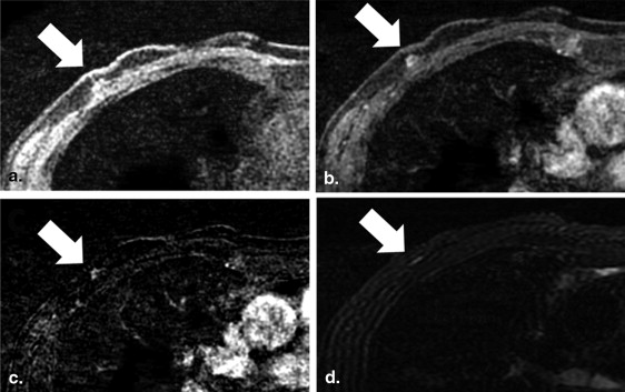
Table 3
Sensitivity for Each Reader and Overall by Imaging Protocol
Protocol Reader 1 (%) Reader 2 (%) Reader 3 (%) Overall (%)P Value Sensitivity Abbreviated 1 92 83 83 86 .22 Abbreviated 2 92 83 92 89 .38 Full 92 92 100 95 Specificity Abbreviated 1 69 56 31 52 1 Abbreviated 2 67 44 25 45 .34 Full 58 58 39 52
Round three was used as the reference standard for P value calculations.
Get Radiology Tree app to read full this article<
Get Radiology Tree app to read full this article<
Table 4
Interpretation Time for Each Reader and Overall by Imaging Protocol
Protocol Reader 1 (SD) Reader 2 (SD) Reader 3 (SD) Overall (SD)P Value Abbreviated 1 3.65 (2.68) 2.25 (0.73) 3.04 (1.34) 2.98 (1.86) <.01 Full 3.58 (2.22) 2.10 (0.67) 3.19 (1.08) 2.95 (1.59)
SD, standard deviation.
A two one-sided test for equivalence was used to calculate the P value.
Get Radiology Tree app to read full this article<
Discussion
Get Radiology Tree app to read full this article<
Get Radiology Tree app to read full this article<
Get Radiology Tree app to read full this article<
Get Radiology Tree app to read full this article<
Get Radiology Tree app to read full this article<
Get Radiology Tree app to read full this article<
Conclusions
Get Radiology Tree app to read full this article<
References
1. Mainiero M.B., Lourenco A., Mahoney M.C., et. al.: ACR appropriateness criteria breast cancer screening. J Am Coll Radiol 2013; 10: pp. 11-14.
2. Saslow D., Boetes C., Burke W., et. al.: American Cancer Society guidelines for breast screening with MRI as an adjunct to mammography. CA Cancer J Clin 2007; 57: pp. 75-89.
3. Morrow M., Waters J., Morris E.: MRI for breast cancer screening, diagnosis, and treatment. Lancet 2011; 378: pp. 1804-1811.
4. Lehman C.D., Gatsonis C., Kuhl C.K., et. al.: MRI evaluation of the contralateral breast in women with recently diagnosed breast cancer. N Engl J Med 2007; 356: pp. 1295-1303.
5. Pilewskie M., King T.A.: Magnetic resonance imaging in patients with newly diagnosed breast cancer: a review of the literature. Cancer 2014; 120: pp. 2080-2089.
6. Hagen A.I., Kvistad K.A., Maehle L., et. al.: Sensitivity of MRI versus conventional screening in the diagnosis of BRCA-associated breast cancer in a national prospective series. Breast 2007; 16: pp. 367-374.
7. Kuhl C.K., Schrading S., Leutner C.C., et. al.: Mammography, breast ultrasound, and magnetic resonance imaging for surveillance of women at high familial risk for breast cancer. J Clin Oncol 2005; 23: pp. 8469-8476.
8. Stout N.K., Nekhlyudov L., Li L., et. al.: Rapid increase in breast magnetic resonance imaging use: trends from 2000 to 2011. JAMA Intern Med 2014; 174: pp. 114-121.
9. Sickles E.A.: The use of breast imaging to screen women at high risk for cancer. Radiol Clin North Am 2010; 48: pp. 859-878.
10. Bevers T.B., Anderson B.O., Bonaccio E., et. al.: NCCN clinical practice guidelines in oncology: breast cancer screening and diagnosis. J Natl Compr Canc Netw 2009; 7: pp. 1060-1096.
11. National Health System: Protocols for the surveillance of women at higher risk of developing breast cancer.2013.
12. 2014. https://www.cancercare.on.ca/cms/one.aspx?portalId=1377&pageId=99638 Accessed August 31, 2014
13. Harvey J.A., Bovbjerg V.E.: Quantitative assessment of mammographic breast density: relationship with breast cancer risk. Radiology 2004; 230: pp. 29-41.
14. Sprague B.L., Gangnon R.E., Burt V., et. al.: Prevalence of mammographically dense breasts in the United States. J Natl Cancer Inst 2014; 106: pp. 1-6.
15. Ho J.M., Jafferjee N., Covarrubias G.M., et. al.: Dense breasts: a review of reporting legislation and available supplemental screening options. AJR Am J Roentgenol 2014; 203: pp. 449-456.
16. Freer P.E.: Mammographic breast density: impact on breast cancer risk and implications for screening. Radiographics 2015; 35: pp. 302-315.
17. Berg W.A., Zhang Z., Lehrer D., et. al.: Detection of breast cancer with addition of annual screening ultrasound or a single screening MRI to mammography in women with elevated breast cancer risk. JAMA 2012; 307: pp. 1394-1404.
18. Kuhl C., Weigel S., Schrading S., et. al.: Prospective multicenter cohort study to refine management recommendations for women at elevated familial risk of breast cancer: the EVA trial. J Clin Oncol 2010; 28: pp. 1450-1457.
19. Warner E., Plewes D.B., Hill K.A., et. al.: Surveillance of BRCA1 and BRCA2 mutation carriers with magnetic resonance imaging, ultrasound, mammography, and clinical breast examination. JAMA 2004; 292: pp. 1317-1325.
20. Riedl C.C., Luft N., Bernhart C., et. al.: Triple-modality screening trial for familial breast cancer underlines the importance of magnetic resonance imaging and questions the role of mammography and ultrasound regardless of patient mutation status, age, and breast density. J Clin Oncol 2015; 33: pp. 1128-1135.
21. Killelea B.K., Long J.B., Chagpar A.B., et. al.: Evolution of breast cancer screening in the Medicare population: clinical and economic implications. J Natl Cancer Inst 2014; 106: pp. 1-8.
22. Ahern C.H., Shih Y.C., Dong W., et. al.: Cost-effectiveness of alternative strategies for integrating MRI into breast cancer screening for women at high risk. Br J Cancer 2014; 111: pp. 1542-1551.
23. Feig S.: Cost-effectiveness of mammography, MRI, and ultrasonography for breast cancer screening. Radiol Clin North Am 2010; 48: pp. 879-891.
24. Griebsch I., Brown J., Boggis C., et. al.: Cost-effectiveness of screening with contrast enhanced magnetic resonance imaging vs X-ray mammography of women at a high familial risk of breast cancer. Br J Cancer 2006; 95: pp. 801-810.
25. Moore S.G., Shenoy P.J., Fanucchi L., et. al.: Cost-effectiveness of MRI compared to mammography for breast cancer screening in a high risk population. BMC Health Serv Res 2009; 9: pp. 9.
26. Pataky R., Armstrong L., Chia S., et. al.: Cost-effectiveness of MRI for breast cancer screening in BRCA1/2 mutation carriers. BMC Cancer 2013; 13: pp. 339.
27. Taneja C., Edelsberg J., Weycker D., et. al.: Cost effectiveness of breast cancer screening with contrast-enhanced MRI in high-risk women. J Am Coll Radiol 2009; 6: pp. 171-179.
28. Kuhl C.K., Schrading S., Strobel K., et. al.: Abbreviated breast magnetic resonance imaging (MRI): first postcontrast subtracted images and maximum-intensity projection-a novel approach to breast cancer screening with MRI. J Clin Oncol 2014; 32: pp. 2304-2310.
29. Mann R.M., Mus R.D., van Zelst J., et. al.: A novel approach to contrast-enhanced breast magnetic resonance imaging for screening: high-resolution ultrafast dynamic imaging. Invest Radiol 2014; 49: pp. 579-585.
30. Trimboli R.M., Verardi N., Cartia F., et. al.: Breast cancer detection using double reading of unenhanced MRI including T1-weighted, T2-weighted STIR, and diffusion-weighted imaging: a proof of concept study. AJR Am J Roentgenol 2014; 203: pp. 674-681.
31. Mango V.L., Morris E.A., David Dershaw D., et. al.: Abbreviated protocol for breast MRI: are multiple sequences needed for cancer detection?. Eur J Radiol 2015; 84: pp. 65-70.
32. Schuirmann D.J.: On hypothesis testing to determine if the mean of a normal distribution is contained in a known interval. Biometrics 1981; 37: pp. 617.
33. Berg W.A., Blume J.D., Cormack J.B., et. al.: Combined screening with ultrasound and mammography vs mammography alone in women at elevated risk of breast cancer. JAMA 2008; 299: pp. 2151-2163.
34. Haas B.M., Kalra V., Geisel J., et. al.: Comparison of tomosynthesis plus digital mammography and digital mammography alone for breast cancer screening. Radiology 2013; 269: pp. 694-700.
35. Warner E., Messersmith H., Causer P., et. al.: Systematic review: using magnetic resonance imaging to screen women at high risk for breast cancer. Ann Intern Med 2008; 148: pp. 671-679.
36. Kriege M., Brekelmans C.T., Boetes C., et. al.: Efficacy of MRI and mammography for breast-cancer screening in women with a familial or genetic predisposition. N Engl J Med 2004; 351: pp. 427-437.
37. Partridge S.C., Stone K.M., Strigel R.M., et. al.: Breast DCE-MRI: influence of postcontrast timing on automated lesion kinetics assessments and discrimination of benign and malignant lesions. Acad Radiol 2014; 21: pp. 1195-1203.