
Rational and Objectives
The increasing importance of imaging for both diagnosis and management in patient care has resulted in a demand for radiology services 7 days a week, 24 hours a day, especially in the emergency department (ED). We hypothesized the resident preliminary reports were better than generalist radiology interpretations, although inferior to subspecialty interpretations.
Materials and Methods
Total radiology volume through our Level I pediatric and adult academic trauma ED was obtained from the radiology information system. We conducted a literature search for error and discordant rates between radiologists of varying experience. For a 2-week prospective period, all preliminary reports generated by the residents and final interpretations were collected. Significant changes in the report were tabulated.
Results
The ED requested 72,886 imaging studies in 2004 (16% of the total radiology department volume). In a 2-week period, 12 of 1929 (0.6%) preliminary reports by residents were discordant to the final subspecialty dictation. In the 15 peer-reviewed publications documenting error rates in radiology, the error rate between American Board of Radiology (ABR)−certified radiologists is greater than that between residents and subspecialists in the literature and in our study. However, the perceived error rate by clinicians outside radiology is significantly higher.
Conclusion
Sixteen percent of the volume of imaging studies comes through the ED. The residents handle off-hours cases with a radiology-detected error rate below the error rate between ABR-certified radiologists. To decrease the perceived clinician-identified error rate, we need to change how academic radiology handles ED cases.
Technological improvements have made imaging essential for patient diagnosis and management. Physicians routinely review imaging studies, especially CT and MR studies, before examining the patient. Nowhere is this more apparent than the emergency department (ED). The physical examination is secondary in many disciplines. Most imaging studies are interpreted during regular office hours, but the need to care for ED patients in a timely fashion demands expedited processing by radiology all hours of the day and all days of the week.
At the time of the study, there was no separate ED radiology division in our academic university health system. Interpretation strategies varied by the day and shift. A radiology junior resident is always present in the ED to review ongoing plain radiographic examinations. During regular working hours, subspecialty faculty interpret the majority of the plain radiographic cases in the radiology reading area in the ED with this resident. Subspecialty faculty during regular working hours interpret cross-sectional imaging studies from a remote location on a PACS.
Get Radiology Tree app to read full this article<
Get Radiology Tree app to read full this article<
Get Radiology Tree app to read full this article<
Materials and methods
Get Radiology Tree app to read full this article<
Get Radiology Tree app to read full this article<
Results
Get Radiology Tree app to read full this article<
Table 1
Distribution of Most Common Imaging Studies Requested Through the ED in 2004
Imaging Study Volume % of Total Plain radiography 47,877 65.7 CT 15,578 21.4 US 4373 6 MRI 947 1.3 Consultations 1020 1.4 Other (GI, GU, angiography, etc.) 3061 4.2 Total 72,886 100
Get Radiology Tree app to read full this article<
Get Radiology Tree app to read full this article<
Table 2
Distribution of Discordant Preliminary Reports
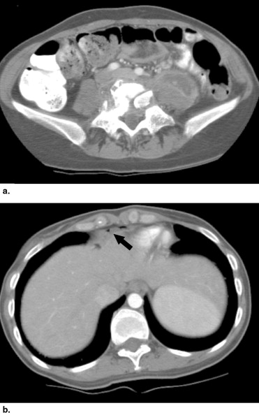
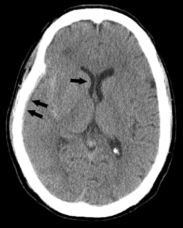
Imaging Study Volume Consensus Discrepant Reports % of Total Plain radiography 934 0 0 Nuclear medicine 41 1 2.4 Adult US 118 0 0 MRI 1 0 0 Neuroradiology 398 3 0.8 Pediatric CT and US 58 0 0 Adult abdomen CT 327 8 2.4 Adult chest CT 52 0 0 Total 1929 12 0.6
Get Radiology Tree app to read full this article<
Get Radiology Tree app to read full this article<
Get Radiology Tree app to read full this article<
Get Radiology Tree app to read full this article<
Get Radiology Tree app to read full this article<
Get Radiology Tree app to read full this article<
Get Radiology Tree app to read full this article<
Discussion
Get Radiology Tree app to read full this article<
Get Radiology Tree app to read full this article<
Get Radiology Tree app to read full this article<
Get Radiology Tree app to read full this article<
Get Radiology Tree app to read full this article<
Get Radiology Tree app to read full this article<
Get Radiology Tree app to read full this article<
Get Radiology Tree app to read full this article<
Get Radiology Tree app to read full this article<
Conclusion
Get Radiology Tree app to read full this article<
Get Radiology Tree app to read full this article<
References
1. Carney E., Kempf J., DeCarvalho V., Yudd A., Nosher J.: Preliminary interpretations of after-hours CT and sonography by radiology residents versus final interpretations by body imaging radiologists at a level 1 trauma center. AJR Am J Roentgenol 2003; 181: pp. 367-373.
2. Wysoki M.G., Nassar C.J., Koenigsberg R.A., Novelline R.A., Faro S.H., Faeber E.N.: Head trauma: CT scan interpretation by radiology residents versus staff radiologists. Radiol 1998; 208: pp. 125-128.
3. Lal N.R., Murray U.M., Eldevik O.P., Desmond J.S.: Clinical consequences of misinterpretations of neuroradiologic CT scans by on-call radiology residents. Am J Neuroradiol 2000; 21: pp. 124-129.
4. Erly W.K., Berger W.G., Krupinski E., Seeger J.F., Guisto J.A.: Radiology resident evaluation of head CT scan orders in the emergency department. AJNR 2002; 23: pp. 103-107.
5. Rhea J.T., Potsaid M.S., DeLuca S.A.: Errors of interpretation as elicited by a quality audit of an emergency radiology facility. Radiology 1979; 132: pp. 277-280.
6. Lowe L.H., Draud K.S., Hernanz-Schulman M., Newton M.R., Heller R.M., Stein S.M., Speroff T.: Nonenhanced limited CT in children suspected of having appendicitis: Prospective comparison of attending and resident interpretations. Radiology 2001; 221: pp. 755-759.
7. Stockbridge H.L., Lewis D., Eisenberg B., Lee M., Schacher S., van Belle G., Keifer M., Brodkin C.A., Buchwald D.: Brain SPECT: A controlled, blinded assessment of interreader and interreader agreement. Nucl Med Commun 2002; 23: pp. 537-544.
8. Chartrand-Lefebvre C., Howarth N., Lucidarme O., et. al.: Contrast-enhanced helical CT for pulmonary embolism detection:inter- and intraobserver agreement among radiologists with variable experience. AJR 1999; 172: pp. 107-112.
9. Markus A.B., Somers S., Franic S.E., Moola C., Stevenson W.: Interobserver variation in the interpretation of abdominal radiographs. Radiology 1989; 171: pp. 69-71.
10. Cascade P.N., Kazerooni E.A., Gross B.H., Quint L.E., Silver T.M., Bowerman R.A., Pernicano P.G., Gebremariam A.: Evaluation of competence in the interpretation of chest radiographs. Acad Radiol 2001; 8: pp. 315-321.
11. Yoon L.S., Haims A.H., Brink J.A., Rabinovici R., Forman H.P.: Evaluation of an emergency radiology quality assurance program at a level I trauma center: Abdominal and pelvic CT studies. Radiology 2002; 242: pp. 42-46.
12. Gollub M.J., Panicek D.M., Bach A.M., Penalver A., Castellino R.A.: Clinical importance of reinterpretation of body CT scans obtained elsewhere in patients referred for care at a tertiary cancer center. Radiology 1999; 210: pp. 109-112.
13. Ginsberg M.S., King V., Panieck D.M.: Comparison of interpretations of CT angiograms in the evaluation of suspected pulmonary embolism by on-call radiology fellows and subsequently by radiology faculty. AJR 2004; 182: pp. 61-66.
14. Elmore J.G., Wells C.K., Lee C.H., Howard D.H., Feinstein A.R.: Variability in radiologists’ interpretations of mammograms. N Engl J Med 1994; 331: pp. 1493-1499.
15. Berg W.A., Campassi C., Langenberg P., Sexton M.J.: Breast imaging reporting and data system. AJR 2000; 174: pp. 1769-1777.
17. Burt CW, McCaig LF. Trends in Hospital Emergency Department Utilization: United States, 1992–99. Vital and Health Statistics 2001; Series 13, Number 150:1–33.
18. Carroll T.J.: Trends in on-call workload in an academic medical center radiology department 1998–2002. Acad Radiol 2003; 10: pp. 1312-1320.
19. Kay C.M., Krishnan M., Smith W.: Results from the problem-solving sessions at the 52nd Annual Meeting of the Association of University Radiologists. Acad Radiol 2005; 12: pp. 526-528.