
Rationale and Objectives
Magnetic resonance (MR) imaging (MRI) provides information that can be used to estimate the symptom onset in patients with wake-up stroke (WUS). Time-resolved MR angiography (MRA) is the fastest available MR sequence technique for vessel assessment, and the different phases acquired can provide information about cerebral perfusion. The aim of this study was to evaluate the diagnostic performance of time-resolved MRA both for the assessment of vessel morphology and for the feasibility of perfusion.
Materials and Methods
Nineteen patients with WUS were included. Image quality and vessel pathologies were evaluated and correlated to time-of-flight–MRA ( n = 14), computed tomography–angiography ( n = 4), sonography ( n = 12), and conventional angiography ( n = 6). The temporal delay of signal enhancement in all pixels of the time-resolved MRA measurement after contrast injection was evaluated and compared to dynamic susceptibility contrast-enhanced (DSC) perfusion imaging ( n = 13).
Results
Time-resolved MRA resulted in the diagnosis of large vessel disease in 14 of 19 patients, involving the internal carotids ( n = 4), the vertebral arteries ( n = 3), and the circle of Willis ( n = 10). All severe vascular pathologies which influence patients’ acute stroke therapy were obtained by time-resolved MRA. Overestimation of stenoses in two of 14 patients resulted in sensitivity and specificity of 100% and 71%, respectively. Time-to-peak (TTP) estimations were hampered by movement artifacts in four patients (31%). Compared to DSC, the area of TTP delay was comparable in size and localization without relevant overestimation or underestimation.
Conclusions
Time-resolved MRA is a valuable technique in patients with WUS with high sensitivity and high negative predictive value. Cerebral perfusion estimation can be performed in selected cases for therapy decision but can be hampered by patient movement.
Noncontrast brain computed tomography (CT) is considered the first line of imaging in patients with suspected acute stroke because of its broad availability and acquisition speed. Because intravenous tissue plasminogen activator (tPA) is the most important therapy for acute ischemic stroke, any contraindications—notably intracranial hemorrhage—have to be ruled out as fast as possible before starting thrombolytic therapy. Noncontrast CT is often combined with CT-angiography (CTA) and CT-perfusion (CTP) imaging to evaluate the underlying stroke cause. Recent technological advances have contributed to more widespread use of new neuroimaging modalities and strategies such as diffusion-weighted (DWI) magnetic resonance (MR) imaging (MRI), cerebral perfusion imaging, and MR angiography. Although DWI is only available in MRI, perfusion imaging and angiography are available in both CT and MRI, which can be beneficial to patient evaluation and management in special cases. MRI is more sensitive than CT for the detection of early infarction, small infarcts, and those in the brain stem and posterior fossa .
In some instances, MRI may be the first imaging test performed, for example, in patients with unknown symptom onset, such as patients with wake-up stroke (WUS). Patients with WUS have often been excluded from thrombolysis because of the unknown time of symptom onset. However, stroke severity is similar in both patient groups, and it has been reported that patients with WUS may benefit from thrombolysis . In WUS, comprehensive stroke evaluation is more important as tPA is still under evaluation in this setting.
Get Radiology Tree app to read full this article<
Get Radiology Tree app to read full this article<
Get Radiology Tree app to read full this article<
Materials and methods
Patient Population
Get Radiology Tree app to read full this article<
Get Radiology Tree app to read full this article<
Imaging Protocol
Get Radiology Tree app to read full this article<
Time-resolved MRA
Get Radiology Tree app to read full this article<
Imaging Parameters of the Additionally Acquired MRI Sequences
Get Radiology Tree app to read full this article<
Get Radiology Tree app to read full this article<
Get Radiology Tree app to read full this article<
Get Radiology Tree app to read full this article<
Get Radiology Tree app to read full this article<
Evaluation of Time-resolved MRA
Get Radiology Tree app to read full this article<
Get Radiology Tree app to read full this article<
Get Radiology Tree app to read full this article<
DSC Time-to-peak Analysis
Get Radiology Tree app to read full this article<
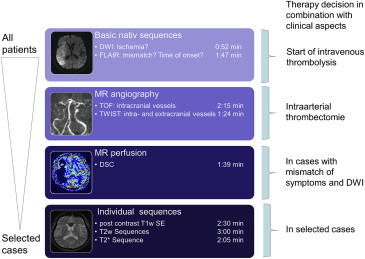
Feasibility of an Ultrafast MR Protocol
Get Radiology Tree app to read full this article<
Statistical Analysis
Get Radiology Tree app to read full this article<
Results
Get Radiology Tree app to read full this article<
Get Radiology Tree app to read full this article<
Get Radiology Tree app to read full this article<
Table 1
Patients’ Characteristics
Patient No. Age, years Sex Clinical Symptoms Stroke Classification MRI Findings FLAIR–DWI Vascular Findings MR Perfusion Analysis Acute Stroke Therapy Clinical Outcome 1 84 F Dysarthria, hemiparesis (left side), hemineglect PACI, TOAST 2 MCA infarction Occlusion MCA PWI/DWI mismatch Mechanical thrombectomy Clinical improvement 2 52 F Dysarthria, hemiparesis (right side) LACI, TOAST 3 Small infarction MCA No stenosis or occlusion No mismatch Conservative Minimal clinical improvement 3 80 M Aphasia, hemiparesis (right side) PACI, TOAST 2 MCA infarction, no mismatch Occlusion MCA (M2; right side) No mismatch Conservative Minimal clinical improvement 4 82 M Facial nerve paresis, somnolent LACI, TOAST 2 Small thalamic infarction, no mismatch Stenosis ICA Not performed Conservative Clinical improvement 5 84 M Hemiparesis (left side) PACI, TOAST 2 Small MCA infarction, no mismatch MCA stenosis (M1; right side) PWI/DWI mismatch MCA stenting Clinical improvement 6 60 M Hemiparesis (left side) LACI, TOAST 3 Small MCA infarction, no mismatch Stenosis MCA (M2) No mismatch Conservative Stable 7 59 M Aphasia, hemiplegia (right side) TACI, TOAST 2 MCA infarction, with FLAIR DWI mismatch Occlusion ICA and MCA PWI/DWI mismatch Stenting ICA, mechanical thrombectomy, i.a. thrombolysis Clinical improvement 8 89 F Hemiparesis (right side), dysarthria PACI, TOAST 5 Mismatch, infarction without FLAIR hyperintensity No stenosis No mismatch i.v. thrombolysis Clinical improvement 9 86 M Dysarthria, hemianopsia, hemiparesis LACI, TOAST 3 Small infarction, no mismatch No stenosis No mismatch Conservative Stable 10 74 F Paresis (hand) LACI, TOAST 3 Small infarction, no mismatch No stenosis Not performed Conservative Clinical improvement 11 75 F Hypoesthesia LACI, TOAST 4 Small infarction, no mismatch MCA stenosis (M1; left side) Not performed Conservative Stable 12 58 F Somnolent, aphasia LACI, TOAST 4 Bleeding due to cerebral venous sinus thrombosis Cerebral venous sinus thrombosis Not performed Mechanical thrombectomy Clinical improvement 13 79 F Hemiplegia, aphasia TACI, TOAST 2 Infarction MCA, mismatch Occlusion ICA, MCA supplied via ACOM PWI/DWI mismatch i.v. thrombolysis Stable 14 37 M Vertigo, nystagmus, emesis PACI, TOAST 2 Small infarction middle cerebellar peduncle (right side), no mismatch Stenosis vertebral artery No mismatch Conservative Complete remission 15 84 F Hemiparesis and aphasia TACI, TOAST 1 Infarction without increase compared to FLAIR Occlusion MCA (M2) PWI/DWI mismatch i.v. thrombolysis Stable 16 62 M Hemiparesis, hemineglect, dysarthria TACI, TOAST 1 No mismatch Occlusion ICA and MCA (right side) No mismatch No lysis, decompressive craniectomy Stable 17 67 M Hemiparesis PACI, TOAST 2 MCA infarction, mismatch Occlusion MCA PWI/DWI mismatch Mechanical thrombectomy Clinical improvement 18 57 F Nystagmus, lateropulsion POCI, TOAST 5 PICA infarction, no mismatch No stenosis No mismatch Conservative Stable 19 77 F Hemiparesis, headache, somnolent PACI, TOAST 1 No mismatch, FLAIR-DWI, intraventricular hemorrhage Occlusion MCA Not performed Conservative Stable
ACOM, anterior communicating artery; DWI, diffusion-weighted imaging; F, female; FLAIR, fluid attenuated inversion recovery; i.a., intra-arterial; ICA, internal carotid artery; i.v., intravenous; LACI, lacunar infarct; M, male; MCA, medial cerebral artery; MR, magnetic resonance; MRI, magnetic resonance imaging; PACI, partial anterior circulation infarct; POCI posterior circulation infarct; PWI, perfusion-weighted imaging; TACI, total anterior circulation infarct; TOAST, Trial of Org 10172 in Acute Stroke Treatment.
Stroke classification: total anterior circulation infarct (TACI), partial anterior circulation infarct (PACI), lacunar infarct (LACI), and posterior circulation infarct (POCI).
TOAST classification: 1) thrombosis or embolism due to atherosclerosis of a large artery, 2) embolism of cardiac origin, 3) occlusion of a small blood vessel, 4) other determined cause, and 5) undetermined cause.
Table 2
Results Time-resolved MRA
Vessel Findings Time-resolved MRA Image Quality Patient No. Result Time-resolved MRA Available Standard Sequence Result of Time-resolved MRA Compared to Standard No significant stenoses 3.15 ± 0.7 Pat. 2 Hypoplasia V4 (left side) TOF, DSA Hypoplasia V4 (left side) Pat. 9 Hypoplasia V4 (right side) TOF Hypoplasia V4 (right side) Pat. 10 Hypoplasia V4 (right side) Sonography Hypoplasia V4 (right side) Pat. 14 No stenoses TOF No stenoses Pat. 18 No stenoses TOF No stenoses ICA 3.18 ± 0.8 Pat. 4 Stenosis >70% Ultrasound Stenosis >70% Pat. 7 Occlusion DSA Stenosis >70% Pat. 13 Occlusion Sonography Occlusion Pat. 16 Occlusion Sonography Occlusion MCA 3.0 ± 0.8 Pat. 6 Stenosis >70% TOF Stenosis >70% Pat. 11 Stenosis >70% TOF, DSA Stenosis >70% Pat. 1 Occlusion TOF, DSA Occlusion Pat. 3 Occlusion TOF, DSA Occlusion Pat. 5 Occlusion TOF, DSA Stenosis >70% Pat. 7 Occlusion TOF, DSA Occlusion Pat. 15 Occlusion TOF Occlusion Pat. 16 Occlusion TOF Occlusion Pat. 17 Occlusion CTA, DSA Occlusion Pat. 19 Occlusion CTA, DSA Occlusion VA 3.08 ± 0.8 Pat. 6 Stenosis >70% TOF Stenosis >70% Pat. 14 Stenosis >70% Sonography Stenosis >70% BA 3.1 ± 0.7 — Dural venous sinus 3.4 ± 0.5 Pat. 12 Occlusion DSA Occlusion
BA, basilar artery; CTA, computed tomography–angiography; DSA, digital subtraction angiography; ICA, internal carotid artery; MCA, medial cerebral artery; MRA, magnetic resonance angiography; Pat., patient; TOF, time-of-flight; VA, vertebral artery.
The table shows the results of time-resolved MRA regarding the main vessels for therapy decision: internal carotid arteries, medial cerebral arteries, vertebral arteries, and the basilar artery.
Get Radiology Tree app to read full this article<
Get Radiology Tree app to read full this article<
Get Radiology Tree app to read full this article<
Get Radiology Tree app to read full this article<
Clinical Therapy Decision
Get Radiology Tree app to read full this article<
Get Radiology Tree app to read full this article<
Get Radiology Tree app to read full this article<
Get Radiology Tree app to read full this article<
Table 3
Feasibility of an Ultrafast MR Protocol
Recommended therapy Proposed Treatment Based on Clinical Symptoms and Ultrafast MRI Proposed Treatment Regarding All Sequences Conservative management 11 11 Intravenous thrombolysis 3 3 Interventional therapy 5 5
DWI, diffusion-weighted imaging; FLAIR, fluid attenuated inversion recovery; MR, magnetic resonance; MRA, magnetic resonance angiography; MRI, magnetic resonance imaging.
The table shows the recommended therapy based on clinical symptoms and the ultrafast MRI on the left side (FLAIR, DWI, and time-resolved MRA) as well as the proposed therapy when all available sequences were considered.
Get Radiology Tree app to read full this article<
Discussion
Get Radiology Tree app to read full this article<
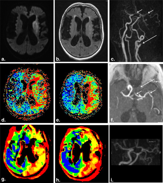
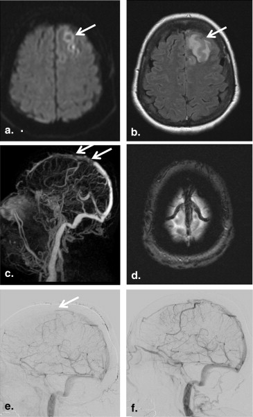
Evaluation of Intracranial and Extracranial Vessel Pathologies
Get Radiology Tree app to read full this article<
Get Radiology Tree app to read full this article<
Get Radiology Tree app to read full this article<
Get Radiology Tree app to read full this article<
Perfusion Analysis
Get Radiology Tree app to read full this article<
Get Radiology Tree app to read full this article<
Get Radiology Tree app to read full this article<
Get Radiology Tree app to read full this article<
Feasibility of an Ultrafast MR Protocol
Get Radiology Tree app to read full this article<
Get Radiology Tree app to read full this article<
Get Radiology Tree app to read full this article<
Get Radiology Tree app to read full this article<
Conclusions
Get Radiology Tree app to read full this article<
Get Radiology Tree app to read full this article<
Get Radiology Tree app to read full this article<
References
1. Latchaw R.E., Alberts M.J., Lev M.H., et. al.: Recommendations for imaging of acute ischemic stroke: a scientific statement from the American Heart Association. Stroke 2009; 40: pp. 3646-3678.
2. Roveri L., La Gioia S., Ghidinelli C., et. al.: Wake-up stroke within 3 hours of symptom awareness: imaging and clinical features compared to standard recombinant tissue plasminogen activator treated stroke. J Stroke Cerebrovasc Dis 2013; 22: pp. 703-708.
3. Thomalla G., Cheng B., Ebinger M., et. al.: DWI-FLAIR mismatch for the identification of patients with acute ischaemic stroke within 4.5 h of symptom onset (PRE-FLAIR): a multicentre observational study. Lancet Neurol 2011; 10: pp. 978-986.
4. Cho A.H., Sohn S.I., Han M.K., et. al.: Safety and efficacy of MRI-based thrombolysis in unclear-onset stroke. A preliminary report. Cerebrovasc Dis 2008; 25: pp. 572-579.
5. Castano C., Dorado L., Guerrero C., et. al.: Mechanical thrombectomy with the Solitaire AB device in large artery occlusions of the anterior circulation: a pilot study. Stroke 2010; 41: pp. 1836-1840.
6. Gralla J., Brekenfeld C., Mordasini P., et. al.: Mechanical thrombolysis and stenting in acute ischemic stroke. Stroke 2012; 43: pp. 280-285.
7. Lim R.P., Shapiro M., Wang E.Y., et. al.: 3D time-resolved MR angiography (MRA) of the carotid arteries with time-resolved imaging with stochastic trajectories: comparison with 3D contrast-enhanced Bolus-Chase MRA and 3D time-of-flight MRA. AJNR Am J Neuroradiol 2008; 29: pp. 1847-1854.
8. Forkert N.D., Kaesemann P., Treszl A., et. al.: Comparison of 10 TTP and Tmax estimation techniques for MR perfusion-diffusion mismatch quantification in acute stroke. AJNR Am J Neuroradiol 2013; 34: pp. 1697-1703.
9. Forkert N.D., Illies T., Moller D., et. al.: Analysis of the influence of 4D MR angiography temporal resolution on time-to-peak estimation error for different cerebral vessel structures. AJNR Am J Neuroradiol 2012; 33: pp. 2103-2109.
10. Ma H., Parsons M.W., Christensen S., et. al.: A multicentre, randomized, double-blinded, placebo-controlled Phase III study to investigate EXtending the time for Thrombolysis in Emergency Neurological Deficits (EXTEND). Int J Stroke 2012; 7: pp. 74-80.
11. Albers G.W., Thijs V.N., Wechsler L., et. al.: Magnetic resonance imaging profiles predict clinical response to early reperfusion: the diffusion and perfusion imaging evaluation for understanding stroke evolution (DEFUSE) study. Ann Neurol 2006; 60: pp. 508-517.
12. Adams H.P., Bendixen B.H., Kappelle L.J., et. al.: Classification of subtype of acute ischemic stroke. Definitions for use in a multicenter clinical trial. TOAST. Trial of Org 10172 in Acute Stroke Treatment. Stroke 1993; 24: pp. 35-41.
13. Serafin Z., Strzesniewski P., Lasek W., et. al.: Time-resolved imaging of contrast kinetics does not improve performance of follow-up MRA of embolized intracranial aneurysms. Med Sci Monit 2012; 18: pp. MT60-MT65.
14. Parmar H., Ivancevic M.K., Dudek N., et. al.: Neuroradiologic applications of dynamic MR angiography at 3 T. Magn Reson Imaging Clin N Am 2009; 17: pp. 63-75.
15. Parmar H., Ivancevic M.K., Dudek N., et. al.: Dynamic MRA with four-dimensional time-resolved angiography using keyhole at 3 tesla in head and neck vascular lesions. J Neuroophthalmol 2009; 29: pp. 119-127.
16. Wu Q., Li M.H.: A comparison of 4D time-resolved MRA with keyhole and 3D time-of-flight MRA at 3.0 T for the evaluation of cerebral aneurysms. BMC Neurol 2012; 12: pp. 50.
17. Lee Y.J., Laub G., Jung S.L., et. al.: Low-dose 3D time-resolved magnetic resonance angiography (MRA) of the supraaortic arteries: correlation with high spatial resolution 3D contrast-enhanced MRA. J Magn Reson Imaging 2011; 33: pp. 71-76.
18. Yigit H., Turan A., Ergun E., et. al.: Time-resolved MR angiography of the intracranial venous system: an alternative MR venography technique. Eur Radiol 2012; 22: pp. 980-989.
19. Schaafsma J.D., Velthuis B.K., Majoie C.B., et. al.: Intracranial aneurysms treated with coil placement: test characteristics of follow-up MR angiography–multicenter study. Radiology 2010; 256: pp. 209-218.
20. Forkert N.D., Fiehler J., Ries T., et. al.: Reference-based linear curve fitting for bolus arrival time estimation in 4D MRA and MR perfusion-weighted image sequences. Magn Reson Med 2011; 65: pp. 289-294.
21. Benner T., Heiland S., Erb G., et. al.: Accuracy of gamma-variate fits to concentration–time curves from dynamic susceptibility-contrast enhanced MRI: influence of time resolution, maximal signal drop and signal-to-noise. Magn Reson Imaging 1997; 15: pp. 307-317.
22. Lu D., Monahan W.G.: Effect of sample numbers on the kinetic data analysis of MR contrast agents. Magn Reson Med 1993; 30: pp. 131-134.
23. Zou Z., Ma L., Cheng L., et. al.: Time-resolved contrast-enhanced MR angiography of intracranial lesions. J Magn Reson Imaging 2008; 27: pp. 692-699.
24. Frolich A.M., Psychogios M.N., Klotz E., et. al.: Angiographic reconstructions from whole-brain perfusion CT for the detection of large vessel occlusion in acute stroke. Stroke 2012; 43: pp. 97-102.
25. Dani K.A., Thomas R.G., Chappell F.M., et. al.: Systematic review of perfusion imaging with computed tomography and magnetic resonance in acute ischemic stroke: heterogeneity of acquisition and postprocessing parameters: a translational medicine research collaboration multicentre acute stroke imaging study. Stroke 2012; 43: pp. 563-566.
26. Wintermark M., Albers G.W., Alexandrov A.V., et. al.: Acute stroke imaging research roadmap. AJNR Am J Neuroradiol 2008; 29: pp. e23-e30.
27. Khan R., Nael K., Erly W.: Acute stroke imaging: what clinicians need to know. Am J Med 2013; 126: pp. 379-386.
28. Aoki J., Kimura K., Iguchi Y., et. al.: FLAIR can estimate the onset time in acute ischemic stroke patients. J Neurol Sci 2010; 293: pp. 39-44.
29. Petkova M., Rodrigo S., Lamy C., et. al.: MR imaging helps predict time from symptom onset in patients with acute stroke: implications for patients with unknown onset time. Radiology 2010; 257: pp. 782-792.
30. Emeriau S., Serre I., Toubas O., et. al.: Can diffusion-weighted imaging-fluid-attenuated inversion recovery mismatch (positive diffusion-weighted imaging/negative fluid-attenuated inversion recovery) at 3 Tesla identify patients with stroke at <4.5 hours?. Stroke 2013; 44: pp. 1647-1651.
31. Serena J., Davalos A., Segura T., et. al.: Stroke on awakening: looking for a more rational management. Cerebrovasc Dis 2003; 16: pp. 128-133.
32. Neumann-Haefelin T., Wittsack H.J., Wenserski F., et. al.: Diffusion- and perfusion-weighted MRI. The DWI/PWI mismatch region in acute stroke. Stroke 1999; 30: pp. 1591-1597.
33. Prosser J., Butcher K., Allport L., et. al.: Clinical-diffusion mismatch predicts the putative penumbra with high specificity. Stroke 2005; 36: pp. 1700-1704.