
Rationale and Objectives
The aim of this study was to evaluate the improved accuracy of radiologic assessment of lung cancer afforded by computer-aided diagnosis (CADx).
Materials and Methods
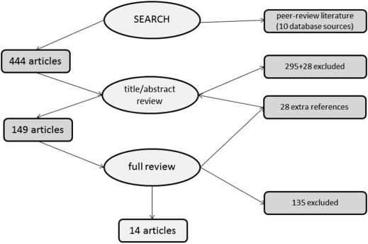
Inclusion/exclusion criteria were formulated, and a systematic inquiry of research databases was conducted. Following title and abstract review, an in-depth review of 149 surviving articles was performed with accepted articles undergoing a Quality Assessment of Diagnostic Accuracy Studies (QUADAS)-based quality review and data abstraction.
Results
A total of 14 articles, representing 1868 scans, passed the review. Increases in the receiver operating characteristic (ROC) area under the curve of .8 or higher were seen in all nine studies that reported it, except for one that employed subspecialized radiologists.
Conclusions
This systematic review demonstrated improved accuracy of lung cancer assessment using CADx over manual review, in eight high-quality observer-performance studies. The improved accuracy afforded by radiologic lung-CADx suggests the need to explore its use in screening and regular clinical workflow.
Introduction
Lung cancer is the second leading cause of death in the United States and among the top 10 worldwide. More Americans die each year from lung cancer than from breast, prostate, and colorectal cancers combined. Annually, lung cancer kills more men than prostate cancer and more women than breast cancer .
Whereas overall cancer incidence rates are declining, lung cancer incidence rates among women are rising. Between 1960 and 1990, deaths from lung cancer among women increased over 400%. It is the second most common cancer among African American men and kills more African Americans than any other cancer. Five-year survival ranges from 70% for stage I disease to less than 5% for stage IV disease. As of 2014, overall 5-year survival is 17%, with only 15% diagnosed at the localized stage .
Get Radiology Tree app to read full this article<
Get Radiology Tree app to read full this article<
Get Radiology Tree app to read full this article<
Get Radiology Tree app to read full this article<
Get Radiology Tree app to read full this article<
Get Radiology Tree app to read full this article<
Get Radiology Tree app to read full this article<
Get Radiology Tree app to read full this article<
Get Radiology Tree app to read full this article<
Materials and Methods
Get Radiology Tree app to read full this article<
Get Radiology Tree app to read full this article<
Get Radiology Tree app to read full this article<
Get Radiology Tree app to read full this article<
Computer-aided (Detection OR Diagnosis) AND (“Lung Neoplasms”[MeSH] OR Lung Nodule) AND (“Radiography”[MeSH] OR “Tomography, X-Ray Computed”[MeSH]) NOT review[PT]
Get Radiology Tree app to read full this article<
Get Radiology Tree app to read full this article<
Get Radiology Tree app to read full this article<
Get Radiology Tree app to read full this article<
Results
Get Radiology Tree app to read full this article<
Get Radiology Tree app to read full this article<
Get Radiology Tree app to read full this article<
Table 1
Description of Lung-CADx Studies
Study/Year Population Type Gold Standard QS # R # SCANS Mode/ST ALG 1 /2003 CA/cont UCAD path 8 N/A 393 LDCT/10 mm LDA 2 /2004 CA only UCAD path 12 N/A 106 LDCT/10 mm MTANN 3 /2005 CA/cont OP path 15 14 27 LDCT/10 mm MTANN 4 /2005 CA/cont OP path 15 16 56 HRCT MTANN 5 /2005 CA/cont OP path 16 8 28 LDCT/3 mm DT 6 /2005 CA/cont UCAD path 10 N/A 81 LDCT/3 mm LDA 7 /2005 CA/cont UCAD path 11 N/A 415 LDCT/10 mm MTANN 8 /2006 CA/cont OP path 18 9 48 CXR LDA 9 /2006 CA/cont OP path 17 10 33 HRCT ANN 10 /2007 CA/cont OP path 18 9 200 LDCT/8 mm ANN 11 /2009 CA only UCAD path 9 N/A 69 LDCT/10 mm MTANN 12 /2010 CA/cont OP path 16 11 60 LDCT/10 mm LDA 13 /2010 CA/cont OP path + hx 15 6 152 LDCT/2 mm LDA 14 /2012 CA/cont OP path + CT 15 10 200 CXR LDA
ALG, algorithm; ANN, artificial neural network; CA/cont, cancer control; CADx, computer-aided diagnosis; CXR, chest radiography; DT, Decision Tree; LDA, linear discriminant analysis; LDCT, low-dose computed tomography; HRCT, high-resolution CT; MT, massive training; MTANN, massive training artificial neural network; OP, observer-performance; QS, Quality Assessment of Diagnostic Accuracy Studies Score; #R, number of raters; SCANS, number of scans total in the study; ST, slice thickness; UCAD, unsupervised computer diagnosis; PATH, pathology review of biopsy; hx, history; N/A, not applicable.
Get Radiology Tree app to read full this article<
Get Radiology Tree app to read full this article<
Get Radiology Tree app to read full this article<
Get Radiology Tree app to read full this article<
Table 2
Lung-CADx Accuracy Measures
Study Sensitivity/CADx Alone FP/Scan ACC A Z Human A Z Machine A Z Both Δ_P_ Value 1 .84 1.0 NR N/A .79 N/A N/A N/A 2 .83 5.8 NR N/A NR N/A N/A N/A 3 .87 3.0 NR .763 NR .854 .091 .002 4 .90 6.5 NR .785 .831 .853 .068 .016 5 .91 (.67 sp) NR 81% .68 NR .81 .13 .020 6 NR NR NR N/A .92 N/A N/A N/A 7 1.00 (.48 sp) NR NR N/A .882 N/A N/A N/A 8 .81 (.70 sp) 1.2 NR .724 NR .778 .054 .008 9 .72 NR 76% .910 .795 .944 .034 .190 10 .93 NR 93% .85 NR .94 .09 .014 11 .84 .5 NR N/A NR N/A N/A N/A 12 NR NR NR .864 NR .924 .060 .010 13 NR NR NR .833 NR .853 .020 .010 14 .87 1.9 NR N/A NR N/A N/A N/A
ACC, machine accuracy calculation (TP + TN)/(P + N), true-positive, true-negative, total-positive, total-negative; A z , receiver operating characteristic-area index; CADx, computer-aided diagnosis; FP, false-positive; NR, not reported; sp, specificity; Δ, difference.
Get Radiology Tree app to read full this article<
Get Radiology Tree app to read full this article<
Discussion
Get Radiology Tree app to read full this article<
Get Radiology Tree app to read full this article<
Get Radiology Tree app to read full this article<
Get Radiology Tree app to read full this article<
Get Radiology Tree app to read full this article<
Get Radiology Tree app to read full this article<
Conclusion
Get Radiology Tree app to read full this article<
Get Radiology Tree app to read full this article<
Acknowledgement
Get Radiology Tree app to read full this article<
References
1. Fry W.A., Menck H.R., Winchester D.P.: The National Cancer Data Base report on lung cancer. Cancer 1996; 77: pp. 1947-1955.
2. American Cancer Society’s 2014 Cancer Facts & Figures Annual Report. Available at: http://www.cancer.org/Research/CancerFactsStatistics/CancerFactsFigures2014/cancer-facts-and-figures-2014.pdf Accessed August 2014
3. Flehinger B.J., Kimmel M., Melamed M.R.: The effect of surgical treatment on survival from early lung cancer. Implications for screening. Chest 1992; 101: pp. 1013-1018.
4. National Lung Screening Trial Research Team, Aberle D.R., Adams A.M., et. al.: Reduced lung-cancer mortality with low-dose computed tomographic screening. N Engl J Med 2011; 365: pp. 395-409.
5. de Hoop B., Schaefer-Prokop C., Gietema H.A., et. al.: Screening for lung cancer with digital chest radiography: sensitivity and number of secondary work-up CT examinations. Radiology 2010; 255: pp. 629-637.
6. McLeod N., Montane G.: The radiologist assistant: the solution to radiology workforce needs. Emerg Radiol 2010; 17: pp. 253-256.
7. Swensen S.J., Viggiano R.W., Midthun D.E., et. al.: Lung nodule enhancement at CT: multicenter study. Radiology 2000; 214: pp. 73-80.
8. van Tulder M., Furlan A., Bombardier C., et. al.: Updated method guidelines for systematic reviews in the Cochrane collaboration back review group. Spine 2003; 28: pp. 1290-1299.
9. Whiting P., Rutjes A.W., Reitsma J.B., et. al.: The development of QUADAS: a tool for the quality assessment of studies of diagnostic accuracy included in systematic reviews. BMC Med Res Methodol 2003; 3: pp. 25.
10. Armato S.G., Altman M.B., Wilkie J., et. al.: Automated lung nodule classification following automated nodule detection on CT: a serial approach. Med Phys 2003; 30: pp. 1188-1197.
11. Arimura H., Katsuragawa S., Suzuki K., et. al.: Computerized scheme for automated detection of lung nodules in low-dose computed tomography images for lung cancer screening. Acad Radiol 2004; 11: pp. 617-629.
12. Li F., Arimura H., Suzuki K., et. al.: Computer-aided detection of peripheral lung cancers missed at CT: ROC analyses w/without localization. Radiology 2005; 237: pp. 684-690.
13. Li Q., Li F., Suzuki K., et. al.: Computer-aided diagnosis in thoracic CT. Semin Ultrasound CT MR 2005; 26: pp. 357-363.
14. Shah S.K., McNitt-Gray M.F., De Zoysa K.R., et. al.: Solitary pulmonary nodule diagnosis on CT: results of an observer study. Acad Radiol 2005; 12: pp. 496-501.
15. Shah S.K., McNitt-Gray M.F., Rogers S.R., et. al.: Computer-aided diagnosis of the solitary pulmonary nodule. Acad Radiol 2005; 12: pp. 570-575.
16. Suzuki K., Li F., Sone S., et. al.: Computer-aided diagnostic scheme for distinction between benign and malignant nodules in thoracic low-dose CT by use of massive training artificial neural network. IEEE Trans Med Imaging 2005; 24: pp. 1138-1150.
17. Shiraishi J., Abe H., Li F., et. al.: Computer-aided diagnosis for the detection and classification of lung cancers on chest radiographs: ROC analysis of radiologists’ performance. Acad Radiol 2006; 13: pp. 995-1003.
18. Awai K., Murao K., Ozawa A., et. al.: Pulmonary nodules: estimation of malignancy at thin-section helical CT—effect of computer-aided diagnosis on performance of radiologists. Radiology 2006; 239: pp. 276-284.
19. Chen H., Wang X.H., Ma D.Q., et. al.: Neural network-based computer-aided diagnosis in distinguishing malignant from benign solitary pulmonary nodules by computed tomography. Chin Med J 2007; 120: pp. 1211-1215.
20. Suzuki K.: A supervised “lesion-enhancement” filter by use of a massive-training artificial neural network (MTANN) in computer-aided diagnosis (CAD). Phys Med Biol 2009; 54: pp. S31-S45.
21. Kusano S., Nakagawa T., Aoki T., et. al.: Efficacy of computer-aided diagnosis in lung cancer screening with low-dose spiral computed tomography: receiver operating characteristic analysis of radiologists’ performance. Jpn J Radiol 2010; 28: pp. 649-655.
22. Way T., Chan H.P., Hadjiiski L., et. al.: Computer-aided diagnosis of lung nodules on CT scans: ROC study of its effect on radiologists’ performance. Acad Radiol 2010; 17: pp. 323-332.
23. Lee K.H., Goo J.M., Park C.M., et. al.: Computer-aided detection of malignant lung nodules on chest radiographs: effect on observers’ performance. Korean J Radiol 2012; 13: pp. 564-571.
24. Chan H.P., Hadjiiski L., Zhou C., et. al.: Computer-aided diagnosis of lung cancer and pulmonary embolism in computed tomography—a review. Acad Radiol 2008; 15: pp. 535-555.
25. Eadie L.H., Taylor P., Gibson A.P.: A systematic review of computer-assisted diagnosis in diagnostic cancer imaging. Eur J Radiol 2012; 81: pp. e70-e76.
26. Freer T.W., Ulissey M.J.: Screening mammography with computer-aided detection: prospective study of 12,860 patients in a community breast center. Radiology 2001; 220: pp. 781-786.
27. Warren Burhenne L.J., Wood S.A., D’Orsi C.J., et. al.: Potential contribution of computer aided detection to the sensitivity of screening mammography. Radiology 2000; 215: pp. 554-562.
28. Phillips M., Cataneo R.N., Cummin A.R., et. al.: Detection of lung cancer with volatile markers in the breath. Chest 2003; 123: pp. 2115-2123.
29. Yu J.K., Zheng S., Tang Y., et. al.: An integrated approach utilizing proteomics and bioinformatics to detect ovarian cancer. J Zhejiang Univ Sci B 2005; 6: pp. 227-231.