
Rationale and Objectives
Carpal tunnel syndrome (CTS) is the most common peripheral nerve entrapment syndrome. Strong pinch or grip with wrist flexion has been considered a risk factor for CTS. Studying median nerve displacement during wrist movements may provide useful information about median nerve kinematic changes in patients with CTS. The purpose of this study was to evaluate the deformability and mobility of the median nerve in patients with CTS compared to healthy subjects.
Materials and Methods
Dynamic ultrasound images were obtained in 20 affected wrists of 13 patients with CTS. Results were compared to complementary data obtained from both wrists of 10 healthy subjects reported in a previous study. Shape and position of initial and final median nerve were measured and analyzed for six defined wrist movements. The deformation ratios for each movement were defined as the median nerve area, perimeter, and circularity of the final position normalized by respective values assessed in the initial position. The median nerve displacement vector and magnitude were also calculated.
Results
The deformation ratio for circularity was significantly less in patients with CTS compared to healthy subjects during wrist flexion ( P < .05). The mean vector of median nerve displacement during wrist flexion was significantly different between patients with CTS and healthy subjects ( P < .05). The displacement magnitude of the median nerve was found to be less in patients with CTS compared to healthy subjects during most movements, with the exception of wrist extension with fingers extended.
Conclusions
Patients with CTS differ from normal subjects with regard to mobility and deformability of the median nerve.
Carpal tunnel syndrome (CTS) is the most common peripheral nerve entrapment syndrome, and its precise etiology remains largely unclear. Like other peripheral nerves, the median nerve is exposed to various mechanical stresses related to limb postures and movements. In the case of the median nerve in the carpal tunnel, the relevant movements are of the fingers and wrist. The median nerve response to these stresses is a combination of displacement and deformation . Both displacement and deformation are known to vary from normal in patients with CTS, because of increased carpal tunnel pressure, and due to fibrosis in the median nerve and the surrounding tissue in the carpal tunnel .
Actuated by finger or wrist motion, the median nerve can displace in three dimensions. Longitudinal sliding of the median nerve in the carpal tunnel has been observed both in vitro and in vivo . Reduced longitudinal excursion of the median nerve at the carpal tunnel has been identified in patients with CTS . Transverse motion of the median nerve has also been studied in the carpal tunnel in normal subjects and in patients with CTS . Nakamichi et al. found reduced transverse motion of the median nerve during passive flexion and extension of the index finger in patients with CTS. Erel et al. studied the transverse motion of the median nerve during passive extension of the digits at the metacarpophalangeal joint from 90° flexion to neutral. Their results showed that in patients with CTS there was a significant reduction in transverse movement on the more symptomatic side compared to the contralateral.
Get Radiology Tree app to read full this article<
Get Radiology Tree app to read full this article<
Get Radiology Tree app to read full this article<
Materials and methods
Get Radiology Tree app to read full this article<
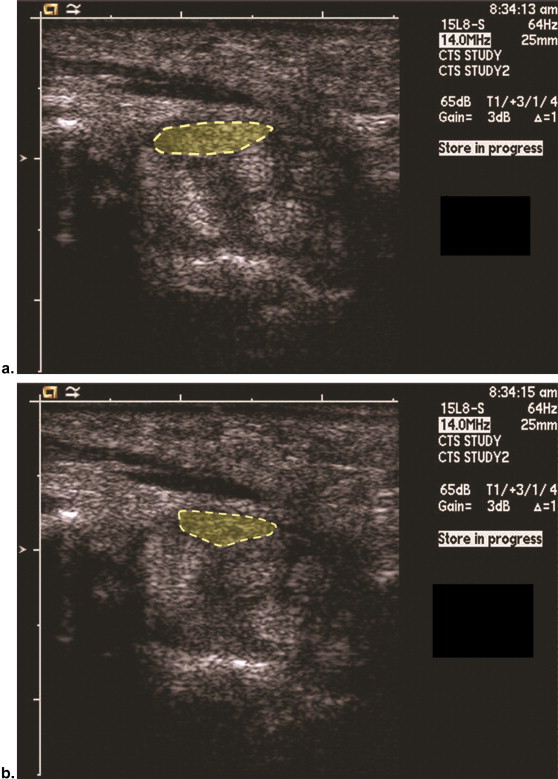
Ultrasound Examination
Get Radiology Tree app to read full this article<
Get Radiology Tree app to read full this article<
Get Radiology Tree app to read full this article<
Get Radiology Tree app to read full this article<
Get Radiology Tree app to read full this article<
Image Analysis
Get Radiology Tree app to read full this article<
circularity=4πareaperimeter2 circularity
=
4
π
area
perimeter
2
Get Radiology Tree app to read full this article<
Get Radiology Tree app to read full this article<
Get Radiology Tree app to read full this article<
Get Radiology Tree app to read full this article<
r=x2+y2−−−−−−√ r
=
x
2
+
y
2
Get Radiology Tree app to read full this article<
Get Radiology Tree app to read full this article<
Normalization of Measurements
Get Radiology Tree app to read full this article<
Statistical Analysis
Get Radiology Tree app to read full this article<
Results
Maximal Wrist Angles during Wrist Movements
Get Radiology Tree app to read full this article<
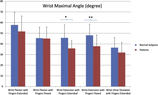
Get Radiology Tree app to read full this article<
Deformation Ratio of the Median Nerve
Get Radiology Tree app to read full this article<
Table 1
Median Nerve Deformation Parameters
Wrist Motions Area Deformation Ratio Perimeter Deformation Ratio Circularity Deformation Ratio Finger flexion Patients 1.00 ± 0.16 0.98 ± 0.08 1.05 ± 0.13 Healthy subjects 0.99 ± 0.12 0.98 ± 0.09 1.09 ± 0.15 Wrist flexion with fingers extended Patients 0.93 ± 0.20 0.87 ± 0.16 1.23 ± 0.16 † Healthy subjects 1.06 ± 0.24 0.86 ± 0.16 1.49 ± 0.38 Wrist flexion with fingers flexed Patients 0.98 ± 0.21 0.89 ± 0.13 ∗ 1.25 ± 0.28 † Healthy subjects 0.94 ± 0.22 0.79 ± 0.15 1.57 ± 0.36 Wrist extension with fingers extended Patients 1.01 ± 0.20 1.02 ± 0.14 0.99 ± 0.20 Healthy subjects 1.03 ± 0.15 0.97 ± 0.11 1.12 ± 0.19 Wrist extension with fingers flexed Patients 0.97 ± 0.20 1.0 ± 0.12 0.99 ± 0.20 Healthy subjects 0.98 ± 0.15 0.98 ± 0.12 1.05 ± 0.22 Wrist ulnar deviation with fingers extended Patients 0.97 ± 0.20 0.90 ± 0.12 1.19 ± 0.16 † Healthy subjects 1.02 ± 0.27 0.93 ± 0.20 1.53 ± 0.32
Get Radiology Tree app to read full this article<
Get Radiology Tree app to read full this article<
Get Radiology Tree app to read full this article<
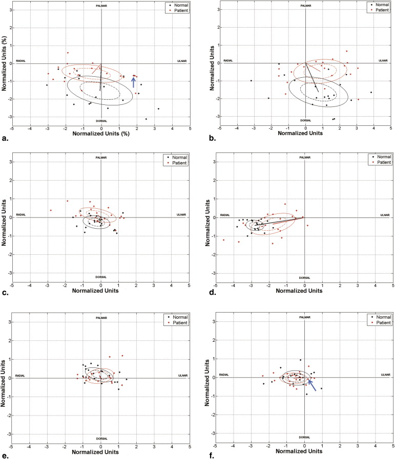
Displacement Vector of the Median Nerve
Get Radiology Tree app to read full this article<
Table 2
Median Nerve Displacement Mean Vector and Confidence Limits in Patients with Carpal Tunnel Syndrome and Healthy Subjects
Mean ρ (NU) Mean θ a (NU) b (NU) ψ Finger flexion Patients 0.1 28.9° 0.5 0.2 9.1° Healthy subjects 0.2 117.3° 0.5 0.2 −16.0° Wrist flexion with fingers extended Patients 0.8 137.3° † 1.0 0.3 −6.2° Healthy subjects 1.5 267.1° 1.2 0.5 −11.9° Wrist flexion with fingers flexed Patients 1.0 331.4° † 1.0 0.4 4.5° Healthy subjects 1.8 295.0° 1.1 0.5 −11.7° Wrist extension with fingers extended Patients 0.2 144.4° ∗ 0.7 0.2 −12° Healthy subjects 0.4 225.6° 0.5 0.2 −7.8° Wrist extension with fingers flexed Patients 0.6 184.4° 0.5 0.2 −0.7° Healthy subjects 0.5 182.0° 0.5 0.2 −2.5° Wrist ulnar deviation with fingers extended Patients 1.8 190.9° † 0.8 0.3 14.6° Healthy subjects 2.8 188.5° 0.3 0.2 −5.2°
ρ = vector length; θ = vector angle; a = major ellipse axes for variation of the confidence limits; b = minor ellipse axes for variation of the confidence limits; ψ = inclination of the ellipse.
One normalized unit (NU) is ∼1.8 mm.
Get Radiology Tree app to read full this article<
Get Radiology Tree app to read full this article<
Get Radiology Tree app to read full this article<
Get Radiology Tree app to read full this article<
Get Radiology Tree app to read full this article<
Normalized Magnitude of the Median Nerve Displacement
Get Radiology Tree app to read full this article<
Table 3
Median Nerve Displacement Normalized Magnitudes
Magnitude of Median Nerve Displacement (NU) Finger flexion Patients 0.75 ± 0.44 Healthy subjects 0.82 ± 0.33 Wrist flexion with fingers extended Patients 1.74 ± 0.78 ∗ Healthy subjects 2.36 ± 0.79 Wrist flexion with fingers flexed Patients 1.71 ± 0.90 † Healthy subjects 2.46 ± 0.84 Wrist extension with fingers extended Patients 0.90 ± 0.68 Healthy subjects 0.77 ± 0.46 Wrist extension with fingers flexed Patients 0.85 ± 0.56 Healthy subjects 0.81 ± 0.58 Wrist ulnar deviation with fingers extended Patients 1.93 ± 1.23 † Healthy subjects 2.86 ± 0.51
Get Radiology Tree app to read full this article<
Get Radiology Tree app to read full this article<
Get Radiology Tree app to read full this article<
Discussion
Get Radiology Tree app to read full this article<
Get Radiology Tree app to read full this article<
Get Radiology Tree app to read full this article<
Get Radiology Tree app to read full this article<
Get Radiology Tree app to read full this article<
Get Radiology Tree app to read full this article<
Get Radiology Tree app to read full this article<
Acknowledgments
Get Radiology Tree app to read full this article<
References
1. Nakamichi K., Tachibana S.: Restricted motion of the median nerve in carpal tunnel syndrome. J Hand Surg Br 1995; 20: pp. 460-464.
2. Ikeda K., Sarnura N., Tornita K.: Segmental carpal canal pressure in patients with carpal tunnel syndrome. J Hand Surg Am 2006; 31: pp. 925-929.
3. Werner C.O., Elmqvist D., Ohlin P.: Pressure and nerve lesion in the carpal tunnel. Acta Orthop Scand 1983; 54: pp. 312-316.
4. Coppieters M.W., Alshami A.M.: Longitudinal excursion and strain in the median nerve during novel nerve gliding exercises for carpal tunnel syndrome. J Orthop Res 2007; 25: pp. 972-980.
5. Hough A.D., Moore A.P., Jones M.P.: Reduced longitudinal excursion of the median nerve in carpal tunnel syndrome. Arch Phys Med Rehabil 2007; 88: pp. 569-576.
6. Szabo R.M., Bay B.K., Sharkey N.A., et. al.: Median nerve displacement through the carpal canal. J Hand Surg Am 1994; 19: pp. 901-906.
7. Tuzuner S., Ozkaynak S., Acikbas C., et. al.: Median nerve excursion during endoscopic carpal tunnel release. Neurosurgery 2004; 54: pp. 1155-1160. discussion 60-1
8. Wright T.W., Glowczewskie F., Wheeler D., et. al.: Excursion and strain of the median nerve. J Bone Joint Surg Am 1996; 78: pp. 1897-1903.
9. Yamaguchi T., Osamura N., Zhao C.F., et. al.: Relative longitudinal motion of the finger flexors, subsynovial connective tissue, and median nerve before and after carpal tunnel release in a human cadaver model. J Hand Surg Am 2008; 33: pp. 888-892.
10. Korstanje J.W., Scheltens-De Boer M., Blok J.H., et. al.: Ultrasonographic assessment of longitudinal median nerve and hand flexor tendon dynamics in carpal tunnel syndrome. Muscle Nerve 2012; 45: pp. 721-729.
11. van Doesburg M.H., Yoshii Y., Villarraga H.R., et. al.: Median nerve deformation and displacement in the carpal tunnel during index finger and thumb motion. J Orthop Res 2010; 28: pp. 1387-1390.
12. Yoshii Y., Villarraga H.R., Henderson J., et. al.: Ultrasound assessment of the displacement and deformation of the median nerve in the human carpal tunnel with active finger motion. J Bone Joint Surg Am 2009; 91: pp. 2922-2930.
13. Erel E., Dilley A., Greening J., et. al.: Longitudinal sliding of the median nerve in patients with carpal tunnel syndrome. J Hand Surg Br 2003; 28: pp. 439-443.
14. de Krom M.C., Kester A.D., Knipschild P.G., et. al.: Risk factors for carpal tunnel syndrome. Am J Epidemiol 1990; 132: pp. 1102-1110.
15. Keir P.J., Wells R.P., Ranney D.A., et. al.: The effects of tendon load and posture on carpal tunnel pressure. J Hand Surg Am 1997; 22: pp. 628-634.
16. Rempel D.M., Keir P.J., Bach J.M.: Effect of wrist posture on carpal tunnel pressure while typing. J Orthop Res 2008; 26: pp. 1269-1273.
17. Ko C., Brown T.D.: A fluid-immersed multi-body contact finite element formulation for median nerve stress in the carpal tunnel. Comput Methods Biomech Biomed Engin 2007; 10: pp. 343-349.
18. Smith E.M., Sonstegard D.A., Anderson W.H.: Carpal tunnel syndrome: contribution of flexor tendons. Arch Phys Med Rehabil 1977; 58: pp. 379-385.
19. Yoshii Y.C., Zhao C., Zhao K.D., et. al.: The effect of wrist position on the relative motion of tendon, nerve, and subsynovial connective tissue within the carpal tunnel in a human cadaver model. J Orthop Res 2008; 26: pp. 1153-1158.
20. Zhao C.F., Ettema A.M., Samura N., et. al.: Gliding characteristics between flexor tendons and surrounding tissues in the carpal tunnel: A biomechanical cadaver study. J Orthop Res 2007; 25: pp. 185-190.
21. Allmann K.H., Horch R., Uhl M., et. al.: MR imaging of the carpal tunnel. Eur J Radiol 1997; 25: pp. 141-145.
22. Goetz J.E., Thedens D.R., Kunze N.M., et. al.: Day-to-day variability of median nerve location within the carpal tunnel. Clin Biomech 2010; 25: pp. 660-665.
23. Wang Y., Zhao C., Passe S.M., et. al.: Quantitative transverse ultrasound to analyze the median nerve deformation and displacement in the human carpal tunnel during wrist movements. Ultrasound Med Biol 2013; doi:pii: S0301–5629(13)01017-X. 10.1016/j.ultrasmedbio.2013.09.009
24. Zeiss J., Skie M., Ebraheim N., et. al.: Anatomic relations between the median nerve and flexor tendons in the carpal tunnel: MR evaluation in normal volunteers. AMR Am J Roentgenol 1989; 153: pp. 533-536.
25. Practice parameter for carpal tunnel syndrome (summary statement). Report of the Quality Standards Subcommittee of the American Academy of Neurology. 1993/11/01 ed. City1993; 2406–2409.
26. Duncan I., Sullivan P., Lomas F.: Sonography in the diagnosis of carpal tunnel syndrome. AJR Am J Roentgenol 1999; 173: pp. 681-684.
27. Clerke A.M., Clerke J.P., Adams R.D.: Effects of hand shape on maximal isometric grip strength and its reliability in teenagers. J Hand Ther 2005; 18: pp. 19-29.
28. Batschelet E.: Circular statistics in biology.1981.Academic PressWaltham, MA, USA
29. Harber P., Bloswick D., Beck J., et. al.: Supermarket checker motions and cumulative trauma risk. J Occup Med 1993; 35: pp. 805-811.
30. Martin S.: Carpal tunnel syndrome: a job-related risk. Am Pharm 1991; NS31: pp. 21-24.
31. Greening J., Lynn B., Leary R., et. al.: The use of ultrasound imaging to demonstrate reduced movement of the median nerve during wrist flexion in patients with non-specific arm pain. J Hand Surg Br 2001; 26: pp. 401-406.
32. Greening J., Smart S., Leary R., et. al.: Reduced movement of median nerve in carpal tunnel during wrist flexion in patients with non-specific arm pain. Lancet 1999; 354: pp. 217-218.
33. van Doesburg M.H., Henderson J., Yoshii Y., et. al.: Median nerve deformation in differential finger motions: ultrasonographic comparison of carpal tunnel syndrome patients and healthy controls. J Orthop Res 2012; 30: pp. 643-648.
34. Skie M., Zeiss J., Ebraheim N.A., et. al.: Carpal tunnel changes and median nerve compression during wrist flexion and extension seen by magnetic resonance imaging. J Hand Surg Am 1990; 15: pp. 934-939.
35. Gamble H.J., Eames R.A.: An electron microscope study of the connective tissues of human peripheral nerve. J Anat 1964; 98: pp. 655-663.
36. Geuna S., Raimondo S., Ronchi G., et. al.: Chapter 3: Histology of the peripheral nerve and changes occurring during nerve regeneration. Int Rev Neurobiol 2009; 87: pp. 27-46.
37. Clark B.D., Barr A.E., Safadi F.F., et. al.: Median nerve trauma in a rat model of work-related musculoskeletal disorder. J Neurotrauma 2003; 20: pp. 681-695.
38. O’Brien J.P., Mackinnon S.E., MacLean A.R., et. al.: A model of chronic nerve compression in the rat. Ann Plast Surg 1987; 19: pp. 430-435.
39. Orman G., Ozben S., Huseyinoglu N., et. al.: Ultrasound elastographic evaluation in the diagnosis of carpal tunnel syndrome: initial findings. Ultrasound Med Biol 2013; 39: pp. 1184-1189.
40. Ettema A.M., Amadio P.C., Zhao C., et. al.: A histological and immunohistochemical study of the subsynovial connective tissue in idiopathic carpal tunnel syndrome. J Bone Joint Surg Am 2004; 86-A: pp. 1458-1466.
41. Lluch A.L.: Thickening of the synovium of the digital flexor tendons: cause or consequence of the carpal tunnel syndrome?. J Hand Surg Br 1992; 17: pp. 209-212.
42. Oh J., Zhao C., Zobitz M.E., et. al.: Morphological changes of collagen fibrils in the subsynovial connective tissue in carpal tunnel syndrome. J Bone Joint Surg Am 2006; 88: pp. 824-831.
43. Visser L.H., Smidt M.H., Lee M.L.: High-resolution sonography versus EMG in the diagnosis of carpal tunnel syndrome. J Neurol Neurosurg Psychiatry 2008; 79: pp. 63-67.
44. Wong S.M., Griffith J.F., Hui A.C., et. al.: Carpal tunnel syndrome: diagnostic usefulness of sonography. Radiology 2004; 232: pp. 93-99.
45. Horch R.E., Allmann K.H., Laubenberger J., et. al.: Median nerve compression can be detected by magnetic resonance imaging of the carpal tunnel. Neurosurgery 1997; 41: pp. 76-82.
46. Yoshioka S., Okuda Y., Tamai K., et. al.: Changes in carpal tunnel shape during wrist joint motion. MRI evaluation of normal volunteers. J Hand Surg Br 1993; 18: pp. 620-623.
47. Keir P.J., Wells R.P.: Changes in geometry of the finger flexor tendons in the carpal tunnel with wrist posture and tendon load: an MRI study on normal wrists. Clin Biomech 1999; 14: pp. 635-645.