
Medical eponyms are ubiquitous, numerous, and at times controversial. They are often useful for succinctly conveying complex concepts, and familiarity with eponyms is important for proper usage and appropriate communication. In this historical review, we identify 18 anatomic eponyms used to describe structures of the brain, cerebral vasculature, and calvarium. For each structure, we first offer a biographical sketch of the individual for whom the structure is named. This is followed by a description of the anatomic structure and a brief discussion of its clinical relevance.
Introduction
During the interpretation of a cross-sectional study of the head, the radiologist evaluates dozens of eponymous neuroanatomic structures, while possibly knowing nothing about the individuals for whom these structures are named. As our understanding of neuroanatomy and our ability to accurately depict it with imaging continue to improve with the passage of time, we become farther removed from and often less familiar with the lives of the historic individuals whose past work forms the foundation of modern medical science. Eponyms are a means of honoring some of these individuals, many of whom led fascinating lives.
It must be acknowledged that eponyms can be a source of confusion, may lack scientific accuracy, often do not reflect the group effort of scientific discovery, and may inappropriately laud unethical or criminal behavior . However, eponyms are extremely commonly used as a means to succinctly convey complex concepts, and eponyms add color to what can at times be a dry science. Therefore, familiarity with eponyms is important for proper usage and appropriate communication.
Previous works published in the radiology literature have covered cardiothoracic , gastrointestinal , musculoskeletal , and vascular eponyms. However, little has been published related to eponyms specific to neuroradiology , and no previous article specifically intended for radiologists has focused on eponymous neuroanatomic structures.
In this historical review, we identify 18 anatomic eponyms used to describe structures of the brain, cerebral vasculature, and calvarium. For each structure, we first offer a biographical sketch of the individual for whom the structure is named. This is followed by a description of the anatomic structure and a brief discussion of its clinical relevance.
Brain
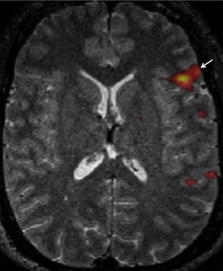
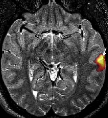
Broca’s Area
Paul Broca (1824–1880) was born near Bordeaux, France and was the son of a former surgeon in Napoleon’s army . Broca obtained his medical degree in Paris in 1848 . Over the course of his career, Broca became involved in the heated academic debate of whether the cerebral hemispheres function as an indivisible unit or whether they have specialized parts . In 1861, Broca assumed care of a patient named Leborgne, who suffered from cellulitis and gangrene and for 20 years produced only the single repetitive syllable “tan” . Broca saw Leborgne’s predicament as a test case for the question of language localization and, upon Leborgne’s death, Broca performed an autopsy demonstrating a lesion of the left frontal lobe third convolution . Broca subsequently demonstrated via at least seven more autopsies of patients with conditions similar to Leborgne’s that expressive aphasia consistently correlated with lesions affecting the posterior inferior left frontal gyrus . Broca married the daughter of Dr. Jean Guillaume Auguste Lugol , who was himself famous for his iodine solution. Broca died suddenly at age 56 of what is presumed to have been a myocardial infarction .
Get Radiology Tree app to read full this article<
Get Radiology Tree app to read full this article<
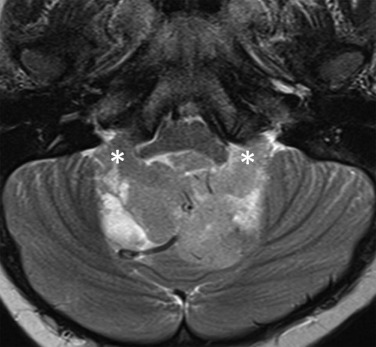
Foramina of Luschka and Magendie
Get Radiology Tree app to read full this article<
Get Radiology Tree app to read full this article<
Get Radiology Tree app to read full this article<
Get Radiology Tree app to read full this article<
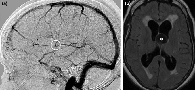
Foramina of Monro
Get Radiology Tree app to read full this article<
Get Radiology Tree app to read full this article<
Get Radiology Tree app to read full this article<
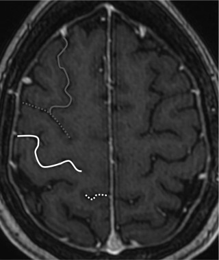
Rolandic Fissure
Get Radiology Tree app to read full this article<
Get Radiology Tree app to read full this article<
Get Radiology Tree app to read full this article<
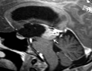
Sylvian Aqueduct
Get Radiology Tree app to read full this article<
Get Radiology Tree app to read full this article<
Get Radiology Tree app to read full this article<
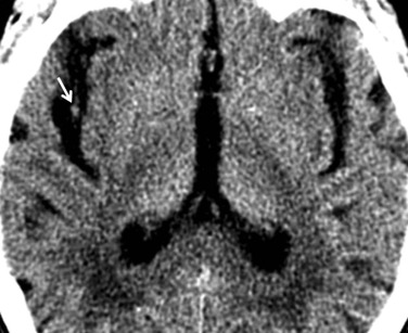
Sylvian Fissure
Get Radiology Tree app to read full this article<
Get Radiology Tree app to read full this article<
Get Radiology Tree app to read full this article<
Virchow-Robin Spaces
Get Radiology Tree app to read full this article<
Get Radiology Tree app to read full this article<
Get Radiology Tree app to read full this article<
Wernicke’s Area
Get Radiology Tree app to read full this article<
Get Radiology Tree app to read full this article<
Get Radiology Tree app to read full this article<
Cerebral Vasculature
Vein of Galen
Get Radiology Tree app to read full this article<
Get Radiology Tree app to read full this article<
Get Radiology Tree app to read full this article<
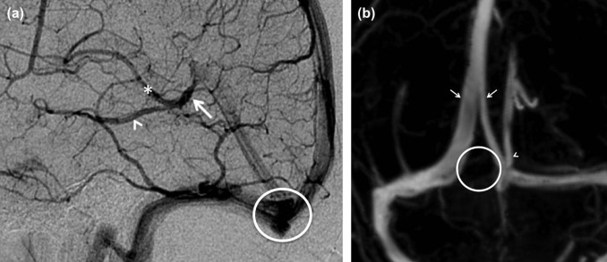
Torcular Herophili
Get Radiology Tree app to read full this article<
Get Radiology Tree app to read full this article<
Artery of Heubner
Get Radiology Tree app to read full this article<
Get Radiology Tree app to read full this article<
Get Radiology Tree app to read full this article<
Vein of Labbé
Get Radiology Tree app to read full this article<
Get Radiology Tree app to read full this article<
Get Radiology Tree app to read full this article<
Artery of Percheron
Get Radiology Tree app to read full this article<
Get Radiology Tree app to read full this article<

Get Radiology Tree app to read full this article<
Basal Vein of Rosenthal
Get Radiology Tree app to read full this article<
Get Radiology Tree app to read full this article<
Vein of Trolard
Get Radiology Tree app to read full this article<
Get Radiology Tree app to read full this article<
Circle of Willis
Get Radiology Tree app to read full this article<
Get Radiology Tree app to read full this article<
Calvarium
Wormian Bones
Get Radiology Tree app to read full this article<
Get Radiology Tree app to read full this article<
Get Radiology Tree app to read full this article<
Conclusion
Get Radiology Tree app to read full this article<
Get Radiology Tree app to read full this article<
References
1. Woywodt A., Matteson E.: Should eponyms be abandoned? Yes. BMJ 2007; 335: pp. 424.
2. Whitworth J.A.: Should eponyms be abandoned? No. BMJ 2007; 335: pp. 425.
3. Harper P.S.: Naming of syndromes and unethical activities: the case of Hallervorden and Spatz. Lancet 1996; 348: pp. 1224-1225.
4. Woywodt A., Haubitz M., Haller H., et. al.: Wegener’s granulomatosis. Lancet 2006; 367: pp. 1362-1366.
5. Wallace D.J., Weisman M.: Should a war criminal be rewarded with eponymous distinction?: the double life of Hans Reiter (1881–1969). J Clin Rheumatol 2000; 6: pp. 49-54.
6. Mohammed T.-L.H., Saettele M.R., Saettele T., et. al.: Eponyms in cardiothoracic radiology: part I. Neoplasms. Curr Probl Diagn Radiol 2014; 43: pp. 91-98.
7. Mohammed T.-L.H., Saettele M.R., Saettele T., et. al.: Eponyms in cardiothoracic radiology—part II: vascular. Curr Probl Diagn Radiol 2014; 43: pp. 219-226.
8. Mohammed T.-L.H., Saettele M.R., Saettele T., et. al.: Eponyms in cardiothoracic radiology: part III—interstitium. Curr Probl Diagn Radiol 2014; 43: pp. 285-293.
9. Kanne J.P., Rohrmann C.A., Lichtenstein J.E.: Eponyms in radiology of the digestive tract: historical perspectives and imaging appearances. Part I. Pharynx, esophagus, stomach, and intestine. Radiographics 2006; 26: pp. 129-142.
10. Kanne J.P., Rohrmann C.A., Lichtenstein J.E.: Eponyms in radiology of the digestive tract: historical perspectives and imaging appearances. Part 2. Liver, biliary system, pancreas, peritoneum, and systemic disease. Radiographics 2006; 26: pp. 465-480.
11. Viteri Jusué A., Eguidazu Elosua J.M., Castillo de Juan J., et. al.: [Eponyms in musculoskeletal radiology: stories of bones, persons, and civilizations]. Radiología 2014; 56: pp. 463-471.
12. Hwang S., Schneider R.: Eponyms of tumors and tumor-like lesions in the musculoskeletal system: who were the people and what are the lesions? Pictorial review. AJR Am J Roentgenol 2010; 195: pp. S50-S61. (Quiz S62)
13. Hunter T.B., Peltier L.F., Lund P.J.: Radiologic history exhibit. Musculoskeletal eponyms: who are those guys?. Radiographics 2000; 20: pp. 819-836.
14. DiPoce J., Jimenez G., Weintraub J.: Historical perspective: eponyms of vascular radiology. Radiographics 2014; 34: pp. 1120-1140.
15. Hoang J.K., Eastwood J.D., Glastonbury C.M.: What’s in a name? Eponyms in head and neck imaging. Clin Radiol 2010; 65: pp. 237-245.
16. Finger S.: Paul Broca (1824–1880). J Neurol 2004; 251: pp. 769-770.
17. Goldstein K.: Paul Broca (1824–1880).Haymaker W.Schiller F.Found. Neurol.1970.Thomas BooksSpringfield, IL:pp. 12-16.
18. Dronkers N.F., Plaisant O., Iba-Zizen M.T., et. al.: Paul Broca’s historic cases: high resolution MR imaging of the brains of Leborgne and Lelong. Brain 2007; 130: pp. 1432-1441.
19. Broca P.: Remarques sur le siège de la faculté du langage articulé; suivies d’une observation d’aphémie (perte de la parole). Bull Société Anat Paris 1861; 6: pp. 330-357. 398–407
20. Broca P.: Sur le siège de la faculté du langage articulé. Bull Société D’Anthropologie 1865; 6: pp. 337-393.
21. Jay V.: Pierre Paul Broca. Arch Pathol Lab Med 2002; 126: pp. 250-251.
22. Binkofski F., Buccino G.: Motor functions of the Broca’s region. Brain Lang 2004; 89: pp. 362-369.
23. Cooper D.L.: Broca’s arrow: evolution, prediction, and language in the brain. Anat Rec B New Anat 2006; 289: pp. 9-24.
24. Stippich C., Rapps N., Dreyhaupt J., et. al.: Localizing and lateralizing language in patients with brain tumors: feasibility of routine preoperative functional MR imaging in 81 consecutive patients. Radiology 2007; 243: pp. 828-836.
25. Saver J.: Approach to the patient with aphasia.Biller J.Pract. Neurol.2012.Wolters Kluwer/Lippincott Williams & Wilkins HeathPhiladelphia, PA:pp. 23-32.
26. Tubbs R.S., Vahedi P., Loukas M., et. al.: Hubert von Luschka (1820–1875): his life, discoveries, and contributions to our understanding of the nervous system. J Neurosurg 2011; 114: pp. 268-272.
27. Dvorak J., Sandler A.: Hubert von Luschka. Pioneer of clinical anatomy. Spine 1994; 19: pp. 2478-2482.
28. Luschka H.: Die Adergeflechte des menschlichen Gehirns.1855.Von Georg ReimerVerl
29. Stahnisch F.W.: François Magendie (1783–1855). J Neurol 2009; 256: pp. 1950-1952.
30. Tubbs R.S., Loukas M., Shoja M.M., et. al.: François Magendie (1783–1855) and his contributions to the foundations of neuroscience and neurosurgery. J Neurosurg 2008; 108: pp. 1038-1042.
31. Magendie F.: Complete Dict Sci Biogr 2008; 9: pp. 6-11.
32. Magendie F.: Menoire physiologique sur le Cerveau.1828.
33. Rogers L.: The foramen of Magendie. J Anat 1931; 65: pp. 457-467.
34. Guerrini A.: Animal experiments and antivivisection debates in the 1820s.Knellwolf C.Goodall J.Frankenstein’s Sci.2008.Ashgate PublishingBurlington, VT:pp. 71-86.
35. Rothschuh K.: Francois Magendie (1783–1855).Haymaker W.Schiller F.Found. Neurol.1970.Thomas BooksSpringfield, IL:pp. 237-240.
36. Spoto G.P., Press G.A., Hesselink J.R., et. al.: Intracranial ependymoma and subependymoma: MR manifestations. AJNR Am J Neuroradiol 1990; 11: pp. 83-91.
37. Poretti A., Meoded A., Huisman T.A.G.M.: Neuroimaging of pediatric posterior fossa tumors including review of the literature. J Magn Reson Imaging 2012; 35: pp. 32-47.
38. Bonneville F., Sarrazin J.L., Marsot-Dupuch K., et. al.: Unusual lesions of the cerebellopontine angle: a segmental approach. Radiographics 2001; 21: pp. 419-438.
39. Wu O.C., Manjila S., Malakooti N., et. al.: The remarkable medical lineage of the Monro family: contributions of Alexander primus, secundus, and tertius. J Neurosurg 2012; 116: pp. 1337-1346.
40. Monro A.: Observations on the structure and functions of the nervous system.1783.Creech and JohnsonEdinburgh
41. Mokri B.: The Monro-Kellie hypothesis: applications in CSF volume depletion. Neurology 2001; 56: pp. 1746-1748.
42. Lin P.M., Mokrohisky J.F., Stauffer H.M., et. al.: The importance of the deep cerebral veins in cerebral angiography, with special emphasis on the orientation of the foramen of Monro through the visualization of the venous angle of the brain. J Neurosurg 1955; 12: pp. 256-277.
43. Maeder P.P., Holtås S.L., Basibüyük L.N., et. al.: Colloid cysts of the third ventricle: correlation of MR and CT findings with histology and chemical analysis. AJNR Am J Neuroradiol 1990; 11: pp. 575-581.
44. Glastonbury C.M., Osborn A.G., Salzman K.L.: Masses and malformations of the third ventricle: normal anatomic relationships and differential diagnoses. Radiographics 2011; 31: pp. 1889-1905.
45. Caputi F., Spaziante R., de Divitiis E., et. al.: Luigi Rolando and his pioneering efforts to relate structure to function in the nervous system. J Neurosurg 1995; 83: pp. 933-937.
46. Sammet K.: Luigi Rolando (1773–1831). J Neurol 2007; 254: pp. 404-405.
47. Manni E.: Luigi Rolando, 1773–1831. Exp Neurol 1973; 38: pp. 1-5.
48. Rolando L.: Saggio sopra la vera struttura del cervello dell’uomo e degli animali e sopra le funzioni del sistema nervosa.1809.
49. Rolando L.: Della struttura degli emisferi cerebrali. Mem Della Regia Accad Delle Sci Torino 1831; 35: pp. 103-145.
50. Naidich T.P., Valavanis A.G., Kubik S.: Anatomic relationships along the low-middle convexity: part I—Normal specimens and magnetic resonance imaging. Neurosurgery 1995; 36: pp. 517-532.
51. Lang J., Belz J.: [Form and measurements of the gyri and sulci of the facies superolateralis and facies inferior hemispherii]. J Hirnforsch 1981; 22: pp. 517-533.
52. Tubbs R.S., Linganna S., Loukas M.: Jacobus Sylvius (1478–1555): physician, teacher, and anatomist. Clin Anat 2007; 20: pp. 868-870.
53. Leite Dos Santos A.R., Fratzoglou M., Perneczky A.: A historical mistake: the aqueduct of Sylvius. Neurosurg Rev 2004; 27: pp. 224-225.
54. Bakkum B.W.: A historical lesson from Franciscus Sylvius and Jacobus Sylvius. J Chiropr Humanit 2011; 18: pp. 94-98.
55. Cinalli G., Spennato P., Nastro A., et. al.: Hydrocephalus in aqueductal stenosis. Childs Nerv Syst 2011; 27: pp. 1621-1642.
56. Matys T., Horsburgh A., Kirollos R.W., et. al.: The aqueduct of Sylvius: applied 3-T magnetic resonance imaging anatomy and morphometry with neuroendoscopic relevance. Neurosurgery 2013; 73: pp. ons132-ons140. discussion ons140
57. Tisell M., Höglund M., Wikkelsø C.: National and regional incidence of surgery for adult hydrocephalus in Sweden. Acta Neurol Scand 2005; 112: pp. 72-75.
58. Tubbs R.S., Linganna S., Loukas M.: Franciscus Sylvius (1614–1672): a historical review. Childs Nerv Syst 2007; 23: pp. 1-2.
59. Bartholin C.: Institutiones anatomicae, novis recentiorum opinionibus et observationibus, quarum innumerae hactenus editae non sunt, figurisque auctae ab auctoris filio Thoma Bartholina.1641.F. HackiusLeiden
60. Anthony J.W., Bideaux R.A., Bladh K.W., et. al.: Handbook of Mineralogy, Mineralogical Society of America, Chantilly, VA 20151-1110, USA. Eds.; Available from: http://www.handbookofmineralogy.org/pdfs/sylvite.pdf Accessed February 13, 2016
61. Roueche B.: Annals of medicine: alcohol—the Christian diversion. New Yorker1960.pp. 32.
62. Quarello E., Stirnemann J., Ville Y., et. al.: Assessment of fetal Sylvian fissure operculization between 22 and 32 weeks: a subjective approach. Ultrasound Obstet Gynecol 2008; 32: pp. 44-49.
63. Correa F.F., Lara C., Bellver J., et. al.: Examination of the fetal brain by transabdominal three-dimensional ultrasound: potential for routine neurosonographic studies. Ultrasound Obstet Gynecol 2006; 27: pp. 503-508.
64. Idowu O.E., Soyemi S., Atobatele K.: Morphometry, asymmetry and variations of the sylvian fissure and sulci bordering and within the pars triangularis and pars operculum: an autopsy study. J Clin Diagn Res 2014; 8: pp. AC11-AC14.
65. Tanriover N., Rhoton A.L., Kawashima M., et. al.: Microsurgical anatomy of the insula and the sylvian fissure. J Neurosurg 2004; 100: pp. 891-922.
66. Gács G., Fox A.J., Barnett H.J., et. al.: CT visualization of intracranial arterial thromboembolism. Stroke 1983; 14: pp. 756-762.
67. Tan S.Y., Brown J.: Rudolph Virchow (1821–1902): “pope of pathology”. Singapore Med J 2006; 47: pp. 567-568.
68. Hajdu S.I.: A note from history: Rudolph Virchow, pathologist, armed revolutionist, politician, and anthropologist. Ann Clin Lab Sci 2005; 35: pp. 203-205.
69. Virchow R.: Ueber die Erweiterung kleinerer Gefaesse. Arch Pathol Anat Physiol Klin Med 1851; 3: pp. 427-462.
70. JAMA. Rudolph Virchow (1821–1902)—anthropologist, archeologist, politician, and pathologist; 1881964.pp. 1080-1081.
71. Robin C.-P.: Complete Dict Sci Biogr 2008; 11: pp. 491-492.
72. Robin C.: Recherches sur quelques particularite’s de la structure des capillaires de l’encephale. J Physiol Homme Anim 1859; 2: pp. 537-548.
73. Salzman K.L., Osborn A.G., House P., et. al.: Giant tumefactive perivascular spaces. AJNR Am J Neuroradiol 2005; 26: pp. 298-305.
74. Kwee R.M., Kwee T.C.: Virchow-Robin spaces at MR imaging. Radiographics 2007; 27: pp. 1071-1086.
75. Goldstein K.: Carl Wernicke (1848–1904).Haymaker W.Schiller F.Found. Neurol.1970.Thomas BooksSpringfield, IL:pp. 531-535.
76. Pillmann F.: Carl Wernicke (1848–1905). J Neurol 2003; 250: pp. 1390-1391.
77. Wernicke C.: Der aphasische Symptomencomplex.1874.Cohn & WeigertBreslau
78. Wernicke C.: Lehrbuch der Gehirnkrankheiten für Ärzte und Studierende. Kassel1881.
79. Fullerton J.B., Silverman M.E.: Claudius Galen of Pergamum: authority of medieval medicine. Clin Cardiol 2009; 32: pp. E82-E83.
80. Toledo-Pereyra L.H.: Claudius Galenus of Pergamum: surgeon of gladiators. Father of experimental physiology. J Invest Surg 2002; 15: pp. 299-301.
81. Chaynes P.: Microsurgical anatomy of the great cerebral vein of Galen and its tributaries. J Neurosurg 2003; 99: pp. 1028-1038.
82. Raybaud C.A., Strother C.M., Hald J.K.: Aneurysms of the vein of Galen: embryonic considerations and anatomical features relating to the pathogenesis of the malformation. Neuroradiology 1989; 31: pp. 109-128.
83. Gailloud P., O’riordan D.P., Burger I., et. al.: Confirmation of communication between deep venous drainage and the vein of Galen after treatment of a vein of Galen aneurysmal malformation in an infant presenting with severe pulmonary hypertension. AJNR Am J Neuroradiol 2006; 27: pp. 317-320.
84. Pearce J.M.S.: The neuroanatomy of herophilus. Eur Neurol 2013; 69: pp. 292-295.
85. Wills A.: Herophilus, Erasistratus, and the birth of neuroscience. Lancet 1999; 354: pp. 1719-1720.
86. Tomey M.I., Komotar R.J., Mocco J.: Herophilus, Erasistratus, Aretaeus, and Galen: ancient roots of the Bell-Magendie Law. Neurosurg Focus 2007; 23: pp. E12.
87. Wiltse L.L., Pait T.G.: Herophilus of Alexandria (325–255 B.C.). The father of anatomy. Spine 1998; 23: pp. 1904-1914.
88. Bay N.S.-Y., Bay B.-H.: Greek anatomist Herophilus: the father of anatomy. Anat Cell Biol 2010; 43: pp. 280-283.
89. Acar F., Naderi S., Guvencer M., et. al.: Herophilus of Chalcedon: a pioneer in neuroscience. Neurosurgery 2005; 56: pp. 861-867. discussion 861–867
90. Moon K., Filis A.K., Cohen A.R.: The birth and evolution of neuroscience through cadaveric dissection. Neurosurgery 2010; 67: pp. 799-809. discussion 809–810
91. Dobson J.F.: Herophilus of Alexandria. Proc R Soc Med 1925; 18: pp. 19-32.
92. Fukusumi A., Okudera T., Takahashi S., et. al.: Anatomical evaluation of the dural sinuses in the region of the torcular Herophili using three dimensional CT venography. Acad Radiol 2010; 17: pp. 1103-1111.
93. Segal S.R., Rosenberg H.K.: Sonographic appearance of the torcular Herophili. AJR Am J Roentgenol 1986; 146: pp. 109-112.
94. Gökçe E., Pınarbaşılı T., Acu B., et. al.: Torcular Herophili classification and evaluation of dural venous sinus variations using digital subtraction angiography and magnetic resonance venographies. Surg Radiol Anat 2014; 36: pp. 527-536.
95. Leach J.L., Fortuna R.B., Jones B.V., et. al.: Imaging of cerebral venous thrombosis: current techniques, spectrum of findings, and diagnostic pitfalls. Radiographics 2006; 26: pp. S19-S41. discussion S42–43
96. Tubbs R.S., Salter G., Oakes W.J.: Superficial surgical landmarks for the transverse sinus and torcular Herophili. J Neurosurg 2000; 93: pp. 279-281.
97. Shekdar K.: Posterior fossa malformations. Semin Ultrasound CT MR 2011; 32: pp. 228-241.
98. Epelman M., Daneman A., Blaser S.I., et. al.: Differential diagnosis of intracranial cystic lesions at head US: correlation with CT and MR imaging. Radiographics 2006; 26: pp. 173-196.
99. Haroun R.I., Rigamonti D., Tamargo R.J.: Recurrent artery of Heubner: Otto Heubner’s description of the artery and his influence on pediatrics in Germany. J Neurosurg 2000; 93: pp. 1084-1088.
100. Heubner J.: Zur Topographie der Er nährungs gebiete der einzelnen Hirnarterien. Zentralbl Med Wiss 1872; 10: pp. 817-821.
101. Aitkens H.: A report on the circulation of the lobar ganglia: made to Dr. James B. Ayer. Boston Med Surg J 1909; 160: pp. 25.
102. Shellshear J.L.: The Basal Arteries of the Forebrain and their functional significance. J Anat 1920; 55: pp. 27-35.
103. Heubner J.: Beobachtungen und Versuche über den Meningokokkus intracellularis (Weichselbaum-Jaeger). Jahrb Kinderheilkd Phys Erzieh 1896; 43: pp. 1-22.
104. Pearce J.M.S.: Heubner’s artery. Eur Neurol 2005; 54: pp. 112-114.
105. Zunon-Kipré Y., Peltier J., Haïdara A., et. al.: Microsurgical anatomy of distal medial striate artery (recurrent artery of Heubner). Surg Radiol Anat 2012; 34: pp. 15-20.
106. El Falougy H., Selmeciova P., Kubikova E., et. al.: The variable origin of the recurrent artery of Heubner: an anatomical and morphometric study. Biomed Res Int 2013; 2013: pp. 873434.
107. Perlmutter D., Rhoton A.L.: Microsurgical anatomy of the distal anterior cerebral artery. J Neurosurg 1978; 49: pp. 204-228.
108. Bartels R.H., van Overbeeke J.J.: Charles Labbé (1851–1889). J Neurosurg 1997; 87: pp. 477-480.
109. Labbé C.: Note sur la circulation veineuse du cerveau et sur le mode de développement des corpuscules de Pacchioni. Arch Physiol Norm Pathol 1879; 6: pp. 135-154.
110. Lustig L.R., Jackler R.K.: The vulnerability of the vein of labbé during combined craniotomies of the middle and posterior fossae. Skull Base Surg 1998; 8: pp. 1-9.
111. Avci E., Dagtekin A., Akture E., et. al.: Microsurgical anatomy of the vein of Labbé. Surg Radiol Anat 2011; 33: pp. 569-573.
112. Agarwal N., Chaudhari A., Hansberry D.R., et. al.: Redefining thalamic vascularization vicariously through Gerald Percheron: a historical vignette. World Neurosurg 2014; 81: pp. 198-201.
113. Percheron G.: The anatomy of the arterial supply of the human thalamus and its use for the interpretation of the thalamic vascular pathology. Z Neurol 1973; 205: pp. 1-13.
114. Kocaeli H., Yilmazlar S., Kuytu T., et. al.: The artery of Percheron revisited: a cadaveric anatomical study. Acta Neurochir (Wien) 2013; 155: pp. 533-539.
115. Griessenauer C.J., Loukas M., Tubbs R.S., et. al.: The artery of Percheron: an anatomic study with potential neurosurgical and neuroendovascular importance. Br J Neurosurg 2014; 28: pp. 81-85.
116. Li X., Agarwal N., Hansberry D.R., et. al.: Contemporary therapeutic strategies for occlusion of the artery of Percheron: a review of the literature. J Neurointerv Surg 2015; 7: pp. 95-98.
117. Lazzaro N.A., Wright B., Castillo M., et. al.: Artery of Percheron infarction: imaging patterns and clinical spectrum. AJNR Am J Neuroradiol 2010; 31: pp. 1283-1289.
118. Binder D.K., Clusmann H., Schaller C.: Friedrich-Christian Rosenthal: surgeon and anatomist. Neurosurgery 2006; 59: pp. 1328-1333.
119. Rosenthal F.: De intimis cerebri venis seu de venae magnae Galeni ramis. Bd XII Teil 1824; 1: pp. 301-312.
120. Tubbs R.S., Loukas M., Louis R.G., et. al.: Surgical anatomy and landmarks for the basal vein of Rosenthal. J Neurosurg 2007; 106: pp. 900-902.
121. Linn J., Pfefferkorn T., Ivanicova K., et. al.: Noncontrast CT in deep cerebral venous thrombosis and sinus thrombosis: comparison of its diagnostic value for both entities. AJNR Am J Neuroradiol 2009; 30: pp. 728-735.
122. Loukas M., Shea M., Shea C., et. al.: Jean Baptiste Paulin Trolard (1842–1910): his life and contributions to neuroanatomy. J Neurosurg 2010; 112: pp. 1192-1196.
123. Trolard P.: Recherches sur l’anatomie du système veineux du crane et de l’encéphale. Arch Générales Méd 1870; 15: pp. 257-271.
124. Tanriverdi T., Al-Jehani H., Poulin N., et. al.: Superficial anastomotic veins: neurosurgical view depending on 251 craniotomies. Can J Neurol Sci 2009; 36: pp. 65-71.
125. Oka K., Rhoton A.L., Barry M., et. al.: Microsurgical anatomy of the superficial veins of the cerebrum. Neurosurgery 1985; 17: pp. 711-748.
126. Chang R., Friedman D.P.: Isolated cortical venous thrombosis presenting as subarachnoid hemorrhage: a report of three cases. AJNR Am J Neuroradiol 2004; 25: pp. 1676-1679.
127. Choudhari K.A., Sharma D., Leyon J.J.: Thomas Willis of the “circle of Willis.”. Neurosurgery 2008; 63: pp. 1185-1190. discussion 1190–1191
128. Feindel W.: Thomas Willis (1621–1675).Haymaker W.Schiller F.Found. Neurol.1970.Thomas BooksSpringfield, IL:pp. 91-95.
129. Ustun C., Uston C.: NEUROwords Dr. Thomas Willis’ famous eponym: the circle of Willis. J Hist Neurosci 2005; 14: pp. 16-21.
130. Rengachary S.S., Xavier A., Manjila S., et. al.: The legendary contributions of Thomas Willis (1621–1675): the arterial circle and beyond. J Neurosurg 2008; 109: pp. 765-775.
131. Willis T.: Cerebri anatome: cui accessit nervorum descriptio et usus.1664.Thomas Roycroft for Joseph Martyn and James AllestryLondon
132. Krabbe-Hartkamp M.J., van der Grond J., de Leeuw F.E., et. al.: Circle of Willis: morphologic variation on three-dimensional time-of-flight MR angiograms. Radiology 1998; 207: pp. 103-111.
133. Riggs H.E., Rupp C.: Variation in form of circle of Willis. The relation of the variations to collateral circulation: anatomic analysis. Arch Neurol 1963; 8: pp. 8-14.
134. Krishnaswamy A., Klein J.P., Kapadia S.R.: Clinical cerebrovascular anatomy. Catheter Cardiovasc Interv 2010; 75: pp. 530-539.
135. Chuang Y.-M., Liu C.-Y., Pan P.-J., et. al.: Anterior cerebral artery A1 segment hypoplasia may contribute to A1 hypoplasia syndrome. Eur Neurol 2007; 57: pp. 208-211.
136. Dimmick S.J., Faulder K.C.: Normal variants of the cerebral circulation at multidetector CT angiography. Radiographics 2009; 29: pp. 1027-1043.
137. Romero-Reveron R., Arraez-Aybar L.: Ole Worm (1588–1654)—anatomist and antiquarian. Eur J Anat 2015; 19: pp. 299-301.
138. Cremin B., Goodman H., Spranger J., et. al.: Wormian bones in osteogenesis imperfecta and other disorders. Skeletal Radiol 1982; 8: pp. 35-38.
139. Jeanty P., Silva S.R., Turner C.: Prenatal diagnosis of wormian bones. J Ultrasound Med 2000; 19: pp. 863-869.
140. Marti B., Sirinelli D., Maurin L., et. al.: Wormian bones in a general paediatric population. Diagn Interv Imaging 2013; 94: pp. 428-432.