
Rationale and Objectives
To retrospectively evaluate the diagnostic performance of texture analysis (TA) for the discrimination of angiomyolipoma (AML) with minimal fat, clear cell renal cell cancer (ccRCC), and papillary renal cell cancer (pRCC) on computed tomography (CT) images and to determine the scanning phase, which contains the strongest discriminative power.
Materials and Methods
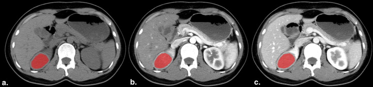
Patients with pathologically proved AMLs ( n = 18) lacking visible macroscopic fat at CT and patients with pathologically proved ccRCCs ( n = 18) and pRCCs ( n = 14) were included. All patients underwent CT scan with three phases (precontrast phase [PCP], corticomedullary phase [CMP], and nephrographic phase [NP]). The selected images were analyzed and classified with TA software (MaZda). Texture classification was performed for 1) minimal fat AML versus ccRCC, 2) minimal fat AML versus pRCC, and 3) ccRCC versus pRCC. The classification results were arbitrarily divided into several levels according to the misclassification rates: excellent (misclassification rates ≤10%), good (10%< misclassification rates ≤20%), moderate (20%< misclassification rates ≤30%), fair (30%< misclassification rates ≤40%), and poor (misclassification rates ≥40%).
Results
Excellent classification results (error of 0.00%–9.30%) were obtained with nonlinear discriminant analysis for all the three groups, no matter which phase was used. On comparison of the three scanning phases, we observed a trend toward better lesion classification with PCP for minimal fat AML versus ccRCC, CMP, and NP images for ccRCC versus pRCC and found similar discriminative power for minimal fat AML versus pRCC.
Conclusions
TA might be a reliable quantitative method for the discrimination of minimal fat AML, ccRCC, and pRCC.
Angiomyolipoma (AML) as the most common benign solid renal tumor is not difficult to be diagnosed when macroscopic fat is appeared, but diagnosis is challenging for AML with minimal fat . Approximately 10%–17% of benign renal tumors are surgically resected , and AMLs account for 18%–59% of the excised benign tumors . In this regard, accurate differential diagnosis of minimal fat AML from renal cell cancer (RCC) is crucial to avoid unnecessary surgery.
In previous studies, investigators have described some imaging features that are highly suggestive of minimal fat AML, such as high attenuation at unenhanced computed tomography (CT) with homogeneous prolonged enhancement , a small renal mass with homogeneous low signal intensity (SI) on T2-weighted images , and the presence of microscopic fat at in- and opposed-phase images ; however, imaging characteristics can be variable while clear cell RCC (ccRCC) often contains microscopic fat with decreasing SI on opposed-phase images compared to in-phase images ; papillary RCC (pRCC) often shows low T2 SI and homogeneous and gradual enhancement at CT or magnetic resonance (MR) images . In a word, there are no reliable imaging features to differentiate minimal fat AML from RCC.
Get Radiology Tree app to read full this article<
Get Radiology Tree app to read full this article<
Material and methods
Get Radiology Tree app to read full this article<
Patient Selection
Get Radiology Tree app to read full this article<
Get Radiology Tree app to read full this article<
CT Examination
Get Radiology Tree app to read full this article<
Conventional Imaging Analysis
CT Characteristics Analysis
Get Radiology Tree app to read full this article<
Get Radiology Tree app to read full this article<
Statistical Analysis
Get Radiology Tree app to read full this article<
TA and Feature Selection
Image Selection
Get Radiology Tree app to read full this article<
ROI Definition
Get Radiology Tree app to read full this article<
Get Radiology Tree app to read full this article<
Texture Feature Calculation and Selection
Get Radiology Tree app to read full this article<
Get Radiology Tree app to read full this article<
Tissue Classification
Get Radiology Tree app to read full this article<
Get Radiology Tree app to read full this article<
Results
Conventional Imaging Analysis
Get Radiology Tree app to read full this article<
Table 1
Attenuation Values and Enhancement Degree of Minimal Fat AML, ccRCC, and pRCC
Parameter Minimal Fat AML ( n = 18) ccRCC ( n = 18) pRCC ( n = 14)P Minimal Fat AML versus ccRCC Minimal Fat AML versus pRCC ccRCC versus pRCC PCP attenuation 43.89 ± 5.49 32.11 ± 6.99 34.00 ± 7.59 .476 .465 .926 CMP attenuation 109.44 ± 39.83 129.22 ± 47.54 53.86 ± 16.18 .260 .008 .000 NP attenuation 95.89 ± 29.48 110.44 ± 29.48 70.86 ± 12.57 .874 .013 .005 Enhancement degree (CMP) 65.56 ± 39.98 97.11 ± 45.77 19.86 ± 14.62 .431 .002 .000 Enhancement degree (NP) 52.00 ± 29.03 78.33 ± 27.75 36.86 ± 10.25 .833 .004 .007
AML, angiomyolipoma; ccRCC, clear cell RCC; CMP, corticomedullary phase; NP, nephrographic phase; pRCC, papillary RCC; PCP, precontrast phase; RCC, renal cell cancer.
Data are means ± standard deviations in Hounsfield units.
Table 2
Subjective Analysis of Tumor Attenuation in Comparison to the Surrounding Renal Parenchyma and Enhancement Pattern
Parameter Minimal Fat AML ( n = 18) ccRCC ( n = 18) pRCC ( n = 14)P Value Minimal Fat AML Versus ccRCC Minimal Fat AML Versus pRCC ccRCC Versus pRCC Attenuation <.001 .048 .244 Hypoattenuation 0 (0.0) 6 (33.3) 4 (28.6) Isoattenuation 3 (16.7) 8 (44.4) 3 (21.4) Hyperattenuation 15 (83.3) 4 (22.2) 7 (50.0) Enhancement pattern .555 .004 .001 Early washout 8 (44.4) 11 (61.1) 0 (0.0) Gradual 3 (16.7) 3 (16.7) 8 (57.1) Prolonged 7 (38.9) 4 (22.2) 6 (42.9)
AML, angiomyolipoma; ccRCC, clear cell RCC; pRCC, papillary RCC; RCC, renal cell cancer.
Data are numbers of patients with a given tumor. Data in parentheses are percentages.
Get Radiology Tree app to read full this article<
Get Radiology Tree app to read full this article<
Get Radiology Tree app to read full this article<
Get Radiology Tree app to read full this article<
TA and Feature Selection
Get Radiology Tree app to read full this article<
Feature Selection
Get Radiology Tree app to read full this article<
Table 3
The Frequency of Each Feature Category to be Selected by all the Three Feature Selection Methods (Fisher, POE + ACC, and MI)
Category Minimal Fat AML Versus ccRCC Minimal Fat AML Versus pRCC ccRCC Versus pRCC PCP CMP NP PCP CMP NP PCP CMP NP Histogram ( n = 11) 17 3 4 14 16 14 2 17 17 Co-occurrence matrix ( n = 220) 11 23 22 12 12 13 22 6 6 Run-length matrix ( n = 20) 0 4 2 1 1 1 0 1 2 Gradient ( n = 5) 0 0 0 0 0 0 1 1 0 Wavelet ( n = 16) 1 0 1 2 0 2 4 1 3 Autoregressive model ( n = 5) 1 0 1 1 1 0 1 3 2
AML, angiomyolipoma; ccRCC, clear cell RCC; CMP, corticomedullary phase; Fisher, Fisher coefficient; MI, mutual information; NP, nephrographic phase; pRCC, papillary RCC; PCP, precontrast phase; POE + ACC, classification error probability combined with average correlation coefficients; RCC, renal cell cancer.
The total number of evaluated texture parameters is 277. The frequencies of each feature category to be selected by Fisher, MI, and POE + ACC from each scanning phase are listed.
Get Radiology Tree app to read full this article<
Tissue Classification
Get Radiology Tree app to read full this article<
Table 4
Intradisease Classification Results in Groups of all the Three Phase Images
Scanning Phase RDA PCA LDA NDA Minimal fat AML versus ccRCC, N = 86, n (%) PCP 4 (4.65) 5 (5.81) 2 (2.33) 0 (0.00) CMP 24 (27.91) 20 (23.26) 11 (12.79) 8 (9.30) NP 8 (9.30) 8 (9.30) 14 (16.28) 6 (6.98) Minimal fat AML versus pRCC, N = 74, n (%) PCP 9 (12.16) 10 (13.51) 9 (12.16) 4 (5.41) CMP 12 (16.22) 12 (16.22) 6 (8.11) 5 (6.76) NP 8 (10.81) 8 (10.81) 2 (2.70) 0 (0.00) ccRCC versus pRCC, N = 80, n (%) PCP 18 (22.50) 19 (23.75) 15 (18.75) 6 (7.50) CMP 13 (16.25) 13 (16.25) 2 (2.50) 5 (6.25) NP 11 (13.75) 11 (13.75) 15 (18.75) 6 (7.50)
AML, angiomyolipoma; ccRCC, clear cell RCC; CMP, corticomedullary phase; LDA, linear discriminant analysis; NDA, nonlinear discriminant analysis; NP, nephrographic phase; pRCC, papillary RCC; PCA, principal component analysis; PCP, precontrast phase; RDA, raw data analysis.
Misclassification results of each computed tomography scanning phase for minimal fat AML versus ccRCC, minimal fat AML versus pRCC, and ccRCC versus pRCC. Number of misclassified images and misclassification % given for RDA, PCA, LDA, and NDA.
Get Radiology Tree app to read full this article<
Minimal fat AML versus ccRCC
Get Radiology Tree app to read full this article<
Minimal fat AML versus pRCC
Get Radiology Tree app to read full this article<
ccRCC versus pRCC
Get Radiology Tree app to read full this article<
Discussion
Get Radiology Tree app to read full this article<
Get Radiology Tree app to read full this article<
Get Radiology Tree app to read full this article<
Get Radiology Tree app to read full this article<
Get Radiology Tree app to read full this article<
Get Radiology Tree app to read full this article<
Get Radiology Tree app to read full this article<
Get Radiology Tree app to read full this article<
References
1. Bosniak M.A., Megibow A.J., Hulnick D.H., et. al.: CT diagnosis of renal angiomyolipoma: the importance of detecting small amounts of fat. AJR Am J Roentgenol 1988; 151: pp. 497-501.
2. Israel G.M., Hindman N., Hecht E., et. al.: The use of opposed-phase chemical shift MRI in the diagnosis of renal angiomyolipomas. AJR Am J Roentgenol 2005; 184: pp. 1868-1872.
3. Kutikov A., Fossett L.K., Ramchandani P., et. al.: Incidence of benign pathologic findings at partial nephrectomy for solitary renal mass presumed to be renal cell carcinoma on preoperative imaging. Urology 2006; 68: pp. 737-740.
4. Fujii Y., Komai Y., Saito K., et. al.: Incidence of benign pathologic lesions at partial nephrectomy for presumed RCC renal masses: Japanese dual-center experience with 176 consecutive patients. Urology 2008; 72: pp. 598-602.
5. Sasiwimonphan K., Takahashi N., Leibovich B.C., et. al.: Small (<4 cm) renal mass: differentiation of angiomyolipoma without visible fat from renal cell carcinoma utilizing MR imaging. Radiology 2012; 263: pp. 160-168.
6. Zhang Y.Y., Luo S., Liu Y., et. al.: Angiomyolipoma with minimal fat: differentiation from papillary renal cell carcinoma by helical CT. Clinical radiology 2013; 68: pp. 365-370.
7. Kim J.K., Park S.Y., Shon J.H., et. al.: Angiomyolipoma with minimal fat: differentiation from renal cell carcinoma at biphasic helical CT. Radiology 2004; 230: pp. 677-684.
8. Hindman N., Ngo L., Genega E.M., et. al.: Angiomyolipoma with minimal fat: can it be differentiated from clear cell renal cell carcinoma by using standard MR techniques?. Radiology 2012; 265: pp. 468-477.
9. Outwater E.K., Blasbalg R., Siegelman E.S., et. al.: Detection of lipid in abdominal tissues with opposed-phase gradient-echo images at 1.5 T: techniques and diagnostic importance. Radiographics : a review publication of the Radiological Society of North America, Inc 1998; 18: pp. 1465-1480.
10. Yang C.W., Shen S.H., Chang Y.H., et. al.: Are there useful CT features to differentiate renal cell carcinoma from lipid-poor renal angiomyolipoma?. AJR Am J Roentgenol 2013; 201: pp. 1017-1028.
11. Kim J.Y., Kim J.K., Kim N., et. al.: CT histogram analysis: differentiation of angiomyolipoma without visible fat from renal cell carcinoma at CT imaging. Radiology 2008; 246: pp. 472-479.
12. Simpfendorfer C., Herts B.R., Motta-Ramirez G.A., et. al.: Angiomyolipoma with minimal fat on MDCT: can counts of negative-attenuation pixels aid diagnosis?. AJR Am J Roentgenol 2009; 192: pp. 438-443.
13. Chaudhry H.S., Davenport M.S., Nieman C.M., et. al.: Histogram analysis of small solid renal masses: differentiating minimal fat angiomyolipoma from renal cell carcinoma. AJR Am J Roentgenol 2012; 198: pp. 377-383.
14. Catalano O.A., Samir A.E., Sahani D.V., et. al.: Pixel distribution analysis: can it be used to distinguish clear cell carcinomas from angiomyolipomas with minimal fat?. Radiology 2008; 247: pp. 738-746.
15. Bahl G., Cruite I., Wolfson T., et. al.: Noninvasive classification of hepatic fibrosis based on texture parameters from double contrast-enhanced magnetic resonance images. Journal of magnetic resonance imaging: JMRI 2012; 36: pp. 1154-1161.
16. Szczypinski P.M., Strzelecki M., Materka A., et. al.: MaZda–a software package for image texture analysis. Computer methods and programs in biomedicine 2009; 94: pp. 66-76.
17. Nachimuthu D.S., Baladhandapani A.: Multidimensional texture characterization: on analysis for brain tumor tissues using MRS and MRI. J Digit Imaging 2014;
18. Holli K., Laaperi A.L., Harrison L., et. al.: Characterization of breast cancer types by texture analysis of magnetic resonance images. Academic radiology 2010; 17: pp. 135-141.
19. Mayerhoefer M.E., Schima W., Trattnig S., et. al.: Texture-based classification of focal liver lesions on MRI at 3.0 Tesla: a feasibility study in cysts and hemangiomas. Journal of magnetic resonance imaging : JMRI 2010; 32: pp. 352-359.
20. Goh V., Ganeshan B., Nathan P., et. al.: Assessment of response to tyrosine kinase inhibitors in metastatic renal cell cancer: CT texture as a predictive biomarker. Radiology 2011; 261: pp. 165-171.
21. Harrison L.C., Luukkaala T., Pertovaara H., et. al.: Non-Hodgkin lymphoma response evaluation with MRI texture classification. J Exp Clin Cancer Res 2009; 28: pp. 87.
22. Chen G., Jespersen S., Pedersen M., et. al.: Evaluation of anti-vascular therapy with texture analysis. Anticancer Res 2005; 25: pp. 3399-3405.
23. Strzelecki M., Szczypinski P., Materka A., et. al.: A software tool for automatic classification and segmentation of 2D/3D medical images. Nuclear Instruments and Methods in Physics Research Section A: Accelerators, Spectrometers, Detectors and Associated Equipment 2013; 702: pp. 137-140.
24. Bata P., Gyebnar J., Tarnoki D.L., et. al.: Clear cell renal cell carcinoma and papillary renal cell carcinoma: differentiation of distinct histological types with multiphase CT. Diagn Interv Radiol 2013; 19: pp. 387-392.
25. Rheinheimer S., Stieltjes B., Schneider F., et. al.: Investigation of renal lesions by diffusion-weighted magnetic resonance imaging applying intravoxel incoherent motion-derived parameters–initial experience. European journal of radiology 2012; 81: pp. e310-e316.
26. Zhang Y.L., Yu B.L., Ren J., et. al.: EADC values in diagnosis of renal lesions by 3.0 T diffusion-weighted magnetic resonance imaging: compared with the ADC values. Appl Magn Reson 2013; 44: pp. 349-363.
27. Sasamori H., Saiki M., Suyama J., et. al.: Utility of apparent diffusion coefficients in the evaluation of solid renal tumors at 3T. Magnetic resonance in medical sciences : MRMS : an official journal of Japan Society of Magnetic Resonance in Medicine 2014;
28. Lanzman R.S., Robson P.M., Sun M.R., et. al.: Arterial spin-labeling MR imaging of renal masses: correlation with histopathologic findings. Radiology 2012; 265: pp. 799-808.
29. Zira A.N., Theocharis S.E., Mitropoulos D., et. al.: (1)H NMR metabonomic analysis in renal cell carcinoma: a possible diagnostic tool. J Proteome Res 2010; 9: pp. 4038-4044.
30. Fruehwald-Pallamar J., Czerny C., Holzer-Fruehwald L., et. al.: Texture-based and diffusion-weighted discrimination of parotid gland lesions on MR images at 3.0 Tesla. NMR in biomedicine 2013; 26: pp. 1372-1379.
31. Harrison L.C., Raunio M., Holli K.K., et. al.: MRI texture analysis in multiple sclerosis: toward a clinical analysis protocol. Academic radiology 2010; 17: pp. 696-707.
32. Zhao X.J., Pu J.X., Ping J.G., et. al.: Angiomyolipoma with minimal fat: differentiation from renal cell carcinoma at helical CT. Chinese medical journal 2013; 126: pp. 991-992.