
Rationale and Objectives
Subcutaneous epidermal cysts and intracranial epidermoid cysts are pathologically identical. Although diffusion-weighted imaging (DWI) studies of intracranial epidermoid cysts have been numerously reported, those of subcutaneous epidermal cysts have not been sufficiently investigated. Our hypothesis for this study is that the apparent diffusion coefficient (ADC) values of subcutaneous epidermal cysts and intracranial epidermoid cysts are not different. This study was intended to evaluate the ADC of subcutaneous epidermal cysts of the head and neck in comparison with that of intracranial epidermoid cysts.
Materials and Methods
The MR studies were performed in 14 patients with head and neck subcutaneous epidermal cysts and 10 patients with intracranial epidermoid cysts using line scan DWI (LSDWI). The ADC was measured and compared between the two types of cyst.
Results
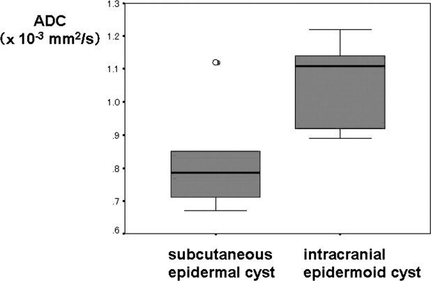
The ADC values (mean ± SD) were 0.81 ± 0.14 × 10 −3 mm 2 /s in subcutaneous epidermal cysts and 1.06 ± 0.12 × 10 −3 mm 2 /s in intracranial epidermoid cysts. A significant difference was found in ADC values between the two types ( P = .0019).
Conclusion
Our preliminary study has shown that the ADC provides useful information regarding tissue characterization of subcutaneous epidermal cysts. However, the ADC of subcutaneous epidermal cysts was significantly lower than that of intracranial epidermoid cysts.
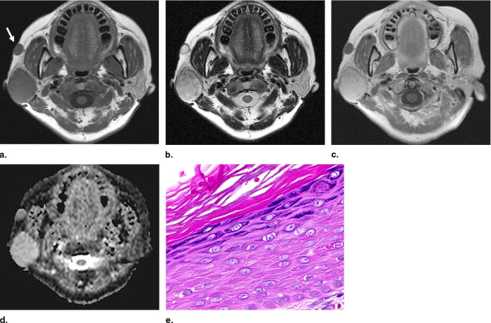
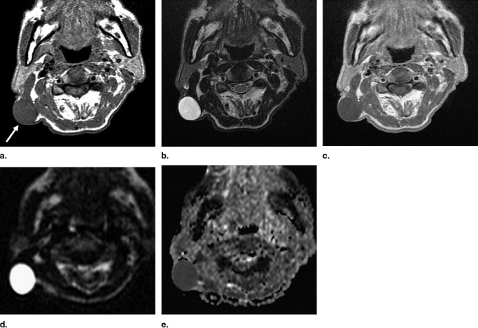
Epidermal cysts are common, benign masses that occur in the skin. The lesions commonly involve the scalp, face, neck, trunk, and back ( ). However, only a few reports regarding MR imaging findings have been issued on subcutaneous epidermal cysts ( ). Typical MR imaging findings of subcutaneous epidermal cysts include a well-circumscribed margin, iso-, or slightly high signal intensity relative to adjacent muscles on T1-weighted images, and very high signal intensity on T2-weighted images ( ). Usually, no apparent enhancement is visible inside the cyst if unruptured ( ).
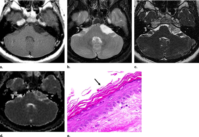
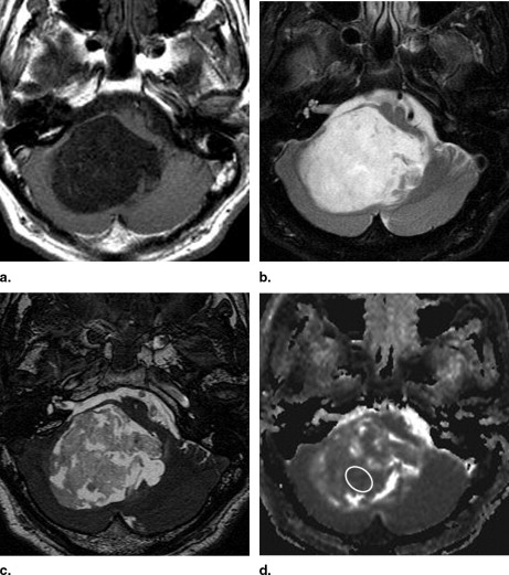
Subcutaneous epidermal cysts and intracranial epidermoid cysts are pathologically identical: cysts filled with keratin debris and bounded by a wall of the stratified squamous epithelium ( ). Unlike subcutaneous epidermal cysts, numerous MR imaging reports have described intracranial epidermoid cysts ( ). Several studies of diffusion-weighted imaging (DWI) and apparent diffusion coefficient (ADC) have had particular impact on the diagnosis of the intracranial epidermoid cysts ( ). However, reports on DWI and ADC of subcutaneous epidermal cysts are insufficient. We hypothesize that ADC values of subcutaneous epidermal cysts and intracranial epidermoid cysts are not different because they are pathologically identical. This study was intended to evaluate the ADC of subcutaneous epidermal cysts of the head and neck in comparison with that of intracranial epidermoid cysts.
Materials and methods
Subjects
Get Radiology Tree app to read full this article<
Get Radiology Tree app to read full this article<
MR Imaging
Get Radiology Tree app to read full this article<
Get Radiology Tree app to read full this article<
Get Radiology Tree app to read full this article<
Imaging Data Analysis
Get Radiology Tree app to read full this article<
Get Radiology Tree app to read full this article<
Get Radiology Tree app to read full this article<
Statistical Analysis
Get Radiology Tree app to read full this article<
Results
Signal Intensities and Contrast Enhancement of the Lesions
Get Radiology Tree app to read full this article<
Table 1
MR Imaging Findings of Subcutaneous Epidermal Cysts and Intracranial Epidermoid Cysts
Imaging Findings Subcutaneous Epidermal Cyst ( n = 14) Intracranial Epidermoid Cyst ( n = 10) Signal intensity (T1-weighted) Low 0 9 High 14 0 Mixed 0 1 Signal intensity (T2-weighted) Low 3 0 High 9 10 Mixed 2 0 Contrast enhancement No enhancement 5 8 Thin and smooth rim 4 0 NA 5 2
NA, not applicable.
Get Radiology Tree app to read full this article<
Get Radiology Tree app to read full this article<
ADC Values
Get Radiology Tree app to read full this article<
Get Radiology Tree app to read full this article<
Get Radiology Tree app to read full this article<
Get Radiology Tree app to read full this article<
Discussion
Get Radiology Tree app to read full this article<
Get Radiology Tree app to read full this article<
Get Radiology Tree app to read full this article<
Get Radiology Tree app to read full this article<
Get Radiology Tree app to read full this article<
Get Radiology Tree app to read full this article<
Get Radiology Tree app to read full this article<
Get Radiology Tree app to read full this article<
Conclusion
Get Radiology Tree app to read full this article<
References
1. Vincent L.M., Parker L.A., Mittelstaedt C.A.: Sonographic appearance of an epidermal inclusion cyst. J Ultrasound Med 1985; 4: pp. 609-611.
2. Sundaram M., McGuire M.H., Herbold D.R., Beshany S.E., Fletcher J.W.: High signal intensity soft tissue masses on T1-weighted pulsing sequences. Skeletal Radiol 1987; 16: pp. 30-36.
3. Krandorf M.J., Jelinek J.S., Moser R.P., et. al.: Soft tissue masses: diagnosis using MR imaging. AJR Am J Roentgenol 1989; 153: pp. 541-547.
4. Fujimoto H., Murakami K., Kashimada A., et. al.: Large epidermal cyst involving the ischiorectal fossa: MR demonstration. Clin Imaging 1993; 17: pp. 146-148.
5. Takano Y., Yokokawa K., Namiki M., Toki K., Okuyama A.: Perineal epidermal cyst. Urol Int 1994; 53: pp. 53-55.
6. Fisher A.R., Mason P.H., Wagenhals K.S.: Ruptured plantar epidermal inclusion cysts. Am J Roentgenol 1998; 171: pp. 1709-1710.
7. Shibata T., Hatori M., Satoh T., Ehara S., Kokubun S.: Magnetic resonance imaging features of epidermoid cyst in the extremities. Arch Orthop Trauma Surg 2003; 123: pp. 239-241.
8. Hong S.H., Chung H.W., Choi J.Y., Koh Y.H., Choi J.A., Kang H.S.: MRI findings of subcutaneous epidermal cysts: emphasis on the presence of rupture. Am J Roentgenol 2006; 186: pp. 961-966.
9. Bullough P.: Orthopedic pathology.2004.MosbyNew York, NY:pp. 445-446.
10. Olson J.J., Beck D.W., Crawford S.C., Menzes A.H.: Comparative evaluation of intracranial epidermoid tumors with computed tomography and magnetic resonance imaging. Neurosurgery 1987; 21: pp. 357-360.
11. Horowitz B.L., Chari M.V., James R., Bryan R.N.: MR of intracranial epidermoid tumors: correlation of in vivo imaging with in vitro 13C spectroscopy. Am J Neuroradiol 1990; 11: pp. 299-302.
12. Tsuruda J.S., Chew W.M., Moseley M.E., Norma D.: Diffusion-weighted MR imaging of the brain: value of differentiating between extraaxial cysts and epidermoid tumors. Am J Neuroradiol 1990; 11: pp. 925-931.
13. Maeda M., Kawamura Y., Tamagawa Y., et. al.: Intravoxel incoherent motion (IVIM) MRI in intracranial, extraaxial tumors and cysts. J Comput Assist Tomogr 1992; 16: pp. 514-518.
14. Ikushima I., Korogi Y., Hirai T., et. al.: MR of epidermoids with various pulse sequences. Am J Neuroradiol 1997; 18: pp. 1359-1363.
15. Chen S., Ikawa F., Kurisu K., Arita K., Takaba J., Kanou Y.: Quantitative MR evaluation of intracranial epidermoid tumors by fast fluid-attenuated inversion recovery imaging and echo-planar diffusion-weighted imaging. Am J Neuroradiol 2001; 22: pp. 1089-1096.
16. Aikele P., Kittner T., Offergeld C., Kaftan H., Huttenbrink K.B., Laniado M.: Diffusion-weighted MR imaging of cholesteatoma in pediatric and adult patients who have undergone middle ear surgery. AJR Am J Roentgenol 2003; 181: pp. 261-265.
17. Hakyemez B., Aksoy U., Yildiz H., Ergin N.: Intracranial epidermoid cysts: diffusion-weighted, FLAIR and conventional MR findings. Eur J Radiol 2005; 54: pp. 214-220.
18. Maier S.E., Gudbjartsson H., Patz S., et. al.: Line scan diffusion imaging: characterization in healthy subjects and stroke patients. AJR Am J Roentgenol 1998; 171: pp. 85-93.
19. Maeda M., Sakuma H., Maier S.E., Takeda K.: Quantitative assessment of diffusion abnormalities in benign and malignant vertebral compression fractures by line scan diffusion-weighted imaging. AJR Am J Roentgenol 2003; 181: pp. 1203-1209.
20. Maeda M., Kato H., Sakuma H., Maier S.E., Takeda K.: Usefulness of the apparent diffusion coefficient in line scan diffusion-weighted imaging for distinguishing between squamous cell carcinomas and malignant lymphomas of the head and neck. Am J Neuroradiol 2005; 26: pp. 1186-1192.
21. Maeda M., Maier S.E., Sakuma H., Ishida M., Takeda K.: Apparent diffusion coefficient in malignant lymphoma and carcinoma involving cavernous sinus evaluated by line scan diffusion-weighted imaging. J Magn Reson Imaging 2006; 24: pp. 543-548.
22. Stejskal E.O., Tanner J.E.: Spin diffusion measurements: spin echoes in the presence of a time-dependent field gradient. J Chem Phys 1965; 42: pp. 288-292.
23. McLendon R.E.: Epidermoid and dermoid tumors.Wilkins R.H.Rengachary S.S.Neurosurgery.1996.McGraw-HillNew York, NY:pp. 959-963.
24. Wand W.A., Labosky D.A.: Ruptured epidermal inclusion cyst of the palm presenting as collar-button abscess. J Hand Surg [Am] 1985; 10: pp. 899-901.
25. Chavez G.D., De Salles A.A., Solberg T.D., Pedroso A., Espinoza D., Villablanca P.: Three-dimensional fast imaging employing steady-state acquisition magnetic resonance imaging for stereotactic radiosurgery of trigeminal neuralgia. Neurosurgery 2005; 56: pp. E628.
26. Yonezawa K., Kim S., Tanaka M., Adachi N., Seto H., Tamaki N.: A case of a giant epidermoid cyst on the occipital scalp. Neurol Surg (Japanese) 1993; 21: pp. 735-738.