
Rationale and Objectives
The study aimed to evaluate the usefulness of an abbreviated protocol (AP) of magnetic resonance imaging (MRI) in comparison to a full diagnostic protocol (FDP) of MRI in the breast cancer screening with dense breast tissue.
Materials and Methods
There are 478 female participants with dense breast tissue and negative mammography results, who were imaged with MRI using AP and FDP. The AP and FDP images were analyzed separately, and the sensitivity and specificity of breast cancer detection were calculated. The chi-square test and receiver operating characteristics curves were used to assess the breast cancer diagnostic capabilities of the two protocols.
Results
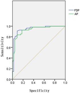
Sixteen cases of breast cancer from 478 patients with dense breasts were detected using the FDP method, with pathologic confirmation of nine cases of ductal carcinoma in situ, six cases of invasive ductal carcinoma, and one case of mucinous carcinoma. Fifteen cases of breast cancer were successfully screened using the AP method. The sensitivity showed no obvious significant difference between AP and FDP (χ 2 = 0.592, P = 0.623), but the specificity showed a statistically significant difference (χ 2 = 4.619, P = 0.036). The receiver operating characteristics curves showed high efficacy of both methods in the detection of breast cancer in dense breast tissue (the areas under the curve were 0.931 ± 0.025 and 0.947 ± 0.024, respectively), and the ability to diagnose breast cancer was not statistically significantly different between the two methods.
Conclusions
The AP of MRI may improve the detection rate of breast cancer in dense breast tissue, and it may be useful in efficient breast cancer screening.
Introduction
Breast cancer, the most common or second most common cancer in women worldwide , has gradually gained prevalence. Early detection of breast cancer can improve the duration of survival and quality of life in patients . Extensive screening can detect breast cancer early, thus screening plays an increasing important role in breast care. Mammography (MG) is the preferred method of screening for breast tissue because it is simple, convenient, affordable, and irreplaceable in the detection of microcalcifications . Studies have shown that, within groups of the same age, there are two to six times higher risk of breast cancer in dense breasts than in non-dense breasts, but MG detection sensitivity in dense breasts is much lower than that in fatty breasts . For example, Mendelson et al. found that the detection rate of breast cancer using MG was only 30% in dense breasts and 80% in fatty breasts . This clearly shows a limitation of using MG in breast screening.
Magnetic resonance imaging (MRI) has outstanding soft tissue resolution and multiplanar imaging capability, thereby offering unique advantages in the detection and diagnosis of breast lesions. The American College of Radiology (ACR) included breast MRI in the fourth edition of the Breast Imaging Reporting and Data System (BI-RADS) in 2003. The use of MRI has been standardized in breast examination . Dynamic contrast-enhanced MRI (DCE-MRI), a more advanced technique for breast MRI, allows an analysis of breast lesions through both morphologic and hemodynamic changes, with a sensitivity of 100% and specificity of up to 97% for breast cancer detection . However, the lengthy inspection time and high medical cost incurred by DCE-MRI have limited its use in breast MRI screening. Currently, it is used as a supplementary examination of certain breast cancers in high-risk populations .
Get Radiology Tree app to read full this article<
Materials and Methods
Participants
Get Radiology Tree app to read full this article<
MRI Examination Techniques
Get Radiology Tree app to read full this article<
Data Analysis
Get Radiology Tree app to read full this article<
Statistical Analysis
Get Radiology Tree app to read full this article<
Results
Get Radiology Tree app to read full this article<
Table 1
Pathology of 41 Benign and Malignant Lesions
Pathology No Malignant ( n = 16) Ductal carcinoma in situ 4 Invasive ductal carcinoma 11 Mucinous carcinoma 1 Benign ( n = 25) Hyperplasia 8 Fibroadenoma 11 Papilloma in situ of duct 2 Cyst 3 Granuloma 1
Get Radiology Tree app to read full this article<
Get Radiology Tree app to read full this article<
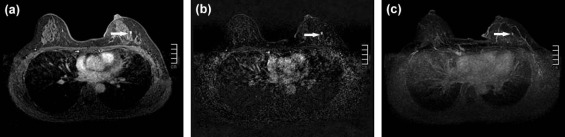
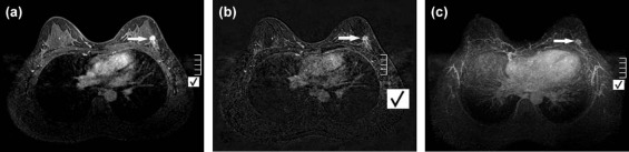
Table 2
Comparison of the Breast Cancer Diagnostic Capabilities of AP and FPD
AP (%) FDP (%) Sensitivity 93.8%(15/16) 100.0%(16/16) Specificity 88.3%(408/462) 94.6%(437/462) PPV 21.7%(15/69) 41.0%(16/39) NPV 99.8%(408/409) 100%(439/439)
AP, abbreviated protocol; FDP, full diagnostic protocol; NPV, negative predictive value; PPV, positive predictive value.
Get Radiology Tree app to read full this article<
Discussion
Get Radiology Tree app to read full this article<
Get Radiology Tree app to read full this article<
Get Radiology Tree app to read full this article<
Get Radiology Tree app to read full this article<
Get Radiology Tree app to read full this article<
Get Radiology Tree app to read full this article<
Get Radiology Tree app to read full this article<
Conclusion
Get Radiology Tree app to read full this article<
References
1. Aberle D.R., Chiles C., Gatsonis C., et. al.: Imaging and cancer: research strategy of the American College of Radiology Imaging Network. Radiology 2005; 235: pp. 741-751.
2. Jemal A., Siegel R., Xu J., et. al.: Cancer statistics, 2010. CA Cancer J Clin 2010; 60: pp. 277-300.
3. Game J.P., Aspegren K., Balldin G., et. al.: Increasing incidence of and declining mortality from breast carcinoma. Trends in Malmö, Sweden, 1961–1992. Cancer 1997; 79: pp. 69-74.
4. Graf O., Berg W.A., Sickle E.A.: Large rodlike calcifications at mammography: analysis of morphologic features. AJR Am J Roentgenol 2013; 200: pp. 299-303.
5. Pollán M., Ascunce N., Ederra M., et. al.: Mammographic density and risk of breast cancer according to tumor characteristics and mode of detection: a Spanish population-based case-control study. Breast Cancer Res 2013; 15: pp. R9.
6. Boyd N.F., Guo H., Martin L.J., et. al.: Mammographic density and the risk and detection of breast cancer. N Engl J Med 2007; 356: pp. 227-236.
7. McCormack V.A., dos Santos Silva I.: Breast density and parenchymal as markers of breast cancer risk: a meta-analysis. Cancer Epidemiol Biomarkers Prev 2006; 15: pp. 1159-1169.
8. Mendelson M.T., Oestreicher N., Porter P.L., et. al.: Breast density as a predictor of mammographic detection: comparison of interval- and screen-detected cancers. J Natl Cancer Inst 2000; 92: pp. 1081-1087.
9. Oeffinger K.C., Fontham E.T., Etzioni R., et. al.: Breast cancer screening for women at average risk: 2015 guideline update from the American Cancer Society. JAMA 2015; 314: pp. 1599-1614.
10. Harnett A., Smallwood J., Titshall V., et. al.: Diagnosis and treatment of early breast cancer, including locally advanced disease-summary of NICE guidance. BMJ 2009; 338: pp. b438.
11. Berg W.A.: How well does supplemental screening magnetic resonance imaging work in high-risk women?. J Clin Oncol 2014; 32: pp. 2193-2196.
12. Kuhl C.K., Schrading S., Strobel K., et. al.: Abbreviated breast magnetic resonance imaging (MRI): first postcontrast subtracted images and maximum-intensity projection-a novel approach to breast cancer screening with MRI. J Clin Oncol 2014; 32: pp. 2304-2310.
13. Morris E.A.: Rethinking breast cancer screening: ultra fast breast magnetic resonance imaging. J Clin Oncol 2014; 32: pp. 2281-2283.
14. Kim S.Y., Kim H.Y., Kim E.K., et. al.: Evaluation of malignancy risk stratification of microcalcifications detected on mammography: a study based on the 5th edition of BI-RADS. Ann Surg Oncol 2015; 22: pp. 2895-2901.
15. Brower V.: Homing in on mechanisms linking breast density to breast cancer risk. J Natl Cancer Inst 2010; 102: pp. 843-845.
16. Kerlikowske K.: The mammogram that cried Wolfe. N Engl J Med 2007; 356: pp. 297-300.
17. Harvey J.A., Bovbjerg V.E.: Quantitative assessment of mammographic breast density: relationship with breast cancer risk. Radiology 2004; 230: pp. 29-41.
18. Boyd N.F., Rommens J.M., Vogt K., et. al.: Mammographic breast density as an intermediate phenotype for breast cancer. Lancet Oncol 2005; 6: pp. 798-808.
19. del Carmen M.G., Halpern E.F., Kopans D.B., et. al.: Mammographic breast density and race. AJR 2007; 188: pp. 1147-1150.
20. Carney P.A., Miglioretti D.L., Yankaskas B.C., et. al.: Individual and combined effects of age, breast density, and hormone replacement therapy use on the accuracy of screening mammography. Ann Intern Med 2003; 138: pp. 168-175.
21. Mandelson M.T., Oestreicher N., Porter P.L., et. al.: Breast density as a predictor of mammographic detection: comparison of interval- and screen-detected cancers. J Natl Cancer Inst 2000; 92: pp. 1081-1087.
22. Kerlikowske K., Grady D., Barclay J., et. al.: Effect of age, breast density, and family history on the sensitivity of first screening mammography. JAMA 1996; 276: pp. 33-38.
23. Kolb T.M., Lichy J., Newhouse J.H.: Comparison of the performance of screening mammography, physical examination, and breast us and evaluation of factors that influence them: an analysis of 27,825 patient evaluations. Radiology 2002; 225: pp. 165-175.
24. Svane G., Azavedo E., Lindman K., et. al.: Clinical experience of photon counting breast tomosynthesis: comparison with traditional mammography. Acta Radiol 2014; 52: pp. 134-142.
25. Gennaro G., Toledano A., di Maggio C., et. al.: Digital breast tomosynthesis versus digital mammography: a clinical performance study. Eur Radiol 2010; 20: pp. 1545-1553.
26. Roubidoux M.A., Bailey J.E., Wray L.A., et. al.: Invasive cancers detected after breast cancer screening yielded a negative result: relationship of mammographic density to tumor prognostic factors. Radiology 2004; 230: pp. 42-48.
27. Clauser P., Carbonaro L.A., Pancot M., et. al.: Additional findings at preoperative breast MRI: the value of second-look digital breast tomosynthesis. Eur Radiol 2015; 25: pp. 2830-2839.
28. Berg W.A., Zhang Z., Lehrer D., et. al.: Detection of breast cancer with addition of annual screening ultrasound or a single screening MRI to mammography in women with elevated breast cancer risk. JAMA 2012; 307: pp. 1394-1404.
29. Tudorica L.A., Oh K.Y., Roy N., et. al.: A feasible high spatiotemporal resolution breast DCE-MRI protocol for clinical settings. Magn Reson Imaging 2012; 30: pp. 1257-1267.
30. Moore S.G., Shenoy P.J., Fanucchi L., et. al.: Cost-effectiveness of MRI compared to mammography for breast cancer screening in a high risk population. BMC Health Serv Res 2009; 9: pp. 9.
31. Heywang S.H., Wolf A., Pruss E., et. al.: MR imaging of the breast with Gd-DTPA: use and limitations. Radiology 1987; 171: pp. 95-103.
32. Varga D., Wöckel A., Wagner J., et. al.: Value of ultrasound in preoperative local staging in early breast cancer. Ultraschall Med 2011; 32: pp. 387-392.
33. U.S. Preventive Services Task Force : Screening for breast cancer: U.S. Preventive Services Task Force recommendation statement. Ann Intern Med 2009; 151: pp. 716-726.