
Rationales and Objectives
Pulmonary hypertension (PH) is a life-threatening condition, characterized by elevated pulmonary arterial pressure, which is confirmed based on invasive right heart catheterization (RHC). Noninvasive examinations may support diagnosis of PH before proceeding to RHC and play an important role in management and treatment of the disease. Although echocardiography is considered a standard tool in diagnosis, recent advances have made computed tomography (CT) and magnetic resonance (MR) imaging promising tools, which may provide morphologic and functional information. In this article, we review image-based assessment of PH with a focus on CT and MR imaging.
Conclusions
CT may provide useful morphologic information for depicting PH and seeking for underlying diseases. With the accumulated technological advancement, CT and MRI may provide practical tools for not only morphologic but also functional assessment of patients with PH.
Pulmonary hypertension (PH) is a life-threatening condition, characterized by elevated pulmonary arterial pressure (PAP) and secondary right ventricular (RV) failure. It has been defined as a mean PAP greater than or equal to 25 mm Hg at rest, based on right heart catheterization (RHC) .
Numerous causes of PH exist. The latest clinical classification—Dana Point classification—comprises five major categories that share pathologic, clinical, and therapeutic features and is intended to standardize diagnosis and treatment and to conduct clinical trials in a well-characterized group of patients ( Table 1 ) .
Table 1
Clinical Classification of Pulmonary Hypertension (Dana Point, 2008)
Adapted from Simonneau et al .
1. Pulmonary arterial hypertension
Get Radiology Tree app to read full this article<
Get Radiology Tree app to read full this article<
Get Radiology Tree app to read full this article<
Get Radiology Tree app to read full this article<
Get Radiology Tree app to read full this article<
Get Radiology Tree app to read full this article<
ALK-1, activin receptor-like kinase 1 gene; BMPR2, bone morphogenetic protein receptor, type 2; HIV, human immunodeficiency virus.
Get Radiology Tree app to read full this article<
Get Radiology Tree app to read full this article<
Get Radiology Tree app to read full this article<
Get Radiology Tree app to read full this article<
Get Radiology Tree app to read full this article<
Computed tomography
Get Radiology Tree app to read full this article<
Get Radiology Tree app to read full this article<
CT Findings of PH
Get Radiology Tree app to read full this article<
Precapillary PH
Get Radiology Tree app to read full this article<
Get Radiology Tree app to read full this article<
Get Radiology Tree app to read full this article<
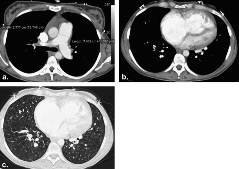
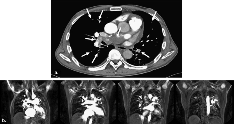
CTEPH (Group 4)
Get Radiology Tree app to read full this article<
Get Radiology Tree app to read full this article<
Get Radiology Tree app to read full this article<
Get Radiology Tree app to read full this article<
Get Radiology Tree app to read full this article<
Postcapillary PH
Get Radiology Tree app to read full this article<
Get Radiology Tree app to read full this article<
Get Radiology Tree app to read full this article<
PVOD and PCH (Group 1′)
Get Radiology Tree app to read full this article<
Evaluation of the Presence and Severity of PH
Get Radiology Tree app to read full this article<
Morphologic evaluations
Evaluation of the pulmonary vasculature
Get Radiology Tree app to read full this article<
Get Radiology Tree app to read full this article<
Get Radiology Tree app to read full this article<
Get Radiology Tree app to read full this article<
Get Radiology Tree app to read full this article<
Get Radiology Tree app to read full this article<
Get Radiology Tree app to read full this article<
Cardiac evaluation
Get Radiology Tree app to read full this article<
Table 2
Approximate Partition Values for Upper Limits of Normal for the Assessment of Cardiac Morphology
Adapted from Hoey et al .
Parameter Value (mm) Diameter of main pulmonary artery 29 Transverse diameter of right ventricle 45 Thickness of right ventricular free wall 3 Transverse diameter of right atrium 35 Thickness of interventricular septum 13 Transverse diameter of left ventricle 55 Thickness of left ventricular free wall 11 Anteroposterior diameter of left atrium 45
Get Radiology Tree app to read full this article<
Get Radiology Tree app to read full this article<
Get Radiology Tree app to read full this article<
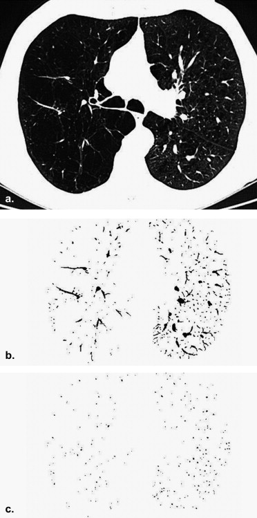
Pulmonary parenchyma
Get Radiology Tree app to read full this article<
Get Radiology Tree app to read full this article<
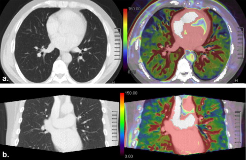
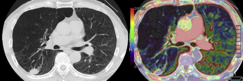
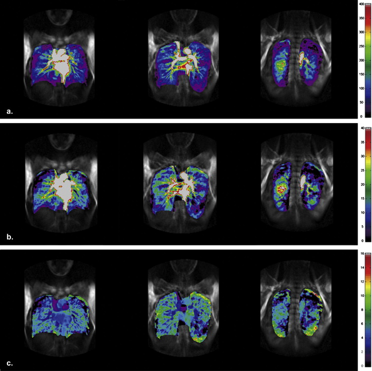
Functional evaluation
Get Radiology Tree app to read full this article<
Get Radiology Tree app to read full this article<
New Investigations and Future Directions
Get Radiology Tree app to read full this article<
Get Radiology Tree app to read full this article<
Get Radiology Tree app to read full this article<
Get Radiology Tree app to read full this article<
Get Radiology Tree app to read full this article<
Magnetic resonance imaging
Get Radiology Tree app to read full this article<
Large Vessels
Get Radiology Tree app to read full this article<
Get Radiology Tree app to read full this article<
Get Radiology Tree app to read full this article<
Get Radiology Tree app to read full this article<
Get Radiology Tree app to read full this article<
Get Radiology Tree app to read full this article<
Get Radiology Tree app to read full this article<
Get Radiology Tree app to read full this article<
Get Radiology Tree app to read full this article<
Get Radiology Tree app to read full this article<
(diastoliccross−sectional area)−(systoliccross−sectional area)systoliccross−sectional area×100 (
diastolic
cross
−
sectional area
)
−
(
systolic
cross
−
sectional area
)
systolic
cross
−
sectional area
×
100
Get Radiology Tree app to read full this article<
Get Radiology Tree app to read full this article<
Get Radiology Tree app to read full this article<
Cardiac Evaluation
Get Radiology Tree app to read full this article<
Get Radiology Tree app to read full this article<
Get Radiology Tree app to read full this article<
Get Radiology Tree app to read full this article<
Get Radiology Tree app to read full this article<
Get Radiology Tree app to read full this article<
Get Radiology Tree app to read full this article<
Get Radiology Tree app to read full this article<
New Investigations and Future Directions
Get Radiology Tree app to read full this article<
Get Radiology Tree app to read full this article<
Get Radiology Tree app to read full this article<
Get Radiology Tree app to read full this article<
Get Radiology Tree app to read full this article<
Get Radiology Tree app to read full this article<
Get Radiology Tree app to read full this article<
Get Radiology Tree app to read full this article<
Get Radiology Tree app to read full this article<
Get Radiology Tree app to read full this article<
Get Radiology Tree app to read full this article<
Conclusion
Get Radiology Tree app to read full this article<
Acknowledgments
Get Radiology Tree app to read full this article<
References
1. Galie N., Hoeper M.M., Humbert M., et. al.: Guidelines for the diagnosis and treatment of pulmonary hypertension. Eur Respir J 2009; 34: pp. 1219-1263.
2. Simonneau G., Robbins I.M., Beghetti M., et. al.: Updated clinical classificaion of pulmonary hypertension. J Am Coll Cardiol 2009; 54: pp. S43-S54.
3. McGoon M., Gutterman D., Steen V., et. al.: Screening, early detection, and diagnosis of pulmonary arterial hypertension: ACCP evidence-based clinical practice guidelines. Chest 2004; 126: pp. 14S-34S.
4. Hoeper M.M., Lee S.H., Voswinckel R., et. al.: Complications of right heart catheterization procedures in patients with pulmonary hypertension in experienced centers. J Am Coll Cardiol 2006; 48: pp. 2546-2552.
5. Ley S., Kreitner K.F., Fink C., et. al.: Assessment of pulmonary hypertension by CT and MR imaging. Eur Radiol 2004; 14: pp. 359-368.
6. Oudiz R.J.: Pulmonary hypertension associated with left-sided heart disease. Clin Chest Med 2007; 28: pp. 233-241.
7. Chatterjee K., De Marco T., Alpert J.S.: Pulmonary hypertension: hemodynamic diagnosis and management. Arch Intern Med 2002; 162: pp. 1925-1933.
8. Groves A.M., Win T., Charman S.C., et. al.: Semi-quantitative assessment of tricuspid regurgitation on contrast-enhanced multidetector CT. Clin Radiol 2004; 59: pp. 715-719.
9. Koito H.: The findings of computed tomography (CT) and magnetic resonance imaging (MRI) in pulmonary arterial hypertension. Nippon Rinsho 2008; 66: pp. 2114-2123.
10. Baque-Juston M.C., Wells A.U., Hansell D.M.: Pericardial thickening or effusion in patients with pulmonary artery hypertension: a CT study. AJR Am J Roentgenol 1999; 172: pp. 361-364.
11. Fischer A., Misumi S., Curran-Everett D., et. al.: Pericardial abnormalities predict the presence of echocardiographically defined pulmonary arterial hypertension in systemic sclerosis-related interstitial lung disease. Chest 2007; 131: pp. 988-992.
12. Lang I.M.: Chronic thromboembolic pulmonary hypertension—not so rare after all. N Engl J Med 2004; 350: pp. 2236-2238.
13. Bailey C.L., Channick R.N., Auger W.R., et. al.: “High probability” perfusion lung scans in pulmonary venoocclusive disease. Am J Respir Crit Care Med 2000; 162: pp. 1974-1978.
14. Wittram C., Maher M.M., Yoo A.J., et. al.: CT angiography of pulmonary embolism: diagnostic criteria and causes of misdiagnosis. Radiographics 2004; 24: pp. 1219-1238.
15. Bergin C.J., Rios G., King M.A., et. al.: Accuracy of high-resolution CT in identifying chronic pulmonary thromboembolic disease. AJR Am J Roentgenol 1996; 166: pp. 1371-1377.
16. King M.A., Ysrael M., Bergin C.J.: Chronic thromboembolic pulmonary hypertension: CT findings. AJR Am J Roentgenol 1998; 170: pp. 955-960.
17. Reichelt A., Hoeper M.M., Galanski M., et. al.: Chronic thromboembolic pulmonary hypertension: evaluation with 64-detector row CT versus digital substraction angiography. Eur J Radiol 2009; 71: pp. 49-54.
18. Ghaye B., Szapiro D., Mastora I., et. al.: Peripheral pulmonary arteries: how far in the lung does multi-detector row spiral CT allow analysis?. Radiology 2001; 219: pp. 629-636.
19. Schoepf U.J., Holzknecht N., Helmberger T.K., et. al.: Subsegmental pulmonary emboli: improved detection with thin-collimation multi-detector row spiral CT. Radiology 2002; 222: pp. 483-490.
20. Falaschi F., Palla A., Formichi B., et. al.: CT evaluation of chronic thromboembolic pulmonary hypertension. J Comp Assisted Tomogr 1992; 16: pp. 897-903.
21. Pitton M.B., Kemmerich G., Herber S., et. al.: [Chronic thromboembolic pulmonary hypertension: diagnostic impact of multislice-CT and selective pulmonary-DSA]. Rofo 2002; 174: pp. 474-479.
22. Azarian R., Wartski M., Collignon M.A., et. al.: Lung perfusion scans and hemodynamics in acute and chronic pulmonary embolism. J Nucl Med 1997; 38: pp. 980-983.
23. Ryan K.L., Fedullo P.F., Davis G.B., et. al.: Perfusion scan findings understate the severity of angiographic and hemodynamic compromise in chronic thromboembolic pulmonary hypertension. Chest 1988; 93: pp. 1180-1185.
24. Sherrick A.D., Swensen S.J., Hartman T.E.: Mosaic pattern of lung attenuation on CT scans: frequency among patients with pulmonary artery hypertension of different causes. AJR Am J Roentgenol 1997; 169: pp. 79-82.
25. Schwickert H.C., Schweden F., Schild H.H., et. al.: Pulmonary arteries and lung parenchyma in chronic pulmonary embolism: preoperative and postoperative CT findings. Radiology 1994; 191: pp. 351-357.
26. Hoeper M.M., Mayer E., Simonneau G., et. al.: Chronic thromboembolic pulmonary hypertension. Circulation 2006; 113: pp. 2011-2020.
27. Perloff J.K., Hart E.M., Greaves S.M., et. al.: Proximal pulmonary arterial and intrapulmonary radiologic features of Eisenmenger syndrome and primary pulmonary hypertension. Am J Cardiol 2003; 92: pp. 182-187.
28. Remy-Jardin M., Duhamel A., Deken V., et. al.: Systemic collateral supply in patients with chronic thromboembolic and primary pulmonary hypertension: assessment with multi-detector row helical CT angiography. Radiology 2005; 235: pp. 274-281.
29. Tadavarthy S.M., Klugman J., Castaneda-Zuniga W.R., et. al.: Systemic-to-pulmonary collaterals in pathological states: a review. Radiology 1982; 144: pp. 55-59.
30. Ashour M.: Hemodynamic alterations in bronchiectasis: a base for a new subclassification of the disease. J Thorac Cardiovasc Surg 1996; 112: pp. 328-334.
31. Shimizu H., Tanabe N., Terada J., et. al.: Dilatation of bronchial arteries correlates with extent of central disease in patients with chronic thromboembolic pulmonary hypertension. Circ J 2008; 72: pp. 1136-1141.
32. Montani D., O’Callaghan D.S., Savale L., et. al.: Pulmonary veno-occlusive disease: Recent progress and current challenges. Resp Med 2010; 104: pp. S23-S32.
33. Frazier A.A., Franks T.J., Mohammed T.L., et. al.: From the Archives of the AFIP: pulmonary veno-occlusive disease and pulmonary capillary hemangiomatosis. Radiographics 2007; 27: pp. 867-882.
34. Resten A., Maitre S., Humbert M., et. al.: Pulmonary hypertension: CT of the chest in pulmonary venoocclusive disease. AJR Am J Roentgenol 2004; 183: pp. 65-70.
35. Montani D., Price L.C., Dorfmuller P., et. al.: Pulmonary veno-occlusive disease. Eur Respir J 2009; 33: pp. 189-200.
36. Resten A., Maitre S., Humbert M., et. al.: Pulmonary arterial hypertension: thin-section CT predictors of epoprostenol therapy failure. Radiology 2002; 222: pp. 782-788.
37. Lawler L.P., Askin F.B.: Pulmonary capillary hemangiomatosis: multidetector row CT findings and clinico-pathologic correlation. J Thorac Imaging 2005; 20: pp. 61-63.
38. El-Gabaly M., Farver C.F., Budev M.A., et. al.: Pulmonary capillary hemangiomatosis imaging findings and literature update. J Comp Assist Tomogr 2007; 31: pp. 608-610.
39. Silva C.J., Massie J., Mandelstam S.A.: Pulmonary capillary haemangiomatosis in a premature infant. Pediatr Radiol 2005; 35: pp. 635-640.
40. Guthaner D.F., Wexler L., Harell G.: CT demonstration of cardiac structures. AJR Am J Roentgenol 1979; 133: pp. 75-81.
41. Edwards P.D., Bull R.K., Coulden R.: CT measurement of main pulmonary artery diameter. Br J Radiol 1998; 71: pp. 1018-1020.
42. Kuriyama K., Gamsu G., Stern R.G., et. al.: CT-determined pulmonary artery diameters in predicting pulmonary hypertension. Invest Radiol 1984; 19: pp. 16-22.
43. Tan R.T., Kuzo R., Goodman L.R., et. al.: Utility of CT scan evaluation for predicting pulmonary hypertension in patients with parenchymal lung disease. Medical College of Wisconsin Lung Transplant Group. Chest 1998; 113: pp. 1250-1256.
44. Schmidt H.C., Kauczor H.U., Schild H.H., et. al.: Pulmonary hypertension in patients with chronic pulmonary thromboembolism: chest radiograph and CT evaluation before and after surgery. Eur Radiol 1996; 6: pp. 817-825.
45. Froelich J.J., Koenig H., Knaak L., et. al.: Relationship between pulmonary artery volumes at computed tomography and pulmonary artery pressures in patients with- and without pulmonary hypertension. Eur J Radiol 2008; 67: pp. 466-471.
46. Haimovici J.B., Trotman-Dickenson B., Halpern E.F., et. al.: Relationship between pulmonary artery diameter at computed tomography and pulmonary artery pressures at right-sided heart catheterization. Massachusetts General Hospital Lung Transplantation Program. Acad Radiol 1997; 4: pp. 327-334.
47. Ng C.S., Wells A.U., Padley S.P.: A CT sign of chronic pulmonary arterial hypertension: the ratio of main pulmonary artery to aortic diameter. J Thorac Imaging 1999; 14: pp. 270-278.
48. Moore N.R., Scott J.P., Flower C.D., et. al.: The relationship between pulmonary artery pressure and pulmonary artery diameter in pulmonary hypertension. Clin Radiol 1988; 39: pp. 486-489.
49. Zisman D.A., Karlamangla A.S., Ross D.J., et. al.: High-resolution chest CT findings do not predict the presence of pulmonary hypertension in advanced idiopathic pulmonary fibrosis. Chest 2007; 132: pp. 773-779.
50. Devaraj A., Wells A.U., Meister M.G., et. al.: The effect of diffuse pulmonary fibrosis on the reliability of CT signs of pulmonary hypertension. Radiology 2008; 249: pp. 1042-1049.
51. Devaraj A., Wells A.U., Meister M.G., et. al.: Detection of pulmonary hypertension with multidetector CT and echocardiography alone and in combination. Radiology 2010; 254: pp. 609-616.
52. Chang S.M., Lin C.C., Hsiao S.H., et. al.: Pulmonary hypertension and left heart function: insights from tissue Doppler imaging and myocardial performance index. Echocardiography 2007; 24: pp. 366-373.
53. Perez-Enguix D., Morales P., Tomas J.M., et. al.: Computed tomographic screening of pulmonary arterial hypertension in candidates for lung transplantation. Transplant Proc 2007; 39: pp. 2405-2408.
54. Matsuoka S., Washko G.R., Dransfield M.T., et. al.: Quantitative CT measurement of cross-sectional area of small pulmonary vessel in COPD: correlations with emphysema and airflow limitation. Acad Radiol 2010; 17: pp. 93-99.
55. Hoey E.T., Gopalan D., Agrawal S.K., et. al.: Cardiac causes of pulmonary arterial hypertension: assessment with multidetector CT. Eur Radiol 2009; 19: pp. 2557-2568.
56. Reid J.H., Murchison J.T.: Acute right ventricular dilatation: a new helical CT sign of massive pulmonary embolism. Clin Radiol 1998; 53: pp. 694-698.
57. van der Meer R.W., Pattynama P.M., van Strijen M.J., et. al.: Right ventricular dysfunction and pulmonary obstruction index at helical CT: prediction of clinical outcome during 3-month follow-up in patients with acute pulmonary embolism. Radiology 2005; 235: pp. 798-803.
58. Collomb D., Paramelle P.J., Calaque O., et. al.: Severity assessment of acute pulmonary embolism: evaluation using helical CT. Eur Radiol 2003; 13: pp. 1508-1514.
59. Yeh B.M., Kurzman P., Foster E., et. al.: Clinical relevance of retrograde inferior vena cava or hepatic vein opacification during contrast-enhanced CT. AJR Am J Roentgenol 2004; 183: pp. 1227-1232.
60. Heinrich M., Uder M., Tscholl D., et. al.: CT scan findings in chronic thromboembolic pulmonary hypertension: predictors of hemodynamic improvement after pulmonary thromboendarterectomy. Chest 2005; 127: pp. 1606-1613.
61. Bergin C.J., Sirlin C., Deutsch R., et. al.: Predictors of patient response to pulmonary thromboendarterectomy. AJR Am J Roentgenol 2000; 174: pp. 509-515.
62. Matsuoka S., Washko G.R., Yamashiro T., et. al.: Pulmonary hypertension and computed tomography measurement of small pulmonary vessels in severe emphysema. Am J Respir Crit Care Med 2010; 181: pp. 218-225.
63. Scharf S.M., Iqbal M., Keller C., et. al.: Hemodynamic characterization of patients with severe emphysema. Am J Respir Crit Care Med 2002; 166: pp. 314-322.
64. Ohno Y., Koyama H., Nogami M., et. al.: Dynamic perfusion MRI: capability for evaluation of disease severity and progression of pulmonary arterial hypertension in patients with connective tissue disease. J Magn Reson Imaging 2008; 28: pp. 887-899.
65. Lawler L.P., Pannu H.K., Fishman E.K.: MDCT evaluation of the coronary arteries, 2004: how we do it—data acquisition, postprocessing, display, and interpretation. AJR Am J Roentgenol 2005; 184: pp. 1402-1412.
66. Juergens K.U., Fischbach R.: Left ventricular function studied with MDCT. Eur Radiol 2006; 16: pp. 342-357.
67. Sundaram B., Patel S., Agarwal P., et. al.: Anatomy and terminology for the interpretation and reporting of cardiac MDCT: part 2, CT angiography, cardiac function assessment, and noncoronary and extracardiac findings. AJR Am J Roentgenol 2009; 192: pp. 584-598.
68. McLaughlin V.V., Archer S.L., Badesch D.B., et. al.: ACCF/AHA 2009 expert consensus document on pulmonary hypertension a report of the American College of Cardiology Foundation Task Force on Expert Consensus Documents and the American Heart Association developed in collaboration with the American College of Chest Physicians; American Thoracic Society, Inc.; and the Pulmonary Hypertension Association. J Am Coll Cardiol 2009; 53: pp. 1573-1619.
69. Kim T.H., Ryu Y.H., Hur J., et. al.: Evaluation of right ventricular volume and mass using retrospective ECG-gated cardiac multidetector computed tomography: comparison with first-pass radionuclide angiography. Eur Radiol 2005; 15: pp. 1987-1993.
70. Dogan H., Kroft L.J., Huisman M.V., et. al.: Right ventricular function in patients with acute pulmonary embolism: analysis with electrocardiography-synchronized multi-detector row CT. Radiology 2007; 242: pp. 78-84.
71. Revel M.P., Faivre J.B., Remy-Jardin M., et. al.: Pulmonary hypertension: ECG-gated 64-section CT angiographic evaluation of new functional parameters as diagnostic criteria. Radiology 2009; 250: pp. 558-566.
72. d’Agostino A.G., Remy-Jardin M., Khalil C., et. al.: Low-dose ECG-gated 64-slices helical CT angiography of the chest: evaluation of image quality in 105 patients. Eur Radiol 2006; 16: pp. 2137-2146.
73. Kim N.H.: Assessment of operability in chronic thromboembolic pulmonary hypertension. Proc Am Thorac Soc 2006; 3: pp. 584-588.
74. Bartalena T., Oboldi D., Guidalotti P.L., et. al.: Lung perfusion in patients with pulmonary hypertension: comparison between MDCT pulmonary angiography with minIP reconstructions and 99mTc-MAA perfusion scan. Invest Radiol 2008; 43: pp. 368-373.
75. Wildberger J.E., Niethammer M.U., Klotz E., et. al.: Multi-slice CT for visualization of pulmonary embolism using perfusion weighted color maps. Rofo 2001; 173: pp. 289-294.
76. Wildberger J.E., Klotz E., Ditt H., et. al.: Multislice computed tomography perfusion imaging for visualization of acute pulmonary embolism: animal experience. Eur Radiol 2005; 15: pp. 1378-1386.
77. Johnson T.R., Krauss B., Sedlmair M., et. al.: Material differentiation by dual energy CT: initial experience. Eur Radiol 2007; 17: pp. 1510-1517.
78. Thieme S.F., Becker C.R., Hacker M., et. al.: Dual energy CT for the assessment of lung perfusion—correlation to scintigraphy. Eur J Radiol 2008; 68: pp. 369-374.
79. Pontana F., Faivre J.B., Remy-Jardin M., et. al.: Lung perfusion with dual-energy multidetector-row CT (MDCT): feasibility for the evaluation of acute pulmonary embolism in 117 consecutive patients. Acad Radiol 2008; 15: pp. 1494-1504.
80. Fink C., Johnson T.R., Michaely H.J., et. al.: Dual-energy CT angiography of the lung in patients with suspected pulmonary embolism: initial results. Rofo 2008; 180: pp. 879-883.
81. Kitajima K., Maeda T., Ohno Y., et. al.: Capability of abdominal 320-detector row CT for small vasculature assessment compared with that of 64-detector row CT. Eur J Radiol 2010; [Epub ahead of print]
82. Salomon E.J., Barfett J., Willems P.W., et. al.: Dynamic CT angiography and CT perfusion employing a 320-detector row CT: protocol and current clinical applications. Klin Neuroradiol 2009; 19: pp. 187-196.
83. Rybicki F.J., Otero H.J., Steigner M.L., et. al.: Initial evaluation of coronary images from 320-detector row computed tomography. Int J Cardiovasc Imaging 2008; 24: pp. 535-546.
84. Bottini P.B., Carr A.A., Prisant L.M., et. al.: Magnetic resonance imaging compared to echocardiography to assess left ventricular mass in the hypertensive patient. Am J Hypertens 1995; 8: pp. 221-228.
85. Grothues F., Smith G.C., Moon J.C., et. al.: Comparison of interstudy reproducibility of cardiovascular magnetic resonance with two-dimensional echocardiography in normal subjects and in patients with heart failure or left ventricular hypertrophy. Am J Cardiol 2002; 90: pp. 29-34.
86. Benza R., Biederman R., Murali S., et. al.: Role of cardiac magnetic resonance imaging in the management of patients with pulmonary arterial hypertension. J Am Coll Cardiol 2008; 52: pp. 1683-1692.
87. McLure L.E., Peacock A.J.: Cardiac magnetic resonance imaging for the assessment of the heart and pulmonary circulation in pulmonary hypertension. Eur Respir J 2009; 33: pp. 1454-1466.
88. Herfkens R.J., Higgins C.B., Hricak H., et. al.: Nuclear magnetic resonance imaging of the cardiovascular system: normal and pathologic findings. Radiology 1983; 147: pp. 749-759.
89. Glazer H.S., Gutierrez F.R., Levitt R.G., et. al.: The thoracic aorta studied by MR imaging. Radiology 1985; 157: pp. 149-155.
90. Higgins C.B., Stark D., McNamara M., et. al.: Multiplane magnetic resonance imaging of the heart and major vessels: studies in normal volunteers. AJR Am J Roentgenol 1984; 142: pp. 661-667.
91. Canter C.E., Gutierrez F.R., Mirowitz S.A., et. al.: Evaluation of pulmonary arterial morphology in cyanotic congenital heart disease by magnetic resonance imaging. Am Heart J 1989; 118: pp. 347-354.
92. Frank H., Globits S., Glogar D., et. al.: Detection and quantification of pulmonary artery hypertension with MR imaging: results in 23 patients. AJR Am J Roentgenol 1993; 161: pp. 27-31.
93. Bouchard A., Higgins C.B., Byrd B.F., et. al.: Magnetic resonance imaging in pulmonary arterial hypertension. Am J Cardiol 1985; 56: pp. 938-942.
94. Murray T.I., Boxt L.M., Katz J., et. al.: Estimation of pulmonary artery pressure in patients with primary pulmonary hypertension by quantitative analysis of magnetic resonance images. J Thorac Imaging 1994; 9: pp. 198-204.
95. Moore E.H., Gamsu G., Webb W.R., et. al.: Pulmonary embolus: detection and follow-up using magnetic resonance. Radiology 1984; 153: pp. 471-472.
96. White R.D., Winkler M.L., Higgins C.B.: MR imaging of pulmonary arterial hypertension and pulmonary emboli. AJR Am J Roentgenol 1987; 149: pp. 15-21.
97. Thickman D., Kressel H.Y., Axel L.: Demonstration of pulmonary embolism by magnetic resonance imaging. AJR Am J Roentgenol 1984; 142: pp. 921-922.
98. von Schulthess G.K., Fisher M.R., Higgins C.B.: Pathologic blood flow in pulmonary vascular disease as shown by gated magnetic resonance imaging. Ann Intern Med 1985; 103: pp. 317-323.
99. Posteraro R.H., Sostman H.D., Spritzer C.E., et. al.: Cine-gradient-refocused MR imaging of central pulmonary emboli. AJR Am J Roentgenol 1989; 152: pp. 465-468.
100. Schiebler M.L., Holland G.A., Hatabu H., et. al.: Suspected pulmonary embolism: prospective evaluation with pulmonary MR angiography. Radiology 1993; 189: pp. 125-131.
101. Wielopolski P., Haacke E.M., Adler L.P.: Evaluation of the pulmonary vasculaure with three-dimensional magnetic resonance imaging technique. MAGMA 1993; 1: pp. 21-34.
102. Gupta A., Frazer C.K., Ferguson J.M., et. al.: Acute pulmonary embolism: diagnosis with MR angiography. Radiology 1999; 210: pp. 353-359.
103. Meaney J.F., Weg J.G., Chenevert T.L., et. al.: Diagnosis of pulmonary embolism with magnetic resonance angiography. N Engl J Med 1997; 336: pp. 1422-1427.
104. Goyen M., Laub G., Ladd M.E., et. al.: Dynamic 3D MR angiography of the pulmonary arteries in under four seconds. J Magn Reson Imaging 2001; 13: pp. 372-377.
105. Maki D.D., Siegelman E.S., Roberts D.A., et. al.: Pulmonary arteriovenous malformations: three-dimensional gadolinium-enhanced MR angiography-initial experience. Radiology 2001; 219: pp. 243-246.
106. Loubeyre P., Revel D., Douek P., et. al.: Dynamic contrast-enhanced MR angiography of pulmonary embolism: comparison with pulmonary angiography. AJR Am J Roentgenol 1994; 162: pp. 1035-1039.
107. Steiner P., McKinnon G.C., Romanowski B., et. al.: Contrast-enhanced, ultrafast 3D pulmonary MR angiography in a single breath-hold: initial assessment of imaging performance. J Magn Reson Imaging 1997; 7: pp. 177-182.
108. Kreitner K.F., Ley S., Kauczor H.U., et. al.: Chronic thromboembolic pulmonary hypertension: pre- and postoperative assessment with breath-hold MR imaging techniques. Radiology 2004; 232: pp. 535-543.
109. Bergin C.J., Hauschildt J., Rios G., et. al.: Accuracy of MR angiography compared with radionuclide scanning in identifying the cause of pulmonary arterial hypertension. AJR Am J Roentgenol 1997; 168: pp. 1549-1555.
110. Wolff K., Bergin C.J., King M.A., et. al.: Accuracy of contrast-enhanced magnetic resonance angiography in chronic thromboembolic disease. Acad Radiol 1996; 3: pp. 10-17.
111. Ley S., Kauczor H.U., Heussel C.P., et. al.: Value of contrast-enhanced MR angiography and helical CT angiography in chronic thromboembolic pulmonary hypertension. Eur Radiol 2003; 13: pp. 2365-2371.
112. Oudkerk M., van Beek E.J., Wielopolski P., et. al.: Comparison of contrast-enhanced magnetic resonance angiography and conventional pulmonary angiography for the diagnosis of pulmonary embolism: a prospective study. Lancet 2002; 359: pp. 1643-1647.
113. Ohno Y., Higashino T., Takenaka D., et. al.: MR angiography with sensitivity encoding (SENSE) for suspected pulmonary embolism: comparison with MDCT and ventilation-perfusion scintigraphy. AJR Am J Roentgenol 2004; 183: pp. 91-98.
114. Ohno Y., Kawamitsu H., Higashino T., et. al.: Time-resolved contrast-enhanced pulmonary MR angiography using sensitivity encoding (SENSE). J Magn Reson Imaging 2003; 17: pp. 330-336.
115. Uematsu H., Dougherty L., Takahashi M., et. al.: Pulmonary MR angiography with contrast agent at 4 Tesla: a preliminary result. Magn Reson Med 2001; 46: pp. 1028-1030.
116. Nael K., Michaely H.J., Kramer U., et. al.: Pulmonary circulation: contrast-enhanced 3.0-T MR angiography—initial results. Radiology 2006; 240: pp. 858-868.
117. Reuben S.R.: Compliance of the human pulmonary arterial system in disease. Circulation Res 1971; 29: pp. 40-50.
118. Bogren H.G., Klipstein R.H., Mohiaddin R.H., et. al.: Pulmonary artery distensibility and blood flow patterns: a magnetic resonance study of normal subjects and of patients with pulmonary arterial hypertension. Am Heart J 1989; 118: pp. 990-999.
119. Gan C.T., Lankhaar J.W., Westerhof N., et. al.: Noninvasively assessed pulmonary artery stiffness predicts mortality in pulmonary arterial hypertension. Chest 2007; 132: pp. 1906-1912.
120. Jardim C., Rochitte C.E., Humbert M., et. al.: Pulmonary artery distensibility in pulmonary arterial hypertension: an MRI pilot study. Eur Respir J 2007; 29: pp. 476-481.
121. Kondo C., Caputo G.R., Masui T., et. al.: Pulmonary hypertension: pulmonary flow quantification and flow profile analysis with velocity-encoded cine MR imaging. Radiology 1992; 183: pp. 751-758.
122. Mousseaux E., Tasu J.P., Jolivet O., et. al.: Pulmonary arterial resistance: noninvasive measurement with indexes of pulmonary flow estimated at velocity-encoded MR imaging–preliminary experience. Radiology 1999; 212: pp. 896-902.
123. Ley S., Mereles D., Puderbach M., et. al.: Value of MR phase-contrast flow measurements for functional assessment of pulmonary arterial hypertension. Eur Radiol 2007; 17: pp. 1892-1897.
124. Reiter G., Reiter U., Kovacs G., et. al.: Magnetic resonance-derived 3-dimensional blood flow patterns in the main pulmonary artery as a marker of pulmonary hypertension and a measure of elevated mean pulmonary arterial pressure. Circulation 2008; 1: pp. 23-30.
125. Ley S., Puderbach M., Fink C., et. al.: Assessment of hemodynamic changes in the systemic and pulmonary arterial circulation in patients with cystic fibrosis using phase-contrast MRI. Eur Radiol 2005; 15: pp. 1575-1580.
126. Biederman R.W.: Cardiovascular magnetic resonance imaging as applied to patients with pulmonary arterial hypertension. Int J Clin Practice 2009; 162: pp. 20-35.
127. Rominger M.B., Bachmann G.F., Pabst W., et. al.: Right ventricular volumes and ejection fraction with fast cine MR imaging in breath-hold technique: applicability, normal values from 52 volunteers, and evaluation of 325 adult cardiac patients. J Magn Reson Imaging 1999; 10: pp. 908-918.
128. Boxt L.M., Katz J., Kolb T., et. al.: Direct quantitation of right and left ventricular volumes with nuclear magnetic resonance imaging in patients with primary pulmonary hypertension. J Am Coll Cardiol 1992; 19: pp. 1508-1515.
129. Doherty N.E., Fujita N., Caputo G.R., et. al.: Measurement of right ventricular mass in normal and dilated cardiomyopathic ventricles using cine magnetic resonance imaging. Am J Cardiol 1992; 69: pp. 1223-1228.
130. Katz J., Whang J., Boxt L.M., et. al.: Estimation of right ventricular mass in normal subjects and in patients with primary pulmonary hypertension by nuclear magnetic resonance imaging. J Am Coll Cardiol 1993; 21: pp. 1475-1481.
131. Grothues F., Moon J.C., Bellenger N.G., et. al.: Interstudy reproducibility of right ventricular volumes, function, and mass with cardiovascular magnetic resonance. Am Heart J 2004; 147: pp. 218-223.
132. Hoeper M.M., Tongers J., Leppert A., et. al.: Evaluation of right ventricular performance with a right ventricular ejection fraction thermodilution catheter and MRI in patients with pulmonary hypertension. Chest 2001; 120: pp. 502-507.
133. Roeleveld R.J., Vonk-Noordegraaf A., Marcus J.T., et. al.: Effects of epoprostenol on right ventricular hypertrophy and dilatation in pulmonary hypertension. Chest 2004; 125: pp. 572-579.
134. Vonk-Noordegraaf A., Marcus J.T., Holverda S., et. al.: Early changes of cardiac structure and function in COPD patients with mild hypoxemia. Chest 2005; 127: pp. 1898-1903.
135. Vonk Noordegraaf A., Marcus J.T., Roseboom B., et. al.: The effect of right ventricular hypertrophy on left ventricular ejection fraction in pulmonary emphysema. Chest 1997; 112: pp. 640-645.
136. Saito H., Dambara T., Aiba M., et. al.: Evaluation of cor pulmonale on a modified short-axis section of the heart by magnetic resonance imaging. Am Rev Resp Dis 1992; 146: pp. 1576-1581.
137. Saba T.S., Foster J., Cockburn M., et. al.: Ventricular mass index using magnetic resonance imaging accurately estimates pulmonary artery pressure. Eur Respir J 2002; 20: pp. 1519-1524.
138. Roeleveld R.J., Marcus J.T., Boonstra A., et. al.: A comparison of noninvasive MRI-based methods of estimating pulmonary artery pressure in pulmonary hypertension. J Magn Reson Imaging 2005; 22: pp. 67-72.
139. Dellegrottaglie S., Sanz J., Poon M., et. al.: Pulmonary hypertension: accuracy of detection with left ventricular septal-to-free wall curvature ratio measured at cardiac MR. Radiology 2007; 243: pp. 63-69.
140. Roeleveld R.J., Marcus J.T., Faes T.J., et. al.: Interventricular septal configuration at MR imaging and pulmonary arterial pressure in pulmonary hypertension. Radiology 2005; 234: pp. 710-717.
141. Gan C.T., Lankhaar J.W., Marcus J.T., et. al.: Impaired left ventricular filling due to right-to-left ventricular interaction in patients with pulmonary arterial hypertension. Am J Physiol 2006; 290: pp. H1528-H1533.
142. Gan C.T., Holverda S., Marcus J.T., et. al.: Right ventricular diastolic dysfunction and the acute effects of sildenafil in pulmonary hypertension patients. Chest 2007; 132: pp. 11-17.
143. Robertson M.B., Kohler U., Hoskins P.R., et. al.: Quantitative analysis of PC MRI velocity maps: pulsatile flow in cylindrical vessels. Magn Res Imaging 2001; 19: pp. 685-695.
144. Kondo C., Caputo G.R., Semelka R., et. al.: Right and left ventricular stroke volume measurements with velocity-encoded cine MR imaging: in vitro and in vivo validation. AJR Am J Roentgenol 1991; 157: pp. 9-16.
145. Marcus J.T., Vonk Noordegraaf A., Roeleveld R.J., et. al.: Impaired left ventricular filling due to right ventricular pressure overload in primary pulmonary hypertension: noninvasive monitoring using MRI. Chest 2001; 119: pp. 1761-1765.
146. Laffon E., Laurent F., Bernard V., et. al.: Noninvasive assessment of pulmonary arterial hypertension by MR phase-mapping method. J Appl Physiol 2001; 90: pp. 2197-2202.
147. Stewart W.J., Currie P.J., Salcedo E.E., et. al.: Evaluation of mitral leaflet motion by echocardiography and jet direction by Doppler color flow mapping to determine the mechanisms of mitral regurgitation. J Am Coll Cardiol 1992; 20: pp. 1353-1361.
148. Nogami M., Ohno Y., Koyama H., et. al.: Utility of phase contrast MR imaging for assessment of pulmonary flow and pressure estimation in patients with pulmonary hypertension: comparison with right heart catheterization and echocardiography. J Magn Reson Imaging 2009; 30: pp. 973-980.
149. van Wolferen S.A., Marcus J.T., Boonstra A., et. al.: Prognostic value of right ventricular mass, volume, and function in idiopathic pulmonary arterial hypertension. Eur Heart J 2007; 28: pp. 1250-1257.
150. Pietra G.G., Capron F., Stewart S., et. al.: Pathologic assessment of vasculopathies in pulmonary hypertension. J Am Coll Cardiol 2004; 43: pp. 25S-32S.
151. Hatabu H., Gaa J., Kim D., et. al.: Pulmonary perfusion: qualitative assessment with dynamic contrast-enhanced MRI using ultra-short TE and inversion recovery turbo FLASH. Magn Reson Med 1996; 36: pp. 503-508.
152. Hatabu H., Gaa J., Kim D., et. al.: Pulmonary perfusion and angiography: evaluation with breath-hold enhanced three-dimensional fast imaging steady-state precession MR imaging with short TR and TE. AJR Am J Roentgenol 1996; 167: pp. 653-655.
153. Hatabu H., Tadamura E., Levin D.L., et. al.: Quantitative assessment of pulmonary perfusion with dynamic contrast-enhanced MRI. Magn Reson Med 1999; 42: pp. 1033-1038.
154. Amundsen T., Kvaerness J., Jones R.A., et. al.: Pulmonary embolism: detection with MR perfusion imaging of lung—a feasibility study. Radiology 1997; 203: pp. 181-185.
155. Berthezene Y., Croisille P., Wiart M., et. al.: Prospective comparison of MR lung perfusion and lung scintigraphy. J Magn Reson Imaging 1999; 9: pp. 61-68.
156. Fink C., Bock M., Puderbach M., et. al.: Partially parallel three-dimensional magnetic resonance imaging for the assessment of lung perfusion—initial results. Invest Radiol 2003; 38: pp. 482-488.
157. Amundsen T., Torheim G., Kvistad K.A., et. al.: Perfusion abnormalities in pulmonary embolism studied with perfusion MRI and ventilation-perfusion scintigraphy: an intra-modality and inter-modality agreement study. J Magn Reson Imaging 2002; 15: pp. 386-394.
158. Nikolaou K., Schoenberg S.O., Attenberger U., et. al.: Pulmonary arterial hypertension: diagnosis with fast perfusion MR imaging and high-spatial-resolution MR angiography—preliminary experience. Radiology 2005; 236: pp. 694-703.
159. Levin D.L., Chen Q., Zhang M., et. al.: Evaluation of regional pulmonary perfusion using ultrafast magnetic resonance imaging. Magn Reson Med 2001; 46: pp. 166-171.
160. Ohno Y., Hatabu H., Murase K., et. al.: Quantitative assessment of regional pulmonary perfusion in the entire lung using three-dimensional ultrafast dynamic contrast-enhanced magnetic resonance imaging: Preliminary experience in 40 subjects. J Magn Reson Imaging 2004; 20: pp. 353-365.
161. Ohno Y., Hatabu H., Murase K., et. al.: Primary pulmonary hypertension: 3D dynamic perfusion MRI for quantitative analysis of regional pulmonary perfusion. AJR Am J Roentgenol 2007; 188: pp. 48-56.
162. Edelman R.R., Siewert B., Adamis M., et. al.: Signal targeting with alternating radiofrequency (STAR) sequences: application to MR angiography. Magn Reson Med 1994; 31: pp. 233-238.
163. Hatabu H., Wielopolski P.A., Tadamura E.: An attempt of pulmonary perfusion imaging utilizing ultrashort echo time turbo FLASH sequence with signal targeting and alternating radio-frequency (STAR). Eur J Radiol 1999; 29: pp. 160-163.
164. Hatabu H., Tadamura E., Prasad P.V., et. al.: Noninvasive pulmonary perfusion imaging by STAR-HASTE sequence. Magn Reson Med 2000; 44: pp. 808-812.
165. Keilholz S.D., Mai V.M., Berr S.S., et. al.: Comparison of first-pass Gd-DOTA and FAIRER MR perfusion imaging in a rabbit model of pulmonary embolism. J Magn Reson Imaging 2002; 16: pp. 168-171.
166. Fan L., Liu S.Y., Sun F., et. al.: Assessment of pulmonary parenchyma perfusion with FAIR in comparison with DCE-MRI—initial results. Eur J Radiol 2009; 70: pp. 41-48.
167. Buxton R.B., Frank L.R., Wong E.C., et. al.: A general kinetic model for quantitative perfusion imaging with arterial spin labeling. Magn Reson Med 1998; 40: pp. 383-396.
168. Levin D.L., Buxton R.B., Spiess J.P., et. al.: Effects of age on pulmonary perfusion heterogeneity measured by magnetic resonance imaging. J Appl Physiol 2007; 102: pp. 2064-2070.
169. Hopkins S.R., Garg J., Bolar D.S., et. al.: Pulmonary blood flow heterogeneity during hypoxia and high-altitude pulmonary edema. Am J Respir Crit Care Med 2005; 171: pp. 83-87.
170. Tadamura E., Hatabu H.: Assessment of pulmonary perfusion using a subtracted HASTE image between diastole and systole. Eur J Radiol 2001; 37: pp. 179-183.
171. Ogasawara N., Suga K., Kawakami Y., et. al.: Assessment of regional lung function impairment in airway obstruction and pulmonary embolic dogs with combined noncontrast electrocardiogram-gated perfusion and gadolinium diethylenetriaminepentaacetic acid aerosol magnetic resonance images. J Magn Reson Imaging 2004; 20: pp. 46-55.
172. Ogasawara N., Suga K., Zaki M., et. al.: Assessment of lung perfusion impairment in patients with pulmonary artery-occlusive and chronic obstructive pulmonary diseases with noncontrast electrocardiogram-gated fast-spin-echo perfusion MR imaging. J Magn Reson Imaging 2004; 20: pp. 601-611.
173. Nakagawa T., Sakuma H., Murashima S., et. al.: Pulmonary ventilation-perfusion MR imaging in clinical patients. J Magn Reson Imaging 2001; 14: pp. 419-424.
174. Mai V.M., Bankier A.A., Prasad P.V., et. al.: MR ventilation-perfusion imaging of human lung using oxygen-enhanced and arterial spin labeling techniques. J Magn Reson Imaging 2001; 14: pp. 574-579.