
Rationale and Objectives
The purpose was to evaluate clinical characteristics associated with low confidence in diagnosis of acute pulmonary embolism (PE) as expressed in computed tomography pulmonary angiography (CTPA) reports and to evaluate the effect of confidence level in PE diagnosis on patient clinical outcomes.
Materials and Methods
This study included radiology reports from 1664 consecutive CTPA considered positive for acute PE (8/2003–5/2010). All reports were retrospectively assessed for the level of confidence in diagnosis. Baseline characteristics and outcomes (therapies related to PE and short-term mortality) were compared between high and low confidence groups. Multivariable logistic and Cox regression analyses were used to analyze the relationship between the confidence level and outcomes.
Results
One-hundred sixty of 1664 (9.6%) reports had language that reflected a low confidence in PE diagnosis. The low confidence group had smaller (segmental and subsegmental) suspected emboli (prevalence, 72.5% vs. 50.7%; P < .001) and more comorbidities. The low confidence group had a lower likelihood of receiving PE-related therapies (adjusted odds ratio [OR], 0.18; 95% confidence interval, 0.10–031, P < .001), but there was no change in the all-cause and PE-related 30-day and/or 90-day mortality (OR of death for low confidence, 0.81–1.13, P values > .5).
Conclusions
Roughly 10% of positive CTPA reports had uncertainty in PE findings, and patients with reports categorized as low confidence had smaller emboli and more comorbidities. Although the low confidence group was less likely to receive PE-related therapies, patients in this group were not associated with higher probability of short-term mortality.
For patients with clinically suspected pulmonary embolism (PE), computed tomography pulmonary angiography (CTPA) is the primary imaging modality based on its speed, strong test characteristics , demonstration of other important thoracic findings, and improved patient outcomes when the results are communicated promptly . Although the in-hospital mortality rate for untreated major PE is 1%–60%, it drops significantly in patients who receive prompt anticoagulation . Low confidence in radiology reporting could, in theory, lead to delay in treatment initiation, increase patient risk through unwarranted anticoagulation , and compromise patient outcomes. Moreover, patients with less confident radiology reports may undergo more frequent repeated imaging with associated costs and radiation exposure.
The referring clinician prefers a definitive impression as opposed to a less confident report . Most referring physicians consider the “Conclusion” or “Impression” to be the single most important part of the radiology report . The use of terms that reflect low confidence (eg appears, suspected, suggestive of, possible, probable, and suspicious) is multifactorial . From the radiologist perspective, lower confidence in reporting arises from image technology limitations, unavailable history and indication for the imaging, variation in level of training, patient noncompliance, anatomic variation, unavailability of standards for reporting, or for protection against litigation .
Get Radiology Tree app to read full this article<
Materials and methods
Get Radiology Tree app to read full this article<
Patient Selection
Get Radiology Tree app to read full this article<
Image Acquisition
Get Radiology Tree app to read full this article<
Confidence of Diagnosis Among CTPA Reports
Get Radiology Tree app to read full this article<
Get Radiology Tree app to read full this article<
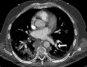
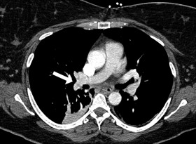
Location of Pulmonary Emboli
Get Radiology Tree app to read full this article<
Baseline Clinical Information
Get Radiology Tree app to read full this article<
Table 1
Baseline Characteristics Among Patients with High Versus Low Confident Positive CTPA Reports
Variables Overall Segmental and Subsegmental Central and Lobar High Confidence (%) Low Confidence (%)P Value High Confidence (%) Low Confidence (%)P Value High Confidence (%) Low Confidence (%)P Value N 1504 160 763 116 741 44 Male 44.2 47.5 .45 37.9 46.6 .08 50.7 50 >.99 Age, mean ± SD, y 57 ± 16 57 ± 17 .17 56 ± 17 56 ± 18 .68 60 ± 16 57 ± 14 .26 Largest reported branch with PE Central, 27; lobar, 23; seg, 38; subseg: 12 Central, 2.5; lobar, 25; seg, 46; subseg, 26 <.001 — — — — — — Cancer 52.6 46.9 .18 52.4 43.1 .07 52.9 56.8 .64 Hypertension 40 46.2 .12 35.2 45.7 .03 44.8 47.7 .75 Diabetes 14.2 21.9 .01 12.2 24.1 .001 16.3 15.9 >.99 CHF 4.8 8.7 .03 5.5 7.8 .39 4 11.4 .04 Surgery 38 41.2 .44 39.3 44 .36 36.6 34.1 .87 COPD 7.6 13.1 .02 8.4 15.5 .02 6.7 6.8 >.99 CRI 2.7 4.4 .21 2.8 4.3 .001 2.7 4.5 .35 CAD 11.7 19.3 .007 10.1 19 .007 13.4 20.4 .17
CAD, coronary artery disease; CHF, congestive heart failure; COPD, chronic obstructive pulmonary disease; CRI, chronic renal insufficiency; CTPA, computed tomography pulmonary angiography; PE, pulmonary embolism; SD, standard deviation.
Get Radiology Tree app to read full this article<
Clinical Outcomes
Get Radiology Tree app to read full this article<
Get Radiology Tree app to read full this article<
Ancillary Studies
Get Radiology Tree app to read full this article<
Statistics
Get Radiology Tree app to read full this article<
Get Radiology Tree app to read full this article<
Get Radiology Tree app to read full this article<
Results
Get Radiology Tree app to read full this article<
Table 2
Keywords for Less Confident Interpretation
Reason Number Motion artifact 63 Insufficient contrast opacification 34 Probable PE 17 Suspicious PE 9 Possible PE 8 Likely (to be PE) 6 Acute versus chronic PE 4 Poor image quality due to large body habitus 4 Poor image quality due to streak artifact 4 Tumor or PE 3 Suggestive of PE 2 Trauma or PE 1 Poor image quality due to scoliosis 1 Poor image quality due to atelectasis 1 Concern of PE 1 PE not excluded 1 Worrisome 1
PE, pulmonary embolism.
Get Radiology Tree app to read full this article<
Baseline Clinical Characteristics Related to Low Confident Reporting
Get Radiology Tree app to read full this article<
Get Radiology Tree app to read full this article<
Get Radiology Tree app to read full this article<
Clinical Outcomes
Get Radiology Tree app to read full this article<
Table 3
Comparison of Outcomes Among Patients with High Versus Low Confident Positive CTPA Reports
Outcome High Confidence, n (%) Low Confidence, n (%)P Value Primary outcomes PE-related 30-day death 70 (4.0) 8 (5.0) ∗ .54 PE-related 90-day death 100 (4.5) 9 (5.6) ∗ .78 Secondary outcomes PE–related therapy (%) 96.4 85.0 <.001 Length of hospital stay, median (IQR) 6 (3–12) 7 (4–13) .033 All-cause 30-day death 173 (11.5) 24 (15.0) .19 All-cause 90-day death 293 (19.5) 34 (21.2) .60
CTPA, computed tomography pulmonary angiography; IQR, interquartile range; PE, pulmonary embolism.
Get Radiology Tree app to read full this article<
Table 4
Results From Multivariable Analyses
Outcome Odds Ratio or Hazard Ratio 95% CI_P_ Value Primary outcomes 30-Day PE-related death 1.09 0.51–2.32 .83 90-Day PE-related death 0.81 0.40–1.64 .56 Secondary outcomes Hospital discharge 0.85 0.72–1.00 .054 PE-related treatment 0.18 0.10–0.31 <.001 30-Day all-cause death 1.13 0.67–1.92 .64 90-Day all-cause death 1.08 0.69–1.71 .75
CI, confidence interval; PE, pulmonary embolism.
Get Radiology Tree app to read full this article<
Ancillary Studies
Get Radiology Tree app to read full this article<
Table 5
Summary of the Follow-Up Radiologic Studies for Low Confident Group
Modality Positive Negative Unclear CTPA 13 8 0 CTPA + CT venography 0 13 3 Ventilation perfusion scan 0 0 0
CTPA, computed tomography pulmonary angiography.
Get Radiology Tree app to read full this article<
Get Radiology Tree app to read full this article<
Discussion
Get Radiology Tree app to read full this article<
Get Radiology Tree app to read full this article<
Get Radiology Tree app to read full this article<
Get Radiology Tree app to read full this article<
Get Radiology Tree app to read full this article<
Get Radiology Tree app to read full this article<
Get Radiology Tree app to read full this article<
Get Radiology Tree app to read full this article<
References
1. Otero H.J., Steigner M.L., Rybicki F.J.: The “post-64” era of coronary CT angiography: understanding new technology from physical principles. Radiol Clin North Am 2009; 47: pp. 79-90.
2. Remy-Jardin M., Remy J., Wattinne L., et. al.: Central pulmonary thromboembolism: diagnosis with spiral volumetric CT with the single-breath-hold technique–comparison with pulmonary angiography. Radiology 1992; 185: pp. 381-387.
3. Garg K., Welsh C.H., Feyerabend A.J., et. al.: Pulmonary embolism: diagnosis with spiral CT and ventilation-perfusion scanning–correlation with pulmonary angiographic results or clinical outcome. Radiology 1998; 208: pp. 201-208.
4. Mayo J.R., Remy-Jardin M., Muller N.L., et. al.: Pulmonary embolism: prospective comparison of spiral CT with ventilation-perfusion scintigraphy. Radiology 1997; 205: pp. 447-452.
5. Hunsaker A.R., Lu M.T., Goldhaber S.Z., et. al.: Imaging in acute pulmonary embolism with special clinical scenarios. Circ Cardiovasc Imaging 2010; 3: pp. 491-500.
6. Stein P.D., Fowler S.E., Goodman L.R., et. al.: Multidetector computed tomography for acute pulmonary embolism. N Engl J Med 2006; 354: pp. 2317-2327.
7. Kumamaru K.K., Hunsaker A.R., Kumamaru H., et. al.: Correlation between early direct communication of positive CT pulmonary angiography findings and improved clinical outcomes. Chest 2013; 144: pp. 1546-1554.
8. Wiener R.S., Schwartz L.M., Woloshin S.: Time trends in pulmonary embolism in the United States: evidence of overdiagnosis. Archives of internal medicine 2011; 171: pp. 831-837.
9. Goldhaber S.Z., Bounameaux H.: Pulmonary embolism and deep vein thrombosis. Lancet 2012; 379: pp. 1835-1846.
10. Berlin L.: Pitfalls of the vague radiology report. AJR American journal of roentgenology 2000; 174: pp. 1511-1518.
11. Espeland A., Baerheim A.: General practitioners’ views on radiology reports of plain radiography for back pain. Scandinavian journal of primary health care 2007; 25: pp. 15-19.
12. Naik S.S., Hanbidge A., Wilson S.R.: Radiology reports: examining radiologist and clinician preferences regarding style and content. AJR American journal of roentgenology 2001; 176: pp. 591-598.
13. Plumb A.A., Grieve F.M., Khan S.H.: Survey of hospital clinicians’ preferences regarding the format of radiology reports. Clinical radiology 2009; 64: pp. 386-394. 95-6
14. Grieve F.M., Plumb A.A., Khan S.H.: Radiology reporting: a general practitioner’s perspective. The British journal of radiology 2010; 83: pp. 17-22.
15. Clinger N.J., Hunter T.B., Hillman B.J.: Radiology reporting: attitudes of referring physicians. Radiology 1988; 169: pp. 825-826.
16. Hall F.M.: Language of the radiology report: primer for residents and wayward radiologists. AJR American journal of roentgenology 2000; 175: pp. 1239-1242.
17. Reiner B.: Uncovering and improving upon the inherent deficiencies of radiology reporting through data mining. J Digit Imaging 2010; 23: pp. 109-118.
18. Wallis A., McCoubrie P.: The radiology report–are we getting the message across?. Clinical radiology 2011; 66: pp. 1015-1022.
19. Abujudeh H.H., Kaewlai R., Farsad K., et. al.: Computed tomography pulmonary angiography: an assessment of the radiology report. Acad Radiol 2009; 16: pp. 1309-1315.
20. ACR practice parameter for communication of diagnostic imaging findings. Available at: http://www.acr.org/~/media/ACR/Documents/PGTS/guidelines/Comm_Diag_Imaging.pdf . Accessed September, 2015.
21. Sobel J.L., Pearson M.L., Gross K., et. al.: Information content and clarity of radiologists’ reports for chest radiography. Acad Radiol 1996; 3: pp. 709-717.
22. Kelly A.M., Patel S., Carlos R.C., et. al.: Multidetector row CT pulmonary angiography and indirect venography for the diagnosis of venous thromboembolic disease in intensive care unit patients. Acad Radiol 2006; 13: pp. 486-495.
23. Hayes S.A., Soff G.A., Zabor E.C., et. al.: Clinical consequences of an indeterminate CT pulmonary angiogram in cancer patients. Clinical imaging 2014; 38: pp. 637-640.
24. Guyatt G.H., Akl E.A., Crowther M., et. al.: Executive summary: Antithrombotic Therapy and Prevention of Thrombosis, 9th ed. American College of Chest Physicians Evidence-Based Clinical Practice Guidelines. Chest 2012; 141: pp. 7S-47S.
25. Eyer B.A., Goodman L.R., Washington L.: Clinicians’ response to radiologists’ reports of isolated subsegmental pulmonary embolism or inconclusive interpretation of pulmonary embolism using MDCT. AJR American journal of roentgenology 2005; 184: pp. 623-628.
26. Torbicki A., Perrier A., Konstantinides S., et. al.: Guidelines on the diagnosis and management of acute pulmonary embolism: the Task Force for the Diagnosis and Management of Acute Pulmonary Embolism of the European Society of Cardiology (ESC). Eur Heart J 2008; 29: pp. 2276-2315.
27. BI-RADS® – MAMMOGRAPHY, 5th ed. American College of Radiology. Available at: http://www.acr.org/Quality-Safety/Resources/BIRADS/Mammography . Accessed September, 2015.