
Rationale and Objectives
To assess the diagnostic value of automated breast volume scanning (ABVS) versus conventional ultrasound (US) in breast cancer screening.
Materials and Methods
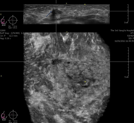
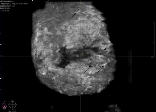
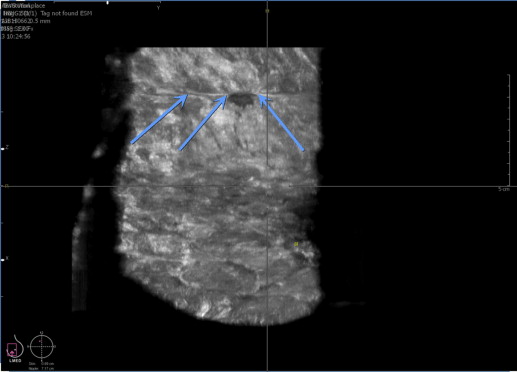
This study retrospectively analyzed the ABVS and US images from 200 women who underwent breast examination and were recommended for biopsy in our health management centers between July 22, 2011, and October 20, 2013. We retrospectively assessed whether breast lesions from 200 women, which were detected and classified by US, could be detected and classified by an independent examiner using only ABVS findings. The sensitivity and specificity of ABVS versus US in determining lesion malignancy were calculated using biopsy as the gold standard.
Results
In the 200 cases, 273 and 194 individual lesions were detected by ABVS and US, respectively. All 194 US-detected lesions were detected by ABVS. Pathologic examination determined that, of the 273 total lesions, 251 lesions were benign and 22 lesions were malignant. US detected 21 of the 22 malignant lesions and ABVS detected all 22 malignant lesions. The sensitivity and specificity of ABVS relative to biopsy (gold standard) were 28.95% and 100%, whereas the sensitivity and specificity of US relative to biopsy were 43.06% and 98.36%.
Conclusions
US displays superior sensitivity to ABVS across all Breast Imaging Reporting and Data System (BI-RADS) density categories while displaying equivalent specificity with the exception of BI-RADS density category 1, in which ABVS displayed a slightly superior specificity. As ABVS possesses several advantages and limitations with respect to US, ABVS may serve as an effective, adjunct, screening tool to mammography and conventional sonography.
Mammography has long been the mainstay of breast cancer detection and is the only screening test proven to reduce mortality . Although mammography remains the gold standard of breast cancer screening, mammography has limitations. First, the sensitivity of mammography is decreased in dense breasts . Second, mammography displays high false-positive rates, resulting in high callback rates and unnecessary biopsies that increase cost, radiation dose, and patient anxiety . Third, mammographic radiation exposure may contribute to an increased incidence of breast cancer in high-risk populations . These concerns may decrease compliance with breast cancer screening recommendations.
As mammographic sensitivity is adversely affected by dense breast tissue, mammography is not particularly suited to women whose breasts are typically more dense . Breast ultrasound (US) has been shown to be an effective adjunct imaging modality in the evaluation of women with dense breast tissue (American College of Radiology’s Breast Imaging Reporting and Data System criteria [ACR BI-RADS] density categories 3 and 4), and mammography combined with US can increase tumor detection rates over mammography alone . However, conventional breast US has provided little practical benefit in cancer detection because of the poor conspicuity of some cancers, the significant operator time and experience necessary for a high-quality screening, and the lack of standardization due to variability in operator skill and experience . Thus, other sonographic methods that adequately address these limitations are needed.
Get Radiology Tree app to read full this article<
Materials and methods
Subject Selection
Get Radiology Tree app to read full this article<
Acquisition of US Data
Get Radiology Tree app to read full this article<
Get Radiology Tree app to read full this article<
Acquisition of ABVS Data
Get Radiology Tree app to read full this article<
Independent Blinded Interpretation of ABVS Data
Get Radiology Tree app to read full this article<
Get Radiology Tree app to read full this article<
Biopsy (Gold Standard)
Get Radiology Tree app to read full this article<
Statistical Analysis
Get Radiology Tree app to read full this article<
Results
Lesion Detection
Get Radiology Tree app to read full this article<
Table 1
Detailed Characteristics of All 273 Lesions
ABVS Lesion (273 Total) Patient ID BI-RADS Breast Density Score Detected by US (Yes or No) BI-RADS Lesion Score by ABVS BI-RADS Lesion Score by US Microcalcifications Detected by ABVS (Yes or No) Microcalcifications Detected by US (Yes or No) Retraction Phenomena Detected by ABVS (Yes or No) Retraction Phenomena Detected by US (Yes or No) Resistance Index Value by US Malignant by Biopsy (Yes or No) 1 1 3 Yes 3 3 No No No No ∗ No 2 2 3 No 3 No No No No ∗ Yes 3 3 2 Yes 2 2 No No No No ∗ No 4 4 4 No 2 No No No No ∗ No 5 5 3 No 2 No No No No ∗ No 6 6 2 Yes 3 3 No No No No 0.51 Yes 7 6 2 Yes 3 3 No No No No ∗ Yes 8 6 2 No 2 No No No No ∗ No 9 7 3 Yes 3 3 No No No No ∗ No 10 7 3 Yes 3 3 No No No No ∗ Yes 11 8 3 Yes 3 3 No No No No ∗ Yes 12 9 1 Yes 3 3 No No No No ∗ Yes 13 10 3 No 2 No No No No ∗ No 14 11 3 Yes 4a 4a No No No No 0.83 Yes 15 12 3 Yes 2 2 No No No No ∗ No 16 13 3 No 3 No No No No ∗ No 17 14 2 Yes 3 3 No No No No ∗ No 18 15 3 No 2 No No No No ∗ No 19 16 2 Yes 3 3 No No No No ∗ No 20 17 3 Yes 3 3 No No No No ∗ No 21 18 2 Yes 3 3 No No No No 0.55 Yes 22 19 1 No 3 No No No No ∗ No 23 20 2 No 2 No No No No ∗ No 24 20 2 Yes 2 2 No No No No ∗ No 25 21 4 Yes 4c 4b No No Yes No 0.8 Yes 26 22 2 Yes 2 2 No No No No ∗ No 27 23 3 Yes 3 3 No No No No ∗ No 28 23 3 No 3 No No No No ∗ No 29 24 2 Yes 2 2 No No No No ∗ No 30 25 3 Yes 3 3 No No No No ∗ No 31 26 2 Yes 4a 4a Yes Yes No No 0.67 Yes 32 26 2 Yes 3 3 No No No No ∗ Yes 33 27 3 Yes 2 2 No No No No ∗ No 34 28 1 No 2 No No No No ∗ No 35 29 4 Yes 2 2 No No No No ∗ No 36 29 4 Yes 3 3 No No No No ∗ Yes 37 30 3 No 2 No No No No ∗ No 38 30 3 Yes 2 2 No No No No ∗ No 39 30 3 Yes 3 3 No No No No 0.63 No 40 30 3 Yes 3 3 No No No No ∗ No 41 31 3 Yes 3 3 No No No No ∗ Yes 42 32 3 No 2 No No No No ∗ No 43 33 1 Yes 3 3 No No No No ∗ No 44 34 3 Yes 5 4a No No Yes No ∗ Yes 45 35 4 Yes 2 2 No No No No ∗ No 46 36 3 No 3 No No No No ∗ No 47 37 2 Yes 3 3 No No No No ∗ No 48 38 3 Yes 3 3 No No No No ∗ No 49 39 3 Yes 3 3 No No No No ∗ No 50 40 3 No 3 No No No No ∗ No 51 41 2 No 3 No No No No ∗ No 52 42 4 Yes 4a 4a Yes No No No 0.75 Yes 53 42 4 No 2 No No No No ∗ No 54 42 4 Yes 2 2 No No No No ∗ Yes 55 43 3 Yes 3 3 No No No No ∗ No 56 44 2 No 2 No No No No ∗ No 57 45 2 Yes 2 2 No No No No ∗ No 58 46 2 Yes 3 3 No No No No ∗ Yes 59 46 2 Yes 3 4a Yes Yes No No 0.68 Yes 60 47 3 Yes 3 3 No No No No ∗ No 61 47 3 No 3 No No No No ∗ No 62 47 3 Yes 3 3 No No No No ∗ No 63 48 1 No 2 No No No No ∗ No 64 49 3 Yes 3 3 No No Yes No ∗ No 65 50 2 Yes 3 3 No No No No ∗ No 66 51 3 No 3 No No No No ∗ Yes 67 52 2 No 2 No No No No ∗ No 68 52 2 Yes 3 3 No No No No 0.51 No 69 53 3 Yes 2 2 No No No No ∗ No 70 54 4 Yes 2 2 No No No No ∗ No 71 55 4 Yes 5 4b Yes Yes Yes No ∗ Yes 72 56 3 Yes 3 4a No No No No ∗ Yes 73 56 3 Yes 2 2 No No No No ∗ No 74 56 3 No 2 No No No No ∗ No 75 57 2 No 2 No No No No ∗ No 76 58 1 Yes 3 3 No No No No 0.54 Yes 77 59 2 Yes 3 4a No No Yes No ∗ Yes 78 60 2 No 2 No No No No ∗ No 79 61 3 Yes 3 3 No No No No ∗ Yes 80 62 2 Yes 2 3 No No No No ∗ Yes 81 62 2 No 2 No No No No ∗ No 82 63 3 Yes 3 3 No No No No ∗ Yes 83 64 3 Yes 2 2 No No No No ∗ No 84 65 3 Yes 4c 4a Yes Yes Yes No ∗ Yes 85 65 3 Yes 2 2 No No No No ∗ No 86 66 1 No 2 No No No No ∗ No 87 67 2 Yes 3 3 No No No No ∗ No 88 68 3 Yes 3 3 No No No No 0.66 Yes 89 69 4 Yes 3 3 No No No No 0.54 Yes 90 70 2 Yes 3 3 No No No No ∗ Yes 91 70 2 No 2 No No Yes No ∗ No 92 71 3 No 3 No No No No ∗ No 93 72 3 Yes 2 2 No No No No ∗ No 94 73 2 Yes 3 3 No No No No ∗ No 95 74 2 Yes 3 3 No No No No ∗ Yes 96 75 3 Yes 3 3 No No No No 0.48 No 97 75 3 No 2 No No No No ∗ No 98 75 3 Yes 5 4c Yes Yes Yes No 0.72 Yes 99 76 2 Yes 2 2 No No No No ∗ No 100 77 4 No 3 No No No No ∗ No 101 77 4 Yes 3 3 No No No No ∗ No 102 78 4 Yes 2 2 No No No No ∗ No 103 79 1 Yes 3 3 No No No No ∗ Yes 104 80 1 Yes 2 2 No No No No ∗ No 105 81 2 No 4a No No Yes No ∗ Yes 106 81 2 No 2 No No No No ∗ No 107 81 2 Yes 2 2 No No No No ∗ No 108 82 3 Yes 2 2 No No No No ∗ No 109 83 3 Yes 3 3 No No No No ∗ Yes 110 84 4 No 2 No No No No ∗ No 111 84 4 Yes 2 2 No No No No ∗ No 112 84 4 No 2 No No No No ∗ No 113 84 4 Yes 3 4a No No No No 0.77 Yes 114 85 2 Yes 2 2 No No No No ∗ No 115 86 2 Yes 2 2 No No No No ∗ No 116 87 3 Yes 2 2 No No No No ∗ No 117 88 1 No 3 No No No No ∗ No 118 89 3 Yes 3 3 Yes No No No ∗ Yes 119 90 2 Yes 3 3 Yes Yes Yes No ∗ Yes 120 90 2 No 2 No No No No ∗ No 121 91 3 Yes 4a 4a No No Yes No ∗ Yes 122 92 4 Yes 3 3 No No No No ∗ No 123 93 3 Yes 3 3 No No No No ∗ No 124 94 3 No 2 No No No No ∗ No 125 95 1 Yes 2 2 No No No No ∗ No 126 95 1 No 3 No No No No ∗ No 127 96 3 Yes 4a 3 No No No No 0.68 Yes 128 97 2 Yes 3 3 No No No No 0.65 Yes 129 97 2 No 2 No No No No ∗ No 130 98 2 Yes 2 2 No No No No ∗ No 131 98 2 No 2 No No No No ∗ No 132 99 1 Yes 3 3 No No No No ∗ Yes 133 100 4 Yes 2 2 No No No No ∗ No 134 101 4 Yes 4c 4b No No Yes No 0.73 Yes 135 102 2 Yes 3 3 No No No No ∗ No 136 102 2 No 3 No No No No ∗ No 137 103 3 Yes 3 3 No No No No 0.63 Yes 138 103 3 No 2 No No No No ∗ No 139 104 2 No 3 No No No No ∗ No 140 105 3 Yes 2 2 No No No No ∗ No 141 106 4 Yes 3 3 No No No No ∗ Yes 142 107 2 Yes 2 2 No No No No ∗ No 143 107 2 No 2 No No No No ∗ No 144 107 2 No 2 No No No No ∗ No 145 108 3 Yes 2 2 No No No No ∗ No 146 109 1 Yes 3 3 No No No No ∗ No 147 110 1 Yes 4a 4a No No Yes No ∗ Yes 148 111 3 Yes 3 3 No No No No ∗ No 149 112 3 No 2 No No No No ∗ No 150 113 3 Yes 3 3 No No No No ∗ No 151 113 3 Yes 3 3 No No No No ∗ No 152 113 3 No 2 No No No No ∗ No 153 114 3 Yes 3 4a Yes Yes No No ∗ Yes 154 115 2 Yes 3 3 No No No No ∗ Yes 155 116 1 No 3 No No No No ∗ No 156 117 1 Yes 3 3 No No No No ∗ No 157 117 1 Yes 3 3 No No No No ∗ No 158 118 4 No 2 No No No No ∗ No 159 119 4 Yes 4a 4b No No No No 0.83 Yes 160 120 3 No 2 No No No No ∗ No 161 121 3 Yes 3 3 No No No No 0.46 Yes 162 122 3 Yes 3 3 No No Yes No ∗ Yes 163 123 2 No 3 No No No No ∗ No 164 123 2 Yes 3 3 No No No No ∗ No 165 124 2 Yes 2 3 No No No No ∗ No 166 125 3 Yes 2 3 No No No No ∗ No 167 126 3 Yes 3 3 No No No No ∗ No 168 126 3 No 3 No No No No ∗ No 169 126 3 No 3 No No No No ∗ No 170 127 3 Yes 4c 4a No No Yes No 0.56 Yes 171 128 2 Yes 3 3 No No No No ∗ Yes 172 129 1 Yes 2 3 No No No No 0.45 Yes 173 129 1 Yes 2 2 No No No No ∗ No 174 130 2 Yes 2 2 No No No No ∗ No 175 130 2 No 3 No No No No ∗ No 176 131 3 Yes 3 3 No No No No ∗ No 177 132 3 Yes 4b 4b No No No No 0.66 Yes 178 133 2 Yes 3 3 No No No No ∗ No 179 134 3 Yes 3 4a No No No No 0.65 No 180 135 2 Yes 3 3 No No No No ∗ Yes 181 135 2 Yes 2 2 No No No No ∗ No 182 135 2 No 2 No No Yes No ∗ No 183 135 2 Yes 2 2 No No No No ∗ No 184 136 3 Yes 3 3 No No No No ∗ No 185 137 3 No 3 No No No No ∗ No 186 138 4 No 2 No No No No ∗ No 187 139 1 Yes 3 3 No No No No ∗ Yes 188 140 2 Yes 3 3 No No No No ∗ Yes 189 141 2 Yes 5 4b Yes No Yes No 0.78 Yes 190 142 2 Yes 3 3 No No No No ∗ No 191 143 3 Yes 3 3 No No No No 0.55 No 192 144 4 No 2 No No No No ∗ No 193 144 4 Yes 3 3 No No No No ∗ No 194 145 3 Yes 2 2 No No No No ∗ No 195 146 1 No 3 No No No No ∗ No 196 147 2 Yes 3 3 No No No No ∗ No 197 148 3 Yes 3 3 No No No No ∗ No 198 149 3 Yes 2 2 No No No No ∗ No 199 149 3 Yes 3 4a No No No No ∗ Yes 200 149 3 No 2 No No No No ∗ No 201 150 2 Yes 3 4a No No No No 0.55 Yes 202 150 2 Yes 4a 4b No No No No 0.85 Yes 203 151 3 Yes 2 2 No No No No ∗ No 204 152 3 Yes 3 3 No No No No ∗ No 205 153 3 Yes 3 3 No No No No ∗ No 206 154 1 Yes 3 3 No No No No ∗ No 207 155 1 No 2 No No No No ∗ No 208 155 1 Yes 2 2 No No No No ∗ No 209 155 1 Yes 3 4a No No No No 0.67 Yes 210 156 3 Yes 3 3 No No No No ∗ No 211 157 2 Yes 3 3 No No No No ∗ No 212 158 3 No 3 No No No No ∗ No 213 158 3 Yes 3 3 No No No No ∗ No 214 159 2 Yes 3 3 No No No No ∗ Yes 215 160 3 Yes 2 2 No No No No ∗ No 216 161 3 No 2 No No No No ∗ No 217 162 2 Yes 3 3 No No Yes No ∗ No 218 162 2 Yes 3 4a No No No No 0.57 Yes 219 163 2 Yes 3 4a No No No No ∗ Yes 220 164 4 No 2 No No No No ∗ No 221 164 4 Yes 2 2 No No No No ∗ No 222 165 4 Yes 4b 4a No No Yes No ∗ Yes 223 166 1 Yes 2 2 No No No No ∗ No 224 166 1 No 2 No No No No ∗ No 225 166 1 No 2 No No No No ∗ No 226 166 1 Yes 2 3 No No No No 0.58 No 227 167 3 Yes 3 3 No No No No ∗ No 228 168 3 No 2 No No No No ∗ No 229 169 3 Yes 2 2 No No No No ∗ No 230 170 3 No 3 No No No No ∗ No 231 171 2 Yes 3 3 No No No No ∗ No 232 172 2 Yes 2 2 No No No No ∗ No 233 173 3 Yes 2 2 No No No No ∗ No 234 173 3 Yes 4a 4a No No No No ∗ Yes 235 174 3 Yes 3 3 No No No No ∗ No 236 174 3 No 3 No No No No ∗ No 237 175 4 Yes 2 2 No No No No ∗ No 238 175 4 Yes 2 3 No No No No ∗ No 239 176 3 Yes 2 2 No No No No ∗ No 240 177 3 Yes 3 3 No No No No ∗ No 241 178 2 Yes 3 3 No No No No ∗ No 242 179 1 Yes 3 2 No No No No ∗ No 243 180 1 No 2 No No No No ∗ No 244 181 3 No 2 No No No No ∗ No 245 181 3 Yes 3 3 No No No No ∗ Yes 246 181 3 Yes 3 4a No No No No 0.6 Yes 247 182 2 Yes 2 2 No No No No ∗ No 248 182 2 Yes 4a 3 Yes No No No ∗ Yes 249 183 3 Yes 3 3 No No No No ∗ No 250 184 1 Yes 3 3 No No No No ∗ No 251 185 2 No 2 No No No No ∗ No 252 186 1 Yes 3 3 No No No No ∗ No 253 187 3 Yes 2 2 No No No No ∗ No 254 187 3 No 3 No No No No ∗ Yes 255 188 4 Yes 2 3 No No No No 0.62 Yes 256 189 3 Yes 2 2 No No No No ∗ No 257 190 2 Yes 3 2 No No No No ∗ No 258 191 2 Yes 3 3 No No No No ∗ Yes 259 192 3 No 3 No No No No ∗ No 260 193 1 Yes 2 2 No No No No ∗ No 261 193 1 No 2 No No No No ∗ No 262 193 1 Yes 3 3 No No No No ∗ No 263 194 2 Yes 2 2 No No No No ∗ No 264 194 2 Yes 3 3 No No Yes No ∗ No 265 195 3 No 3 No No No No ∗ No 266 196 3 Yes 3 3 No No No No ∗ No 267 197 2 Yes 2 2 No No No No ∗ No 268 197 2 Yes 4a 4a Yes Yes No No ∗ Yes 269 198 1 Yes 3 4a No No No No 0.45 No 270 199 4 No 3 No No No No ∗ No 271 200 3 Yes 2 2 No No No No ∗ No 272 200 3 Yes 3 4a No No No No ∗ Yes 273 200 3 No 2 No No No No ∗ No
ABVS, automated breast volume scanning; BI-RADS, Breast Imaging Reporting and Data System; US, conventional ultrasound.
Get Radiology Tree app to read full this article<
Get Radiology Tree app to read full this article<
Lesion Characteristics
Get Radiology Tree app to read full this article<
Get Radiology Tree app to read full this article<
Table 2
Sensitivity and Specificity of ABVS versus US Relative to Biopsy (Gold Standard)
BI-RADS Breast Density ABVS US Sensitivity (%) Specificity (%) AUC 95% CI Sensitivity (%) Specificity (%) AUC 95% CI 1 12.50 100 0.7188 0.5537–0.8837 25.00 94.12 0.7500 0.5997–0.9000 2 22.22 100 0.8273 0.7565–0.8979 34.62 100 0.8410 0.7709–0.9110 3 31.03 100 0.8137 0.7548–0.8725 50.00 100 0.8393 0.7685–0.9107 4 50.00 100 0.8598 0.7235–0.9961 58.33 100 0.8889 0.7655–1.0000 Total 28.95 100 0.7864 0.7345–0.8384 43.06 98.36 0.8309 0.7854–0.8764
ABVS, automated breast volume scanning; AUC; area under the receiver operating characteristic curve; CI, confidence interval; US, conventional ultrasound.
Get Radiology Tree app to read full this article<
Discussion
Get Radiology Tree app to read full this article<
Advantages of ABVS Over US in Detecting Breast Lesions
Get Radiology Tree app to read full this article<
Advantages of ABVS Over US in Detecting Malignant Breast Lesions
Get Radiology Tree app to read full this article<
Get Radiology Tree app to read full this article<
Get Radiology Tree app to read full this article<
Get Radiology Tree app to read full this article<
Limitations of ABVS
Get Radiology Tree app to read full this article<
Get Radiology Tree app to read full this article<
Study Limitations
Get Radiology Tree app to read full this article<
Conclusions
Get Radiology Tree app to read full this article<
Get Radiology Tree app to read full this article<
References
1. Drukteinis J.S., Mooney B.P., Flowers C.I., et. al.: Beyond mammography: new frontiers in breast cancer screening. The American journal of medicine 2013;
2. Kolb T.M., Lichy J., Newhouse J.H.: Comparison of the performance of screening mammography, physical examination, and breast us and evaluation of factors that influence them: an analysis of 27,825 patient evaluations. Radiology 2002; 225: pp. 165-175.
3. Hou M.F., Chuang H.Y., Ou-Yang F., et. al.: Comparison of breast mammography, sonography and physical examination for screening women at high risk of breast cancer in Taiwan. Ultrasound in medicine & biology 2002; 28: pp. 415-420.
4. Feig S.A.: Adverse effects of screening mammography. Radiologic Clinics of North America 2004; 42: pp. 807-819.
5. Kaplan S.S.: Clinical utility of bilateral whole-breast US in the evaluation of women with dense breast tissue. Radiology 2001; 221: pp. 641-649.
6. Mendelson E.B., Tobin C.E.: Critical pathways in using breast US. Radiographics 1995; 15: pp. 935-945.
7. Flobbe K., Bosch A.M., Kessels A.G., et. al.: The additional diagnostic value of ultrasonography in the diagnosis of breast cancer. Archives of internal medicine 2003; 163: pp. 1194.
8. Drukteinis J.S., Mooney B.P., Flowers C.I., et. al.: Beyond mammography: new frontiers in breast cancer screening. The American journal of medicine 2013;
9. Wojcinski S., Farrokh A., Hille U., et. al.: The automated breast volume scanner (ABVS): initial experiences in lesion detection compared with conventional handheld B-mode ultrasound: a pilot study of 50 cases. International journal of women’s health 2011; 3: pp. 337.
10. Kim Y.W., Kim S.K., Youn H.J., et. al.: The clinical utility of automated breast volume scanner: a pilot study of 139 cases. Journal of breast cancer 2013; 16: pp. 329-334.
11. Tozaki M., Isobe S., Yamaguchi M., et. al.: Optimal scanning technique to cover the whole breast using an automated breast volume scanner. Japanese journal of radiology 2010; 28: pp. 325-328.
12. CHEN L., CHEN Y., PANG Y., et. al.: Automated breast volume scanner of ultrasound in initial application of breast occupation diseases. [J] Chinese Journal of Medical Imaging Technology 2011; 7, 021
13. Zhang Q., Hu B., Li W.B.: Detection of breast lesions using an automated breast volume scanner system. Journal of International Medical Research 2012; 40: pp. 300-306.
14. ZHANG Q., HU B., HU B., et. al.: Clinical application of automated breast volume scanner. [J] Chinese Journal of Interventional Imaging and Therapy 2011; 1, 023
15. Kotsianos-Hermle D., Hiltawsky K.M., Wirth S., et. al.: Analysis of 107 breast lesions with automated 3D ultrasound and comparison with mammography and manual ultrasound. European journal of radiology 2009; 71: pp. 109-115.
16. Golatta M., Franz D., Harcos A., et. al.: Interobserver reliability of automated breast volume scanner (ABVS) interpretation and agreement of ABVS findings with hand held breast ultrasound (HHUS), mammography and pathology results. European journal of radiology 2013;
17. Isobe S., Tozaki M., Yamaguchi M., et. al.: Detectability of breast lesions under the nipple using an automated breast volume scanner: comparison with handheld ultrasonography. Japanese Journal of Radiology 2011; 29: pp. 361-365.
18. Shin H.J., Kim H.H., Cha J.H., et. al.: Automated ultrasound of the breast for diagnosis: interobserver agreement on lesion detection and characterization. American Journal of Roentgenology 2011; 197: pp. 747-754.
19. Moskowitz M.: The predictive value of certain mammographic signs in screening for breast cancer. Cancer 1983; 51: pp. 1007-1011.
20. Zhu Q.Q., Bao L.Y., Zhu L.Q., et. al.: ABVS aided diagnosis of breast ultrasound image based on BI-RADS in diagnosis of breast ductal carcinoma in situ. Yixue Yingxiangxue Zazhi (Journal of Medical Imaging) 2012; 22:
21. Watermann D.O., Földi M., Hanjalic-Beck A., et. al.: Three-dimensional ultrasound for the assessment of breast lesions. Ultrasound in obstetrics & gynecology 2005; 25: pp. 592-598.
22. Isobe S., Tozaki M., Yamaguchi M., et. al.: Detectability of breast lesions under the nipple using an automated breast volume scanner: comparison with handheld ultrasonography. [J] Jpn Radiol 2011; 29: pp. 361-365.
23. Wanga H.Y., Jianga Y.X., Zhua Q.-L., et. al.: Differentiation of benign and malignant breast lesions: a comparison between automatically generated breast volume scans and handheld ultrasound examinations. European Journal of Radiology 2012; 81: pp. 3190-3200.
24. Chen C-J., Zhang Y., Shi X-y., et. al.: Ultrasonic automated breast volume scanning and Doppler ultrasonography imaging in different breast lesions. [J] Clinical Focus 2012; 27: pp. 106-107.
25. Wojcinski S., Farrokh A.: The automated breast volume scanner (ABVS): initial experiences in lesion detection compared with conventional handheld B-mode ultrasound: a pilot study of 50 cases. International Journal of Women’s Health 2011; 3: pp. 337-346.
26. Kotsianos-Hermle a D., Wirth a S., Fischer a T., et. al.: First clinical use of a standardized three-dimensional ultrasound for breast imaging. European Journal of Radiology 2009; 71: pp. 102-108.