
Rationale and Objectives
Today, lung volumes can be easily calculated from chest computed tomography (CT) scans. Modern postprocessing workstations allow automated volume measurement of data sets acquired. However, there are challenges in the use of lung volume as an indicator of pulmonary disease when it is obtained from routine CT. Intra-individual variation and methodologic aspects have to be considered. Our goal was to assess the reliability of volumetric measurements in routine CT lung scans.
Materials and Methods
Forty adult cancer patients whose lungs were unaffected by the disease underwent routine chest CT scans in 3-month intervals, resulting in a total number of 302 chest CT scans. Lung volume was calculated by automatic volumetry software. On average of 7.2 CT scans were successfully evaluable per patient (range 2–15). Intra-individual changes were assessed.
Results
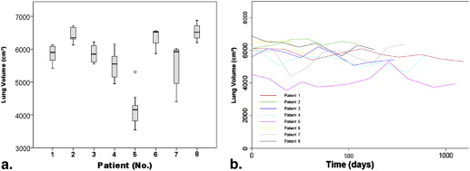
In the set of patients investigated, lung volume was approximately normally distributed, with a mean of 5283 cm 3 (standard deviation = 947 cm 3 , skewness = −0.34, and curtosis = 0.16). Between different scans in one and the same patient the median intra-individual standard deviation in lung volume was 853 cm 3 (16% of the mean lung volume).
Conclusions
Automatic lung segmentation of routine chest CT scans allows a technically stable estimation of lung volume. However, substantial intra-individual variations have to be considered. A median intra-individual deviation of 16% in lung volume between different routine scans was found.
Computed tomography (CT) is a routine tool in clinical practice. CT imaging has been shown superior to plain imaging and magnetic resonance imaging in assessment of lung disease .
State-of-the-art postprocessing workstations allow automated lung-volume measurement from CT data sets. These measurements are of interest in assessing pulmonary conditions that involve alterations in lung volume . Emphysema or chronic allergic asthma can cause an increase in lung volume, cystic fibrosis or sclerodermia can reduce lung volume. In these diseases, lung volume and its time are appropriate markers of disease activity and disease progression .
Get Radiology Tree app to read full this article<
Get Radiology Tree app to read full this article<
Materials and methods
Subjects
Get Radiology Tree app to read full this article<
Procedures and Techniques
Get Radiology Tree app to read full this article<
Data Collection and Validation
Get Radiology Tree app to read full this article<
Get Radiology Tree app to read full this article<
Statistical Tests
Get Radiology Tree app to read full this article<
Results
Study Group
Get Radiology Tree app to read full this article<
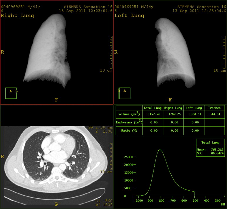
Segmentation
Get Radiology Tree app to read full this article<
Lung Volumes
Get Radiology Tree app to read full this article<

Table 1
Calculated Lung Volumes (Philips Extended Brilliance Workspace, Philips Medical Systems, Netherlands)
Lung Volume Total (cm 3 ) Right Lung (cm 3 ) Left Lung (cm 3 ) Mean 5283 2869 2414 Standard deviation 947 506 480 Median 5457 2962 2468
Get Radiology Tree app to read full this article<
Get Radiology Tree app to read full this article<
Get Radiology Tree app to read full this article<
Discussion
Get Radiology Tree app to read full this article<
Get Radiology Tree app to read full this article<
Get Radiology Tree app to read full this article<
Get Radiology Tree app to read full this article<
Get Radiology Tree app to read full this article<
Get Radiology Tree app to read full this article<
Conclusions
Get Radiology Tree app to read full this article<
Acknowledgments
Get Radiology Tree app to read full this article<
Get Radiology Tree app to read full this article<
Get Radiology Tree app to read full this article<
References
1. Maki D.D., Gefter W.B., Alavi A.: Recent advances in pulmonary imaging. Chest 1999; 116: pp. 1388-1402.
2. Vij R., Strek M.E.: Diagnosis and treatment of connective tissue disease-associated interstitial lung disease. Chest 2013; 143: pp. 814-824.
3. Loutfi S., Khankan A., Al Ghanim S.: Guidelines for multimodality radiological staging of lung cancer. J Infect Public Health 2012; 5: pp. S14-S21.
4. Porcel J.M., Leung C.C., Restrepo M.I., et. al.: Year in review 2012: lung cancer, respiratory infections, tuberculosis, pleural diseases, bronchoscopic intervention and imaging. Respirology 2013; 18: pp. 573-583.
5. de Jong P.A., Lindblad A., Rubin L., et. al.: Progression of lung disease on computed tomography and pulmonary function tests in children and adults with cystic fibrosis. Thorax 2006; 61: pp. 80-85.
6. Garfield J.L., Marchetti N., Gaughan J.P., et. al.: Total lung capacity by plethysmography and high-resolution computed tomography in COPD. Int J Chron Obstruct Pulmon Dis 2012; 7: pp. 119-126.
7. Perez At, Coxson H.O., Hogg J.C., et. al.: Use of CT morphometry to detect changes in lung weight and gas volume. Chest 2005; 128: pp. 2471-2477.
8. Jones P.W., Agusti A.G.: Outcomes and markers in the assessment of chronic obstructive pulmonary disease. Eur Respir J 2006; 27: pp. 822-832.
9. Kumar P., Leonidas J.C., Ashtari M., et. al.: Comparison of lung area by chest radiograph, with estimation of lung volume by helium dilution during prone and supine positioning in mechanically ventilated preterm infants: a pilot study. Pediatr Pulmonol 2005; 40: pp. 219-222.
10. Clausen J.: Measurement of absolute lung volumes by imaging techniques. Eur Respir J 1997; 10: pp. 2427-2431.
11. Bafadhel M., Umar I., Gupta S., et. al.: The role of CT scanning in multidimensional phenotyping of COPD. Chest 2011; 140: pp. 634-642.
12. Chen F., Kubo T., Shoji T., et. al.: Comparison of pulmonary function test and computed tomography volumetry in living lung donors. J Heart Lung Transplant 2011; 30: pp. 572-575.
13. Date H., Shiraishi T., Sugimoto S., et. al.: Outcome of living-donor lobar lung transplantation using a single donor. J Thorac Cardiovasc Surg 2012; 144: pp. 710-715.
14. Iwano S., Okada T., Satake H., et. al.: 3D-CT volumetry of the lung using multidetector row CT: comparison with pulmonary function tests. Acad Radiol 2009; 16: pp. 250-256.
15. Matsuo K., Iwano S., Okada T., et. al.: 3D-CT lung volumetry using multidetector row computed tomography: pulmonary function of each anatomic lobe. J Thorac Imaging 2012; 27: pp. 164-170.
16. Zaporozhan J., Ley S., Gast K.K., et. al.: Functional analysis in single-lung transplant recipients: a comparative study of high-resolution CT, 3He-MRI, and pulmonary function tests. Chest 2004; 125: pp. 173-181.
17. Archer D.C., Coblentz C.L., deKemp R.A., et. al.: Automated in vivo quantification of emphysema. Radiology 1993; 188: pp. 835-838.
18. Bae K.T., Slone R.M., Gierada D.S., et. al.: Patients with emphysema: quantitative CT analysis before and after lung volume reduction surgery. Work in progress. Radiology 1997; 203: pp. 705-714.
19. Palumbo A.A., Luccichenti G., Belgrano M., et. al.: Three-dimensional quantitative assessment of lung parenchyma in cystic fibrosis: preliminary results. Radiol Med 2007; 112: pp. 21-30.
20. Sluimer I., Schilham A., Prokop M., et. al.: Computer analysis of computed tomography scans of the lung: a survey. IEEE Trans Med Imaging 2006; 25: pp. 385-405.
21. Coxson H.O., Mayo J.R., Behzad H., et. al.: Measurement of lung expansion with computed tomography and comparison with quantitative histology. J Appl Physiol 1995; 79: pp. 1525-1530.
22. Kauczor H.U., Heussel C.P., Fischer B., et. al.: Assessment of lung volumes using helical CT at inspiration and expiration: comparison with pulmonary function tests. AJR Am J Roentgenol 1998; 171: pp. 1091-1095.
23. Kendrick A.H.: Comparison of methods of measuring static lung volumes. Monaldi Arch Chest Dis 1996; 51: pp. 431-439.
24. O’Donnell C.R., Bankier A.A., Stiebellehner L., et. al.: Comparison of plethysmographic and helium dilution lung volumes: which is best for COPD?. Chest 2010; 137: pp. 1108-1115.
25. Stanescu D.C.: How to measure lung volume?. Chest 2010; 138: pp. 1280-1281. author reply 1281-1282
26. Patroniti N., Bellani G., Manfio A., et. al.: Lung volume in mechanically ventilated patients: measurement by simplified helium dilution compared to quantitative CT scan. Intensive Care Med 2004; 30: pp. 282-289.
27. Zompatori M., Fasano L., Pacilli A.M., et. al.: [Automatic evaluation of total lung capacity and of emphysema involvement with spiral computerized tomography (CT) in obstructive pneumonia]. Radiol Med 2001; 101: pp. 18-24.
28. Bartlett M.: Properties of sufficiency and statistical tests. Proceedings of the Royal Society Series A 1937; 160: pp. 268-282.
29. Molho M., Shulimzon T., Benzaray S., et. al.: Importance of inspiratory load in the assessment of severity of airways obstruction and its correlation with CO 2 retention in chronic obstructive pulmonary disease. Am Rev Respir Dis 1993; 147: pp. 45-49.
30. Taube C., Kanniess F., Gronke L., et. al.: Reproducibility of forced inspiratory and expiratory volumes after bronchodilation in patients with COPD or asthma. Respir Med 2003; 97: pp. 568-577.
31. Coxson H.O., Rogers R.M., Whittall K.P., et. al.: A quantification of the lung surface area in emphysema using computed tomography. Am J Respir Crit Care Med 1999; 159: pp. 851-856.
32. AARC clinical practice guideline. Body plethysmography. American Association for Respiratory Care. Respir Care 1994; 39: pp. 1184-1190.
33. Wanger J., Clausen J.L., Coates A., et. al.: Standardisation of the measurement of lung volumes. Eur Respir J 2005; 26: pp. 511-522.
34. Quanjer P.H., Tammeling G.J., Cotes J.E., et. al.: Lung volumes and forced ventilatory flows. Report Working Party Standardization of Lung Function Tests, European Community for Steel and Coal. Official Statement of the European Respiratory Society. Eur Respir J Suppl 1993; 16: pp. 5-40.
35. Pelzer A.M., Thomson M.L.: Effect of age, sex, stature, and smoking habits on human airway conductance. J Appl Physiol 1966; 21: pp. 469-476.
36. Denison D.M., Morgan M.D., Millar A.B.: Estimation of regional gas and tissue volumes of the lung in supine man using computed tomography. Thorax 1986; 41: pp. 620-628.
37. Coxson H.O., Hogg J.C., Mayo J.R., et. al.: Quantification of idiopathic pulmonary fibrosis using computed tomography and histology. Am J Respir Crit Care Med 1997; 155: pp. 1649-1656.
38. Crapo R.O., Montague T., Armstrong J.: Inspiratory lung volume achieved on routine chest films. Invest Radiol 1979; 14: pp. 137-140.
39. Kilburn K.H., Warshaw R.H., Thornton J.C., et. al.: Predictive equations for total lung capacity and residual volume calculated from radiographs in a random sample of the Michigan population. Thorax 1992; 47: pp. 519-523.