
Rationale and Objectives
Correlation of imaging studies and reference standard outcomes is a significant challenge in radiology. This study evaluates the effectiveness of a new communication tool by assessing the ability of this system to correctly match the imaging studies to arthroscopy reports and qualitatively assessing radiologist behavior before and after the implementation of this system.
Materials and Methods
Using a commercially available communication or educational tool and applying a novel matching rule algorithm, radiology and arthroscopy reports were matched from January 17, 2017 to March 1, 2017 based on anatomy. The interpreting radiologist was presented with email notifications containing the impression of the imaging report and the entire arthroscopy report. Total correlation rate of appropriate report pairings, modality-specific correlation rate, and the anatomy-specific correlation rate were calculated. Radiologists using the system were given a survey.
Results
Overall correlation rate for all musculoskeletal imaging was 83.1% (433 or 508). Low correlation was found in fluoroscopic procedures at 74.4%, and the highest correlation was found with ultrasound at 88.4%. Anatomic location varied from 51.6% for spine to 98.8% for hips and pelvis studies.
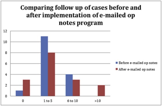
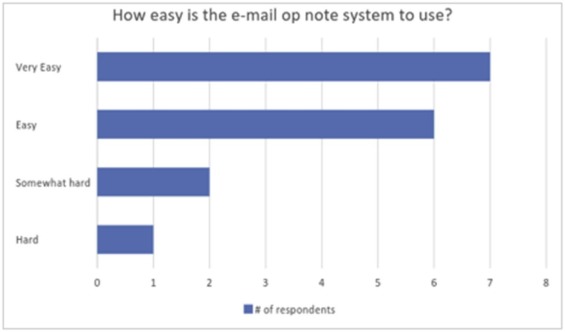
Survey results revealed 87.5% of the respondents reporting being either satisfied or very satisfied with the new communication tool. The survey also revealed that some radiologists reviewed more cases than before.
Conclusions
Matching of radiology and arthroscopy reports by anatomy allows for excellent report correlation (83.1%). Automated correlation improves the quality and efficiency of feedback to radiologists, providing important opportunities for learning and improved accuracy.
Introduction
Correlation of imaging findings with histopathology results or operative findings is essential for attaining expertise in radiology . Although correlation to a reference standard has been a hallmark of radiology teaching for years, some areas within radiology are poorly suited for this type of learning . Correlations for musculoskeletal (MSK) imaging, aside from pathology results for bone and soft tissue tumors, can be difficult to perform because the process of obtaining the reference standard, the arthroscopy report, can be arduous and requires dedication of the radiologist to manually correlate the arthroscopy report with the imaging studies . Report correlation can be more difficult when the patient does not undergo surgery for days or weeks, if ever, following the interpretation of the relevant imaging study. Although imaging-arthroscopy correlation conferences and tumor boards are valuable, only a small number of select cases are reviewed, limiting the number of cases discussed for each individual radiologist.
Get Radiology Tree app to read full this article<
Get Radiology Tree app to read full this article<
Get Radiology Tree app to read full this article<
Methods
Radiology—Operative Report Matching Module Development
Get Radiology Tree app to read full this article<
Get Radiology Tree app to read full this article<
Subjects
Get Radiology Tree app to read full this article<
Radiology—Arthroscopy Report Correlation Assessment
Get Radiology Tree app to read full this article<
Survey of Radiologist Behavior
Get Radiology Tree app to read full this article<
Statistics
Get Radiology Tree app to read full this article<
Get Radiology Tree app to read full this article<
Results
Radiology—Arthroscopy Report Correlation Assessment
Get Radiology Tree app to read full this article<
Get Radiology Tree app to read full this article<
TABLE 1
Correlative Results of the Radiology-Operative Note Module Separated by Specific Modality
Body Part Total ( n ) % Correlative Spine 95 51.6 Elbow, forearm, wrist, and hand 75 86.7 Shoulder and humerus 82 87.8 Knee and femur 108 93.4 Ankle or foot and lower extremity 79 93.7 Pelvis and hips 82 98.8
Get Radiology Tree app to read full this article<
Get Radiology Tree app to read full this article<
TABLE 2
Correlative Results of the Radiology—Operative Note Module Separated by Specific Anatomic Location
Body Part Total ( n ) % Correlative Fluoroscopy 35 71.4 CT 177 79.7 Ultrasound 43 88.4 MRI 266 86.1
CT, computed tomography; MRI, magnetic resonance imaging.
Get Radiology Tree app to read full this article<
Survey Results
Get Radiology Tree app to read full this article<
Get Radiology Tree app to read full this article<
Get Radiology Tree app to read full this article<
Get Radiology Tree app to read full this article<
Get Radiology Tree app to read full this article<
Get Radiology Tree app to read full this article<
TABLE 3
Level of Satisfaction of Follow-up Before and After the OpNote System
Manual Follow-up Automated OpNote Follow-up Very unsatisfied 13% (2/16) 6% (1/16) Unsatisfied 25% (4/16) 6% (1/16) Indifferent 44% (7/16) 0% (0/16) Satisfied 13% (2/16) 50% (8/16) Very satisfied 6% (1/16) 38% (6/16)
Get Radiology Tree app to read full this article<
Get Radiology Tree app to read full this article<
Get Radiology Tree app to read full this article<
Discussion
Get Radiology Tree app to read full this article<
Get Radiology Tree app to read full this article<
Get Radiology Tree app to read full this article<
Get Radiology Tree app to read full this article<
Get Radiology Tree app to read full this article<
Get Radiology Tree app to read full this article<
Conclusion/Summary
Get Radiology Tree app to read full this article<
Supplementary Data
Get Radiology Tree app to read full this article<
Appendix S1
Get Radiology Tree app to read full this article<
Get Radiology Tree app to read full this article<
References
1. Murphey M.D., Madewell J.E., Olmsted W.W., et. al.: A history of radiologic pathology correlation at the Armed Forces Institute of Pathology and its evolution into the American Institute for Radiologic Pathology. Radiology 2012; 262: pp. 623-634.
2. Delic J.A., Fuhrman C.R., Trejo Bittar H.E.: Pulmonary alveolar microlithiasis: AIRP best cases in radiologic-pathologic correlation. Radiographics 2016; 36: pp. 1334-1338.
3. Kantartzis S.N., Dacic S., Strollo D.C.: AIRP best cases in radiologic-pathologic correlation: pulmonary sarcoidosis complicated by aspergilloma formation. Radiographics 2012; 32: pp. 469-473.
4. Walton W.J., Flores R.R.: Desmoplastic small round cell tumor of the kidney: AIRP best cases in radiologic-pathologic correlation. Radiographics 2016; 36: pp. 1533-1538.
5. Hartman M., Silverman J., Spruill L., et. al.: Radiologic-pathologic correlation—an advanced fourth-year elective: how we do it. Acad Radiol 2016; 23: pp. 889-893.
6. Syed M.: Black box thinking: why most people never learn from their mistakes—but some do.2015.PenguinNew York, NY
7. Gyftopoulos S., Strauss E.J.: MRI-arthroscopy correlation for shoulder anatomy and pathology: a teaching guide. AJR Am J Roentgenol 2015; 204: pp. W684-W694. Review
8. Griffith J.F., Yung P.S., Antonio G.E., et. al.: CT compared with arthroscopy in quantifying glenoid bone loss. AJR Am J Roentgenol 2007; 189: pp. 1490-1493. PMID 18029890
9. Naqvi G.A., Jadaan M., Harrington P.: Accuracy of ultrasonography and magnetic resonance imaging for detection of full thickness rotator cuff tears. Int J Shoulder Surg 2009; 3: pp. 94-97.
10. Sharma G., Bhandrari S., Khandige G., et. al.: MR imaging of rotator cuff tears: correlation with arthroscopy. J Clin Diagn Res 2017; 11: pp. TC24-TC27. Epub 2017 May 1; PMID 28658874 PubMed Central PMCID: PMC5483776
11. Kelehan L., Kalaria A., Filice R.: Keeping Abreast of Breast Imagers: Radiology Pathology Correlation for the Rest of Us. SIIM Scientific Session2016.