
Rationale and Objectives
To develop an automated computed tomography angiography (CTA) imaging protocol that allows visualization of the whole brain vasculature and evaluate the clinical usefulness of the technique for delineation of intracranial vessels in patients with cerebrovascular disorders.
Materials and Methods
We prospectively included 100 patients who underwent automated subtraction CTA for suspected cerebrovascular disorders. The nonenhanced and contrast enhanced scans were obtained with the same table feeding speed. The x-ray tube start angles of the two scans were matched to enable accurate registration and subtraction of the CTA datasets. Subtracted CTA datasets were reformatted as three-dimensional volume rendering and maximum intensity projection images for further review. Two independent readers assessed the quality of subtraction and delineation of intracranial vessels. The visibility of ophthalmic arteries was also assessed.
Results
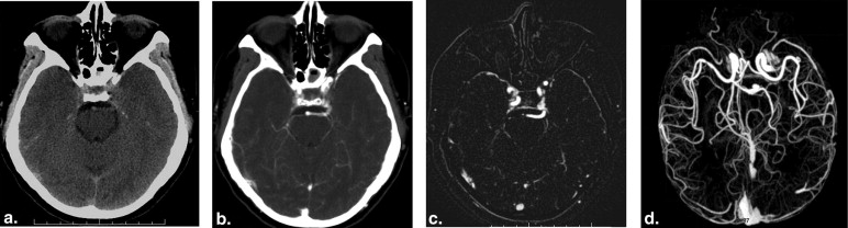
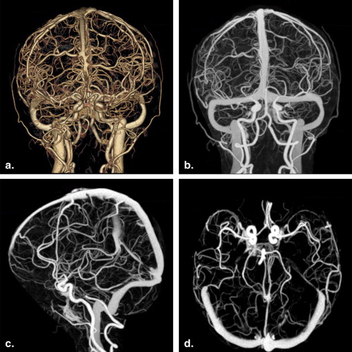
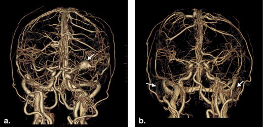
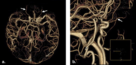
Subtraction was successful in all patients. The image quality of bone removal was rated excellent in 95 patients, with no or minimal bone remnants. Incomplete bone removal was observed in five patients because of severe motions between the scans. In 97 of 100 patients, arterial segments at the circle of Willis could be clearly visualized. Excellent delineation of bilateral ophthalmic arteries was possible in 81 of 100 patients.
Conclusions
The whole brain vasculature would be clearly visualized by using the optimized automated CTA protocol. Our automated, single-source, dual-energy subtraction CTA protocol is a fully automated subtraction method that is capable of delineating major intracranial vessels as well as very small arteries.
Imaging of cerebrovascular structures is important because it provides necessary information for the diagnosis and treatment of central nervous system disorders . Digital subtraction angiography (DSA) has long been considered the reference method for the diagnostic workup of patients with cerebrovascular disorders. Nevertheless, it is an invasive and time-consuming technique that is associated with a 0.5% risk of permanent neurological complications . There is an increasing need for a fast and noninvasive imaging method that could be widely implemented in clinical practice. Magnetic resonance angiography (MRA) is an alternative noninvasive imaging method that allows direct visualization of the intracranial vessels . However, the low spatial resolution and prolonged examination time makes it a less than ideal imaging method in the diagnostic workup of patients with acute stroke Another advantage of CTA is whole brain coverage, which is not yet practical with MRA within a reasonable amount of time. In contrast to DSA and MRA, computed tomography angiography (CTA) has the advantage of high spatial resolution and short examination time.
Recent technical innovations have greatly advanced the role of CTA in screening and follow-up study of patients with cerebrovascular disorders . However, CTA may be less useful than DSA for evaluation of vascular structures because of the presence of bone structures . In previous studies, several attempts, such as the section-by-section subtraction algorithm and the matched mask bone elimination technique, have been developed to eliminate bone structures . However, these techniques are operator-dependent and require specially adapted equipment or subtraction software. More recently, dual-energy CTA that allows automated subtraction of bone structures were increasingly been used in evaluation of intracranial vessels . Compared to standard CTA, dual-energy CTA have considerably improved the subtraction of bony structures at the skull base. However, the technique requires the use of dual-source CT scanners, which is unavailable in many institutions. The purpose of our study was to develop a fully automated, single-source, dual-energy subtraction CTA protocol and evaluate its clinical usefulness for visualization of the whole brain vasculature.
Materials and methods
Phantom Study
Get Radiology Tree app to read full this article<
Patient Population
Get Radiology Tree app to read full this article<
Data Acquisition
Get Radiology Tree app to read full this article<
Get Radiology Tree app to read full this article<
Get Radiology Tree app to read full this article<
Get Radiology Tree app to read full this article<
Evaluation of Images
Get Radiology Tree app to read full this article<
Get Radiology Tree app to read full this article<
Results
Get Radiology Tree app to read full this article<
Get Radiology Tree app to read full this article<
Get Radiology Tree app to read full this article<
Get Radiology Tree app to read full this article<
Get Radiology Tree app to read full this article<
Get Radiology Tree app to read full this article<
Get Radiology Tree app to read full this article<
Get Radiology Tree app to read full this article<
Discussion
Get Radiology Tree app to read full this article<
Get Radiology Tree app to read full this article<
Get Radiology Tree app to read full this article<
Get Radiology Tree app to read full this article<
Get Radiology Tree app to read full this article<
Get Radiology Tree app to read full this article<
Conclusions
Get Radiology Tree app to read full this article<
Acknowledgments
Get Radiology Tree app to read full this article<
Get Radiology Tree app to read full this article<
References
1. Wintermark M., Albers G.W., Alexandrov A.V., et. al.: Acute stroke imaging research roadmap. Stroke 2008; 39: pp. 1621-1628.
2. Willinsky R.A., Taylor S.M., TerBrugge K., et. al.: Neurologic complications of cerebral angiography: prospective analysis of 2,899 procedures and review of the literature. Radiology 2003; 227: pp. 522-528.
3. Anzalone N., Scomazzoni F., Cirillo M., et. al.: Follow-up of coiled cerebral aneurysms: comparison of three-dimensional time-of-flight magnetic resonance angiography at 3 Tesla with three-dimensional time-of-flight magnetic resonance angiography and contrast-enhanced magnetic resonance angiography at 1.5 Tesla. Invest Radiol 2008; 43: pp. 559-567.
4. Villablanca J.P., Nael K., Habibi R., et. al.: 3 T contrast-enhanced magnetic resonance angiography for evaluation of the intracranial arteries: comparison with time-of-flight magnetic resonance angiography and multislice computed tomography angiography. Invest Radiol 2006; 41: pp. 799-805.
5. Li Q., Lv F., Li Y., et. al.: Evaluation of 64-section CT angiography for detection and treatment planning of intracranial aneurysms by using DSA and surgical findings. Radiology 2009; 252: pp. 808-815.
6. Lell M.M., Anders K., Uder M., et. al.: New techniques in CT angiography. Radiographics 2006; 26: pp. S45-S62.
7. Jayaraman M.V., Mayo-Smith W.W., Tung G.A., et. al.: Detection of intracranial aneurysms: multi-detector row CT angiography compared with DSA. Radiology 2004; 230: pp. 510-518.
8. Imakita S., Onishi Y., Hashimoto T., et. al.: Subtraction CT angiography with controlled-orbit helical scanning for detection of intracranial aneurysms. AJNR Am J Neuroradiol 1998; 19: pp. 291-295.
9. Venema H.W., Hulsmans F.J., den Heeten G.J.: CT angiography of the circle of Willis and intracranial internal carotid arteries: maximum intensity projection with matched mask bone elimination-feasibility study. Radiology 2001; 218: pp. 893-898.
10. Schulz B., Kuehling K., Kromen W., et. al.: Automatic bone removal technique in whole-body dual-energy CT angiography: performance and image quality. AJR Am J Roentgenol 2012; 199: pp. W646-W650.
11. Lell M.M., Kramer M., Klotz E., et. al.: Carotid computed tomography angiography with automated bone suppression: a comparative study between dual energy and bone subtraction techniques. Invest Radiol 2009; 44: pp. 322-328.
12. Morhard D., Fink C., Graser A., et. al.: Cervical and cranial computed tomographic angiography with automated bone removal: dual energy computed tomography versus standard computed tomography. Invest Radiol 2009; 44: pp. 293-297.
13. White P.M., Wardlaw J.M., Easton V.: Can noninvasive imaging accurately depict intracranial aneurysms? A systematic review. Radiology 2000; 217: pp. 361-370.
14. Villablanca J.P., Martin N., Jahan R., et. al.: Volume-rendered helical computerized tomography angiography in the detection and characterization of intracranial aneurysms. J Neurosurg 2000; 93: pp. 254-264.
15. Villablanca J.P., Jahan R., Hooshi P., et. al.: Detection and characterization of very small cerebral aneurysms by using 2D and 3D helical CT angiography. AJNR Am J Neuroradiol 2002; 23: pp. 1187-1198.
16. Lubicz B., Levivier M., Francois O., et. al.: Sixty-four-row multisection CT angiography for detection and evaluation of ruptured intracranial aneurysms: interobserver and intertechnique reproducibility. AJNR Am J Neuroradiol 2007; 28: pp. 1949-1955.
17. Tomandl B.F., Kostner N.C., Schempershofe M., et. al.: CT angiography of intracranial aneurysms: a focus on postprocessing. Radiographics 2004; 24: pp. 637-655.
18. Gorzer H., Heimberger K., Schindler E.: Spiral CT angiography with digital subtraction of extra- and intracranial vessels. J Comput Assist Tomogr 1994; 18: pp. 839-841.
19. Jayakrishnan V.K., White P.M., Aitken D., et. al.: Subtraction helical CT angiography of intra- and extracranial vessels: technical considerations and preliminary experience. AJNR Am J Neuroradiol 2003; 24: pp. 451-455.
20. Romijn M., Gratama van H.A.F., Andel , et. al.: Diagnostic accuracy of CT angiography with matched mask bone elimination for detection of intracranial aneurysms: comparison with digital subtraction angiography and 3D rotational angiography. AJNR Am J Neuroradiol 2008; 29: pp. 134-139.
21. Majoie C.B., van Straten M., Venema H.W., et. al.: Multisection CT venography of the dural sinuses and cerebral veins by using matched mask bone elimination. AJNR Am J Neuroradiol 2004; 25: pp. 787-791.
22. Gratama van Andel H.A., Venema H.W., Streekstra G.J., et. al.: Removal of bone in CT angiography by multiscale matched mask bone elimination. Med Phys 2007; 34: pp. 3711-3723.
23. Sakamoto S., Kiura Y., Shibukawa M., et. al.: Subtracted 3D CT angiography for evaluation of internal carotid artery aneurysms: comparison with conventional digital subtraction angiography. AJNR Am J Neuroradiol 2006; 27: pp. 1332-1337.
24. Tomandl B.F., Hammen T., Klotz E., et. al.: Bone-subtraction CT angiography for the evaluation of intracranial aneurysms. AJNR Am J Neuroradiol 2006; 27: pp. 55-59.
25. Lell M.M., Ruehm S.G., Kramer M., et. al.: Cranial computed tomography angiography with automated bone subtraction: a feasibility study. Invest Radiol 2009; 44: pp. 38-43.
26. Lell M., Anders K., Klotz E., et. al.: Clinical evaluation of bone-subtraction CT angiography (BSCTA) in head and neck imaging. Eur Radiol 2006; 16: pp. 889-897.
27. Morhard D., Fink C., Becker C., et. al.: Value of automatic bone subtraction in cranial CT angiography: comparison of bone-subtracted vs. standard CT angiography in 100 patients. Eur Radiol 2008; 18: pp. 974-982.
28. Venema H.W., den Heeten G.J.: Subtraction helical CT angiography of intra and extracranial vessels: technical considerations and preliminary experience–rediscovery of matched mask bone elimination?. AJNR Am J Neuroradiol 2003; 24: pp. 1491-1492.
29. Lell M.M., Ditt H., Panknin C., et. al.: Bone-subtraction CT angiography: evaluation of two different fully automated image-registration procedures for interscan motion compensation. AJNR Am J Neuroradiol 2007; 28: pp. 1362-1368.
30. Burgstahler C., Reimann A., Brodoefel H., et. al.: Quantitative parameters to compare image quality of non-invasive coronary angiography with 16-slice, 64-slice and dual-source computed tomography. Eur Radiol 2009; 19: pp. 584-590.