
Rationale and Objectives
The objective of our study was to calculate mammographic breast density with a fully automated volumetric breast density measurement method and to compare it to breast imaging reporting and data system (BI-RADS) breast density categories assigned by two radiologists.
Materials and Methods
A total of 476 full-field digital mammography examinations with standard mediolateral oblique and craniocaudal views were evaluated by two blinded radiologists and BI-RADS density categories were assigned. Using a fully automated software, mean fibroglandular tissue volume, mean breast volume, and mean volumetric breast density were calculated. Based on percentage volumetric breast density, a volumetric density grade was assigned from 1 to 4.
Results
The weighted overall kappa was 0.895 (almost perfect agreement) for the two radiologists’ BI-RADS density estimates. A statistically significant difference was seen in mean volumetric breast density among the BI-RADS density categories. With increased BI-RADS density category, increase in mean volumetric breast density was also seen ( P < 0.001). A significant positive correlation was found between BI-RADS categories and volumetric density grading by fully automated software (ρ = 0.728, P < 0.001 for first radiologist and ρ = 0.725, P < 0.001 for second radiologist). Pairwise estimates of the weighted kappa between Volpara density grade and BI-RADS density category by two observers showed fair agreement (κ = 0.398 and 0.388, respectively).
Conclusions
In our study, a good correlation was seen between density grading using fully automated volumetric method and density grading using BI-RADS density categories assigned by the two radiologists. Thus, the fully automated volumetric method may be used to quantify breast density on routine mammography.
Introduction
Mammography is the recommended screening method for breast cancer in the general population . However, the sensitivity of screening mammography is reduced in women with dense breasts because of obscuration of underlying lesion by dense and heterogenous breast tissue. Because of this, chances of missing breast cancer are increased . At the same time, increased breast density has been considered as an independent risk factor for the development of breast cancer. Various studies have shown that women with high breast density compared to women with low breast density are four to six times more likely to get breast cancer . Bertrand et al. reported that women with breast density more than 50% have approximately twofold risk of developing breast cancer than women with breast density of 11–23%. The authors also reported a positive correlation between breast density and larger tumor size, positive lymph nodes, and negative estrogen receptor status .
The American College of Radiology (ACR) recommends annual screening mammography in women more than 40 years of age. However, in a subset of population having increased risk for breast cancer, ACR not only recommends screening at earlier age but also emphasizes the need of supplemental screening methods like ultrasound and magnetic resonance imaging . ACR has also recommended supplemental screening in women with genetic predisposition to the disease and in women with dense breasts . Thus, breast density determines the need for supplemental screening and can prove to be an important factor in risk stratification.
Get Radiology Tree app to read full this article<
Get Radiology Tree app to read full this article<
Materials and Methods
Patients and Mammography
Get Radiology Tree app to read full this article<
BI-RADS Density Classification
Get Radiology Tree app to read full this article<
Get Radiology Tree app to read full this article<
Mammographic Density Analysis by Fully Automated Volumetric Software
Get Radiology Tree app to read full this article<
Get Radiology Tree app to read full this article<
Data and Statistical Analysis
Get Radiology Tree app to read full this article<
Results
Get Radiology Tree app to read full this article<
Get Radiology Tree app to read full this article<
Get Radiology Tree app to read full this article<
TABLE 1
Table Showing Age of the Patients and Various Parameters Obtained by Automated Breast Density Calculation Using Volpara Software (Version 1.4.5)
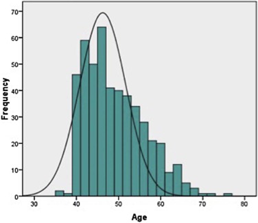
Age (mean ± SD) 48.8 ± 7.07 Volume of fibroglandular tissue (mean ± SD) 3.999 ± 2.529 Breast volume (mean ± SD) 57.905 ± 25.325 Volumetric breast density (mean ± SD) 7.676 ± 4.891
Get Radiology Tree app to read full this article<
Get Radiology Tree app to read full this article<
Get Radiology Tree app to read full this article<
TABLE 2
Table Showing Number of Patients in Each Grade According to Volpara Density Grade (VDG) and BI-RADS Grading by Two Observers
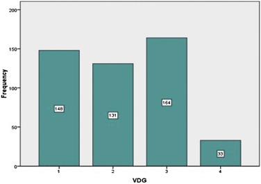
VDG Grade Number of Patients (Percentage) BI-RADS Grade Observer 1 Observer 2 1 148 (31.1) 1 159 (33.4) 163 (34.2) 2 131 (27.5) 2 212 (44.5) 220 (46.2) 3 164 (34.5) 3 98 (20.6) 90 (18.9) 4 33 (6.9) 4 7 (1.5) 3 (0.6)
BI-RADS, breast imaging reporting and data system.
Get Radiology Tree app to read full this article<
Get Radiology Tree app to read full this article<
TABLE 3
Table Showing Correlation Between Results of Automated Volumetric Breast Density Calculation and BI-RADS Grading (By Agreement of Both Observers)
BI-RADS Grade (By Agreement of Both Observers) Number Volume of Fibroglandular Tissue Breast Volume Volumetric Breast Density_P_ Value <0.001P Value <0.001P Value <0.001 1 156 2.664 ± 3.764 47.933 ± 24.311 3.853 ± 1.635 2 202 4.336 ± 3.261 74.391 ± 26.004 8.268 ± 4.410 3 83 5.858 ± 2.860 50.055 ± 23.286 12.432 ± 4.606 4 3 8.500 ± 6.477 32.617 ± 14.167 23.417 ± 9.994
Get Radiology Tree app to read full this article<
Get Radiology Tree app to read full this article<
TABLE 4
Results of Automated Volumetric Breast Density Calculation According to Age of Patients
Age Number Volume of Fibroglandular Tissue Breast Volume Volumetric Breast Density_P_ Value <0.001P Value <0.001P Value <0.001 <40 49 4.981 ± 3.208 52.043 ± 26.701 10.510 ± 5.336 41–50 254 4.641 ± 3.616 71.420 ± 23.195 8.799 ± 5.573 51–60 141 3.281 ± 1.692 62.180 ± 25.121 5.945 ± 3.624 >60 32 2.227 ± 1.051 58.364 ± 25.138 4.389 ± 2.561
Get Radiology Tree app to read full this article<
Get Radiology Tree app to read full this article<
Discussion
Get Radiology Tree app to read full this article<
Get Radiology Tree app to read full this article<
Get Radiology Tree app to read full this article<
Get Radiology Tree app to read full this article<
Get Radiology Tree app to read full this article<
Get Radiology Tree app to read full this article<
Get Radiology Tree app to read full this article<
Get Radiology Tree app to read full this article<
Get Radiology Tree app to read full this article<
References
1. Mainiero M.B., Lourenco A., Mahoney M.C., et. al.: ACR Appropriateness Criteria breast cancer screening. J Am Coll Radiol 2013; 10: pp. 11-14.
2. Boyd N.F., Guo H., Martin L.J., et. al.: Mammographic density and the risk and detection of breast cancer. N Engl J Med 2007; 356: pp. 227-236.
3. Van Gils C., Otten J.D., Verbeek A.L., et. al.: Mammographic breast density and risk of breast cancer: masking bias or causality?. Eur J Epidemiol 1998; 14: pp. 315-320.
4. Mandelson M.T., Oestreicher N., Porter P.L., et. al.: Breast density as a predictor of mammographic detection: comparison of interval- and screen-detected cancers. J Natl Cancer Inst 2000; 92: pp. 1081-1087. Review
5. Boyd N.F., Martin L.J., Yaffe M.J., et. al.: Mammographic density and breast cancer risk: current understanding and future prospects. Breast Cancer Res 2011; 13: pp. 223.
6. McCormack V.A., dos Santos Silva I.: Breast density and parenchymal patterns as markers of breast cancer risk: a meta-analysis. Cancer Epidemiol Biomarkers Prev 2006; 15: pp. 1159-1169.
7. Vachon C.M., van Gils C.H., Sellers T.A., et. al.: Mammographic density, breast cancer risk and risk prediction. Breast Cancer Res 2007; 9: pp. 217.
8. Bertrand K.A., Tamimi R.M., Scott C.G., et. al.: Mammographic density and risk of breast cancer by age and tumour characteristics. Breast Cancer Res 2013; 15: pp. R104.
9. Kerlikowske K., Grady D., Barclay J., et. al.: Variability and accuracy in mammographic interpretation using the American College of Radiology Breast Imaging Reporting and Data System. J Natl Cancer Inst 1998; 90: pp. 1801-1809.
10. Berg W.A., Campassi C., Langenberg P., et. al.: Breast Imaging Reporting and Data System: inter- and intraobserver variability in feature analysis and final assessment. AJR Am J Roentgenol 2000; 174: pp. 1769-1777.
11. Martin K.E., Helvie M.A., Zhou C., et. al.: Mammographic density measured with quantitative computer-aided method: comparison with radiologists’ estimates and BI-RADS categories. Radiology 2006; 240: pp. 656-665.
12. Spayne M.C., Gard C.C., Skelly J., et. al.: Reproducibility of BI-RADS breast density measures among community radiologists: a prospective cohort study. Breast J 2012; 18: pp. 326-333.
13. Byng J.W., Boyd N.F., Fishell E., et. al.: The quantitative analysis of mammographic densities. Phys Med Biol 1994; 39: pp. 1629-1638.
14. Sivaramakrishna R., Obuchowski N.A., Chilcote W.A., et. al.: Automatic segmentation of mammographic density. Acad Radiol 2001; 8: pp. 250-256.
15. Prevrhal S., Shepherd J., Smith-Bindman R., et. al.: Accuracy of mammographic breast density analysis: results of formal operator training. Cancer Epidemiol Biomarkers Prev 2002; 11: pp. 1389-1393.
16. Shepherd J.A., Kerlikowske K., Ma L., et. al.: Volume of mammographic density and risk of breast cancer. Cancer Epidemiol Biomarkers Prev 2011; 20: pp. 1473-1482.
17. Highnam R., Pan X., Warren R., et. al.: Breast composition measurements using retrospective standard mammogram form (SMF). Phys Med Biol 2006; 51: pp. 2695-2713.
18. Jeffreys M., Warren R., Highnam R., et. al.: Initial experiences of using an automated volumetric measure of breast density: the standard mammogram form. Br J Radiol 2006; 79: pp. 378-382.
19. Highnam R.P., Brady J.M., Shepstone B.J.: Mammographic image analysis. Eur J Radiol 1997; 24: pp. 20-32.
20. D’Orsi C.J., Sickles E.A., Mendelson E.B., et. al.: ACR BI-RADS—breast imaging atlas, breast imaging reporting and data system.2013.American College of RadiologyReston, VA
21. Landis J.R., Koch G.G.: The measurement of observer agreement for categorical data. Biometrics 1977; 33: pp. 159-174.
22. Harvey J.A., Bovbjerg V.E.: Quantitative assessment of mammographic breast density: relationship with breast cancer risk. Radiology 2004; 230: pp. 29-41.
23. Gweon H.M., Youk J.H., Kim J.A., et. al.: Radiologist assessment of breast density by BI-RADS categories versus fully automated volumetric assessment. AJR Am J Roentgenol 2013; 201: pp. 692-697.
24. Seo J.M., Ko E.S., Han B.K., et. al.: Automated volumetric breast density estimation: a comparison with visual assessment. Clin Radiol 2013; 68: pp. 690-695.
25. McEntee M.F., Damases C.N.: Mammographic density measurement: a comparison of automated volumetric density measurement to BIRADS. Proc SPIE Int Soc Opt Eng 2014;
26. Ko S.Y., Kim E.K., Kim M.J., et. al.: Mammographic density estimation with automated volumetric breast density measurement. Korean J Radiol 2014; 15: pp. 313-321.
27. Tagliafico A., Tagliafico G., Tosto S., et. al.: Mammographic density estimation: comparison among BI-RADS categories, a semi-automated software and a fully automated one. Breast 2009; 18: pp. 35-40.
28. Gubern-Mérida A., Kallenberg M., Platel B., et. al.: Volumetric breast density estimation from full field digital mammograms: a validation study. PLoS ONE 2014; 9: pp. e85952. eCollection 2014
29. Wang J., Aziz A., Newitt D., et. al.: Comparison of Hologic’s Quantra volumetric assessment to MRI breast density.Proceedings of the 11th International Conference on Breast Imaging, IWDM‘12.2012.Springer-Verlag.Berlin, Heidelberg:pp. 619-626.
30. van Engeland S., Snoeren P.R., Huisman H., et. al.: Volumetric breast density estimation from full-field digital mammograms. IEEE Trans Med Imaging 2006; 25: pp. 273-282.
31. Jeffreys M., Harvey J., Highnam R.: Comparing a new volumetric breast density method (Volpara™) to Cumulus.Martı’ J.IWDM’10: Proceedings of the 10th International Workshop on Digital Mammography.2010.Springer-VerlagBerlin, Heidelberg:pp. 408-413.
32. Bernardi D.1., Pellegrini M., Di Michele S., et. al.: Interobserver agreement in breast radiological density attribution according to BI-RADS quantitative classification. Radiol Med 2012; 117: pp. 519-528.
33. Eng A., Gallant Z., Shepherd J., et. al.: Digital mammographic breast density and breast cancer risk: a case control study of six alternative density assessment methods. Breast Cancer Res 2014; 16: pp. 439.
34. Stone J., Warren R.M., Pinney E., et. al.: Determinants of percentage and area measures of mammographic density. Am J Epidemiol 2009; 170: pp. 1571-1578.
35. Aitken Z., McCormack V.A., Highnam R.P., et. al.: Screen-film mammographic density and breast cancer risk: a comparison of the volumetric standard mammogram form and the interactive threshold measurement methods. Cancer Epidemiol Biomarkers Prev 2010; 19: pp. 418-428.
36. Jeffreys M., Warren R., Highnam R., et. al.: Breast cancer risk factors and a novel measure of volumetric breast density: cross-sectional study. Br J Cancer 2008; 98: pp. 210-216.