
Rationale and Objectives
The study aimed to determine the outcome of patients presenting for evaluation of abnormal breast thermography.
Materials and Methods
Following Institutional Review Board approval, retrospective search identified 38 patients who presented for conventional breast imaging following a thermography-detected abnormality. Study criteria included women who had mammogram and/or breast ultrasound performed for evaluation of a thermography-detected abnormality between January 1, 2000, and December 31, 2015. Patients whose mammograms and ultrasounds were initiated at an outside institution or who did not have imaging at our institution were excluded. Records were reviewed for clinical history, thermography results, mammogram and/or ultrasound findings, and pathology. Mammograms and ultrasounds were prospectively interpreted by one of 14 Mammography Quality Standards Act–certified breast imaging radiologists with 3–30 years of experience. Patient outcomes were determined by biopsy or at least 1 year of follow-up. Patient ages ranged from 23 to 70 years (mean = 50 years).
Results
Ninety-five percent (36 of 38) of patients did not have breast cancer. The two patients diagnosed with breast cancer had suspicious clinical symptoms including palpable mass and erythema. No asymptomatic woman had breast cancer. Negative predictive value was 100%. Of 38 patients, 79% (30 of 38) had Breast Imaging Reporting and Data System (BI-RADS) 1 or 2 assessments; 5% (2 of 38) had BI-RADS 3; and 16% (6 of 38) had BI-RADS 4 ( n = 5) or BI-RADS 5 ( n = 1) assessments. Two of six patients with biopsy recommendations were diagnosed with breast cancer (Positive predictive value 2 = 33.3%). All findings recommended for biopsy were ipsilateral to the reported thermography abnormality.
Conclusions
No cancer was diagnosed among asymptomatic women. The 5% of patients diagnosed with cancer had co-existing suspicious clinical findings. Mammogram and/or ultrasound were useful in accurately characterizing patients with abnormal thermography.
Introduction
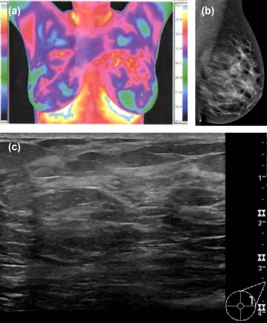
Breast cancer is one of the leading causes of death among women worldwide. Screening mammography is the most thoroughly researched and widely utilized examination for breast cancer detection. Screening mammography has repeatedly been shown to contribute to decreased breast cancer–associated mortality . Supplemental screening with ultrasound, tomosynthesis, and magnetic resonance imaging (MRI) are performed as clinically indicated, especially for higher risk women; these imaging modalities have all demonstrated effectiveness and safety in the detection of breast cancer. However, patients may seek alternative breast cancer screening methods such as breast thermography. Less is known about the efficacy of thermography for breast cancer detection. Breast thermography was originally developed in the late 1950s in Canada . Thermography was implemented as a breast cancer screening study in the 1960s. In 1977, Feig et al. compared mammography and thermography screening in 16,000 women and found thermography to have a sensitivity of 39% . Based on this low sensitivity, Feig concluded that thermography was not practical as a breast cancer screening tool. Following the results of the study by Feig, breast thermography was largely abandoned .
Since that time, thermal imaging technology has improved and breast thermography is regarded as an adequate method for breast cancer screening in some medical communities, which describe it as offering earlier breast cancer diagnosis relative to conventional imaging modalities and clinical examinations . Although the US Food and Drug Administration (FDA) has not approved thermography as a stand-alone modality for breast cancer screening or diagnosis , patients concerned with mammographic radiation or compression may seek thermography in lieu of screening mammography.
Get Radiology Tree app to read full this article<
Materials and Methods
Get Radiology Tree app to read full this article<
Get Radiology Tree app to read full this article<
Get Radiology Tree app to read full this article<
Results
Get Radiology Tree app to read full this article<
Get Radiology Tree app to read full this article<
Get Radiology Tree app to read full this article<
TABLE 1
Suspicious Findings on Conventional Breast Imaging and Outcomes following Abnormal Thermography Results
Age BI-RADS Thermography Abnormality Mammogram Finding Imaging Finding Size (mm) Biopsy Method Pathology Result Clinical Symptoms 41 4 Right Right—calcifications 60 Stereotactic Fibrocystic No 54 4 Left Left—calcifications 7 Surgical Fibrocystic No 47 4 Left Left—calcifications 30 Stereotactic PASH No 68 4 Right Right—mass 9 Patient declined N/A—Mass stable for 2 years No 70 4 Right Right—mass 15 Ultrasound IDC Palpable right breast mass 48 5 Right Right—mass and calcifications Multifocal Ultrasound IDC Palpable breast mass and erythematous breast
IDC, invasive ductal carcinoma; N/A, not applicable; PASH, pseudoangiomatous stromal hyperplasia.
Get Radiology Tree app to read full this article<
Get Radiology Tree app to read full this article<
Get Radiology Tree app to read full this article<
Get Radiology Tree app to read full this article<
TABLE 2
Conventional Breast Imaging Results and Patient Risk Factors
Patients Referred for Abnormal Thermogram
n = 38
Number and (%) of Cases BI-RADS 1 26 (68.4) 2 4 (10.5) 3 2 (5.3) 4 5 (13.2) 5 1 (2.6) First-degree family history of breast cancer Negative 35 (92.1) Positive 2 (5.3) BRCA mutation 1 (2.6) Personal history of breast cancer Negative 32 (84.2) Positive 5 (13.2) High-risk (LCIS) 1 (2.6) Menopausal status Premenopausal 19 (50) Postmenopausal 19 (50) Clinical symptoms Yes 13 (34.2) No 25 (65.8) Mammogram density_n_ = 33 Almost entirely fatty 2 (6.1) Scattered areas of fibroglandular densities 24 (72.7) Heterogeneously dense 6 (18.2) Extremely dense 1 (3)
BI-RADS, Breast Imaging Reporting and Data System; LCIS, lobular carcinoma in situ.
Get Radiology Tree app to read full this article<
Discussion
Get Radiology Tree app to read full this article<
Get Radiology Tree app to read full this article<
Get Radiology Tree app to read full this article<
Get Radiology Tree app to read full this article<
Get Radiology Tree app to read full this article<
Get Radiology Tree app to read full this article<
Get Radiology Tree app to read full this article<
Get Radiology Tree app to read full this article<
Get Radiology Tree app to read full this article<
Get Radiology Tree app to read full this article<
Acknowledgments
Get Radiology Tree app to read full this article<
Get Radiology Tree app to read full this article<
References
1. Tabar L., Fagerberg C.J., Gad A., et. al.: Reduction in mortality from breast cancer after mass screening with mammography. Randomised trial from the Breast Cancer Screening Working Group of the Swedish National Board of Health and Welfare. Lancet 1985; 1: pp. 829-832.
2. Hendrick R.E., Smith R.A., Rutledge J.H., et. al.: Benefit of screening mammography in women aged 40–49: a new meta-analysis of randomized controlled trials. J Natl Cancer Inst Monogr 1997; 22: pp. 87-92.
3. Duffy S.W., Tabar L., Chen H.H., et. al.: The impact of organized mammography service screening on breast carcinoma mortality in seven Swedish counties. Cancer 2002; 95: pp. 458-469.
4. Tabar L., Yen M.F., Vitak B., et. al.: Mammography service screening and mortality in breast cancer patients: 20-year follow-up before and after introduction of screening. Lancet 2003; 361: pp. 1405-1410.
5. Otto S.J., Fracheboud J., Looman C.W., et. al.: Initiation of population-based mammography screening in Dutch municipalities and effect on breast-cancer mortality: a systematic review. Lancet 2003; 361: pp. 1411-1417.
6. Tabar L., Vitak B., Chen T.H., et. al.: Swedish two-county trial: impact of mammographic screening on breast cancer mortality during 3 decades. Radiology 2011; 260: pp. 658-663.
7. Fraser J.: Hot bodies; cold war: the forgotten history of breast thermography. CMAJ 2017; 189: pp. E573-E575.
8. Lawson R.: Thermography; a new tool in the investigation of breast lesions. Can Serv Med J 1957; 8: pp. 517-524.
9. Feig S.A., Shaber G.S., Schwartz G.F., et. al.: Thermography, mammography, and clinical examination in breast cancer screening. Review of 16,000 studies. Radiology 1977; 122: pp. 123-127.
10. Foster K.R.: Thermographic detection of breast cancer. IEEE Eng Med Biol Mag 1998; 17: pp. 10-14.
11. American College of Clinical Thermology : What is Breast Thermography?. Available at: http://www.thermologyonline.org/Breast/breast_thermography_what.htm Date posted: Not available
12. U.S. Food and Drug Administration : Thermogram No Substitute for Mammography. Available at: https://www.fda.gov/MedicalDevices/Safety/AlertsandNotices/ucm257499.htm Date posted: 6/2/2011
13. D’Orsi C.J., Sickles E.A., Mendelson E.B., et. al.: ACR BI-RADS® Atlas, Breast Imaging Reporting and Data System. Reston, VA, American College of Radiology2013.
14. National Comprehensive Cancer Network : Breast Cancer Screening and Diagnosis (Version 1.2017). Available at: https://www.nccn.org/professionals/physician_gls/pdf/breast-screening.pdf Date posted: 6/2/2017
15. American College of Clinical Thermology : Overview of Digital Infrared Thermal Imaging. Available at: http://www.thermologyonline.org/Patients/patients_overview.htm Date posted: Not available
16. American College of Thermology : Indications for Thermographic Evaluation. Available at: http://www.thermologyonline.org/Patients/patients_indications.htm Date posted: Not available
17. U.S. Food and Drug Administration : Breast Cancer Screening—Thermography Is Not an Alternative to Mammography: FDA Safety Communication. Available at: https://www.fda.gov/NewsEvents/Newsroom/PressAnnouncements/ucm257633.htm Date posted: 6/2/2011
18. Health Canada : Thermography Machines Not Authorized to Screen for Breast Cancer. Available at: http://www.healthycanadians.gc.ca/recall-alert-rappel-avis/hc-sc/2012/15920a-eng.php Date posted 11/28/2012
19. American College of Clinical Thermology : ACCT Approved Thermography Clinics. Available at: http://www.thermologyonline.org/Breast/breast_thermography_clinics.htm Date posted: Not available
20. Amalric R., Giraud D., Altschuler C., et. al.: Does infrared thermography truly have a role in present-day breast cancer management?. Prog Clin Biol Res 1982; 107: pp. 269-278.
21. Arora N., Martins D., Ruggerio D., et. al.: Effectiveness of a noninvasive digital infrared thermal imaging system in the detection of breast cancer. Am J Surg 2008; 196: pp. 523-526.
22. Wishart G.C., Campisi M., Boswell M., et. al.: The accuracy of digital infrared imaging for breast cancer detection in women undergoing breast biopsy. Eur J Surg Oncol 2010; 36: pp. 535-540.
23. Kennedy D.A., Lee T., Seely D.: A comparative review of thermography as a breast cancer screening technique. Integr Cancer Ther 2009; 8: pp. 9-16.
24. Lehman C.D., Wellman R.D., Buist D.S., et. al.: Diagnostic accuracy of digital screening mammography with and without computer-aided detection. JAMA Intern Med 2015; 175: pp. 1828-1837.
25. Kontos M., Wilson R., Fentiman I.: Digital infrared thermal imaging (DITI) of breast lesions: sensitivity and specificity of detection of primary breast cancers. Clin Radiol 2011; 66: pp. 536-539.
26. Moskowitz M., Milbrath J., Gartside P., et. al.: Lack of efficacy of thermography as a screening tool for minimal and stage I breast cancer. N Engl J Med 1976; 295: pp. 249-252.
27. Omranipour R., Kazemian A., Alipour S., et. al.: Comparison of the accuracy of thermography and mammography in the detection of breast cancer. Breast Care (Basel) 2016; 11: pp. 260-264.
28. Threatt B., Norbeck J.M., Ullman N.S., et. al.: Thermography and breast cancer an analysis of a blind reading. Ann N Y Acad Sci 1980; 335: pp. 501-527.
29. Vreugdenburg T.D., Willis C.D., Mundy L., et. al.: A systematic review of elastography, electrical impedance scanning, and digital infrared thermography for breast cancer screening and diagnosis. Breast Cancer Res Treat 2013; 137: pp. 665-676.
30. Meller M.T., Cox J.E.M., Callanan K.W.R.: Incidental detection of breast lesions with computed tomography. Clin Breast Cancer 2007; 7: pp. 634-637.
31. Monzawa S., Washio T., Yasuoka R., et. al.: Incidental detection of clinically unexpected breast lesions by computed tomography. Acta Radiol 2013; 54: pp. 374-379.
32. Moyle P., Sonoda L., Britton P., et. al.: Incidental breast lesions detected on CT: what is their significance?. Br J Radiol 2010; 83: pp. 233-240.
33. Prabhu1 V., Chhor C.M., Ego-Osuala I.O., et. al.: Frequency and outcomes of incidental breast lesions detected on abdominal MRI over a 7-year period. AJR Am J Roentgenol 2017; 208: pp. 107-113.
34. Shin K.M., Kim H.J., Jung S.J., et. al.: Incidental breast lesions identified by 18F-FDG PET/CT: which clinical variables differentiate between benign and malignant breast lesions?. J Breast Cancer 2015; 18: pp. 73-79.
35. Kang B.J., Lee J.H., Yoo I.R., et. al.: Clinical significance of incidental finding of focal activity in the breast at 18F-FDG PET/CT. AJR Am J Roentgenol 2011; 197: pp. 341-347.
36. Litmanovich D.1., Gourevich K., Israel O., et. al.: Unexpected foci of 18F-FDG uptake in the breast detected by PET/CT: incidence and clinical significance. Eur J Nucl Med Mol Imaging 2009; 36: pp. 1558-1564.