
Rationale and Objectives
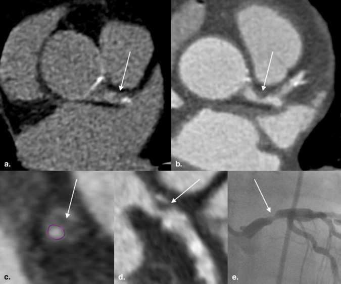
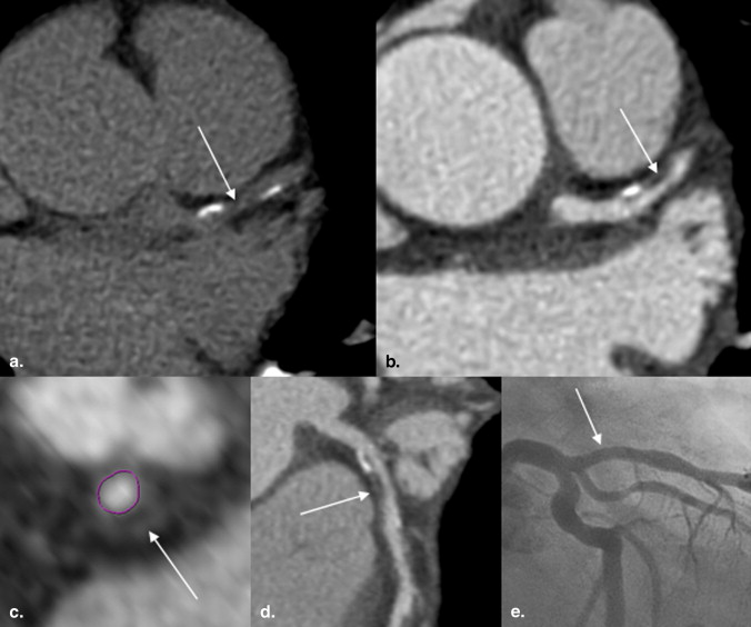
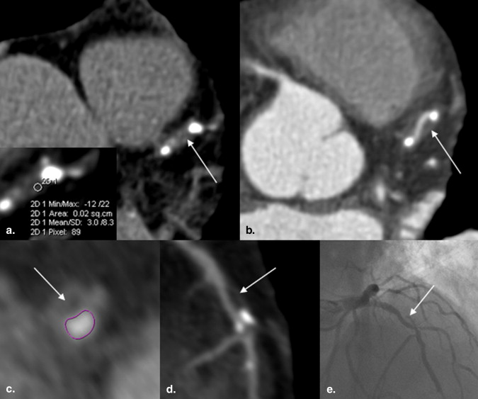
Coronary computed tomographic (CT) angiography has been shown to detect noncalcified coronary artery plaque. Depending on tissue composition, noncalcified plaque differs in CT attenuation from blood and epicardial fat. The aim of this study was to determine whether noncalcified plaque can be visually detected on non-contrast-enhanced CT calcium scoring studies.
Materials and Methods
A total of 106 patients (40 women; mean age, 59 years) who underwent coronary calcium scoring, coronary CT angiography, and quantitative catheter angiography were prospectively investigated. Two blinded observers independently reviewed calcium scoring studies for positive vascular remodeling and hypoattenuation within the vessel wall, suggestive of noncalcified plaque. Findings on calcium scoring studies were compared with those on coronary CT angiography and quantitative catheter angiography.
Results
The mean Agatston score was 515.8 ± 826.8. Overall interobserver agreement for the identification of noncalcified lesions was substantial (κ = 0.69). Observer 1 and observer 2 identified 21 and 17 patients, respectively, with 38 and 35 lesions suggestive of noncalcified plaque. Coronary CT angiography confirmed noncalcified plaque in 33 of 38 (86.8%; observer 1) and 31 of 35 (88.6%; observer 2) lesions. Thus, the overall positive predictive value for correct identification of noncalcified plaque on calcium scoring studies was 0.88, although overall sensitivity was low at 0.39.
Conclusions
Noncalcified plaque can be visually detected on calcium scoring studies. Review of calcium scoring studies for features of noncalcified plaque may enhance the identification of patients with more active disease and higher cardiovascular risk.
Computed tomographic (CT) coronary calcium scoring has been proposed to provide incremental prognostic information in addition to conventional risk factor assessment for predicting cardiac events in intermediate-risk populations . Although there is no good relationship between the degree of coronary artery calcification and stenotic coronary artery disease (CAD) , the amount of coronary artery calcium is thought to reflect the overall coronary atherosclerotic plaque burden , albeit in an imperfect manner .
There is increasing evidence that the acute coronary syndrome (unstable angina, ST-segment elevation and non–ST-segment elevation myocardial infarction, sudden cardiac death) frequently has its surrogate in the sudden rupture of noncalcified or mixed (ie, containing both calcified and noncalcified components) coronary artery plaques . Therefore, efforts in primary disease prevention increasingly focus on the prospective identification of patients with such “vulnerable” lesions .
Get Radiology Tree app to read full this article<
Get Radiology Tree app to read full this article<
Materials and methods
Patients
Get Radiology Tree app to read full this article<
Get Radiology Tree app to read full this article<
Table 1
Characteristics of the Study Population
Variable Value Age (y) 59 ± 12 (27–79) Men/women 66 (62%)/40 (38%) Body mass index (kg/m2) 31 ± 7 <25 20 (18.9%) 25–30 32 (30.2%) >30 54 (50.9%) Hypertension 85 (80.2%) Hyperlipidemia 29 (27.4%) Current or former smoker 54 (50.9%) Diabetes mellitus 21 (19.8%) Family history of CAD 60 (56.6%)
Data are expressed as mean ± standard deviation (range) or as number (percentage).
Get Radiology Tree app to read full this article<
CT Procedures
Get Radiology Tree app to read full this article<
Get Radiology Tree app to read full this article<
QCA
Get Radiology Tree app to read full this article<
Data Analysis
Get Radiology Tree app to read full this article<
Get Radiology Tree app to read full this article<
Statistical Analysis
Get Radiology Tree app to read full this article<
Results
Get Radiology Tree app to read full this article<
Table 2
Interobserver Agreement for Detecting Noncalcified Coronary Artery Plaque on Non-contrast-enhanced Calcium Scoring Studies by Vessel and Overall
Vessel Observer 1 Observer 2 Interobserver Agreement (κ) LM 4 5 0.86 LAD 21 17 ∗ CX 5 4 0.58 RCA 8 9 0.51 Total 38 35 0.69
CX, circumflex coronary artery; LAD, left anterior descending coronary artery; LM, left main coronary artery; RCA, right coronary artery.
Get Radiology Tree app to read full this article<
Get Radiology Tree app to read full this article<
Get Radiology Tree app to read full this article<
Table 3
Performance Indices for Detecting Noncalcified Coronary Artery Plaque on Non-contrast-enhanced Calcium Scoring Studies
Performance Index LM LAD CX RCA Total Observer 1 Observer 2 Observer 1 Observer 2 Observer 1 Observer 2 Observer 1 Observer 2 Observer 1 Observer 2 Sensitivity 0.27 0.33 0.49 0.48 0.40 0.38 0.30 0.28 0.39 0.38 Specificity 1.00 1.00 0.96 0.99 0.99 0.99 0.99 0.98 0.99 0.99 PPV 1.00 1.00 0.86 0.94 0.80 0.75 0.88 0.78 0.88 0.89 NPV 0.89 0.90 0.80 0.82 0.94 0.95 0.84 0.84 0.87 0.88
CX, circumflex coronary artery; LAD, left anterior descending coronary artery; LM, left main coronary artery; NPV, negative predictive value; PPV, positive predictive value; RCA, right coronary artery.
Get Radiology Tree app to read full this article<
Get Radiology Tree app to read full this article<
Get Radiology Tree app to read full this article<
Get Radiology Tree app to read full this article<
Discussion
Get Radiology Tree app to read full this article<
Get Radiology Tree app to read full this article<
Get Radiology Tree app to read full this article<
Get Radiology Tree app to read full this article<
Get Radiology Tree app to read full this article<
Conclusions
Get Radiology Tree app to read full this article<
Get Radiology Tree app to read full this article<
References
1. Oudkerk M., Stillman A.E., Halliburton S.S., et. al.: Coronary artery calcium screening: current status and recommendations from the European Society of Cardiac Radiology and North American Society for Cardiovascular Imaging. Int J Cardiovasc Imaging 2008; 24: pp. 645-671.
2. O’Rourke R.A., Brundage B.H., Froelicher V.F., et. al.: American College of Cardiology/American Heart Association expert consensus document on electron-beam computed tomography for the diagnosis and prognosis of coronary artery disease. Circulation 2000; 102: pp. 126-140.
3. Frick M., Karakolcu F., Gschnitzer H., et. al.: Calcium score as assessed by multi-slice computed tomography does not predict maximum plaque burden: an in vitro study. Heart 2004; 90: pp. 1057-1058.
4. Hoffmann U., Moselewski F., Nieman K., et. al.: Noninvasive assessment of plaque morphology and composition in culprit and stable lesions in acute coronary syndrome and stable lesions in stable angina by multidetector computed tomography. J Am Coll Cardiol 2006; 47: pp. 1655-1662.
5. Rumberger J.A., Simons D.B., Fitzpatrick L.A., et. al.: Coronary artery calcium area by electron-beam computed tomography and coronary atherosclerotic plaque area. A histopathologic correlative study. Circulation 1995; 92: pp. 2157-2162.
6. Schoepf U.J., Becker C.R., Ohnesorge B.M., et. al.: CT of coronary artery disease. Radiology 2004; 232: pp. 18-37.
7. Achenbach S., Moselewski F., Ropers D., et. al.: Detection of calcified and noncalcified coronary atherosclerotic plaque by contrast-enhanced, submillimeter multidetector spiral computed tomography: a segment-based comparison with intravascular ultrasound. Circulation 2004; 109: pp. 14-17.
8. Becker C.R., Nikolaou K., Muders M., et. al.: Ex vivo coronary atherosclerotic plaque characterization with multi-detector-row CT. Eur Radiol 2003; 13: pp. 2094-2098.
9. Schmid M., Achenbach S., Ropers D., et. al.: Assessment of changes in non-calcified atherosclerotic plaque volume in the left main and left anterior descending coronary arteries over time by 64-slice computed tomography. Am J Cardiol 2008; 101: pp. 579-584.
10. Sun J., Zhang Z., Lu B., et. al.: Identification and quantification of coronary atherosclerotic plaques: a comparison of 64-MDCT and intravascular ultrasound. AJR Am J Roentgenol 2008; 190: pp. 748-754.
11. Leber A.W., Knez A., Becker A., et. al.: Accuracy of multidetector spiral computed tomography in identifying and differentiating the composition of coronary atherosclerotic plaques: a comparative study with intracoronary ultrasound. J Am Coll Cardiol 2004; 43: pp. 1241-1247.
12. Dey D., Callister T., Slomka P., et. al.: Computer-aided detection and evaluation of lipid-rich plaque on noncontrast cardiac CT. AJR Am J Roentgenol 2006; 186: pp. S407-S413.
13. Diamond G.A., Forrester J.S.: Analysis of probability as an aid in the clinical diagnosis of coronary-artery disease. N Engl J Med 1979; 300: pp. 1350-1358.
14. Flohr T., Stierstorfer K., Raupach R., et. al.: Performance evaluation of a 64-slice CT system with z-flying focal spot. Rofo 2004; 176: pp. 1803-1810.
15. Austen W.G., Edwards J.E., Frye R.L., et. al.: A reporting system on patients evaluated for coronary artery disease. Report of the Ad Hoc Committee for Grading of Coronary Artery Disease, Council on Cardiovascular Surgery, American Heart Association. Circulation 1975; 51: pp. 5-40.
16. Schmid M., Pflederer T., Jang I.K., et. al.: Relationship between degree of remodeling and CT attenuation of plaque in coronary atherosclerotic lesions: an in-vivo analysis by multi-detector computed tomography. Atherosclerosis 2008; 197: pp. 457-464.
17. Achenbach S., Ropers D., Hoffmann U., et. al.: Assessment of coronary remodeling in stenotic and nonstenotic coronary atherosclerotic lesions by multidetector spiral computed tomography. J Am Coll Cardiol 2004; 43: pp. 842-847.
18. Landis J.R., Koch G.G.: The measurement of observer agreement for categorical data. Biometrics 1977; 33: pp. 159-174.
19. Greenland P., Bonow R.O., Brundage B.H., et. al.: ACCF/AHA 2007 clinical expert consensus document on coronary artery calcium scoring by computed tomography in global cardiovascular risk assessment and in evaluation of patients with chest pain: a report of the American College of Cardiology Foundation Clinical Expert Consensus Task Force (ACCF/AHA Writing Committee to Update the 2000 Expert Consensus Document on Electron Beam Computed Tomography) developed in collaboration with the Society of Atherosclerosis Imaging and Prevention and the Society of Cardiovascular Computed Tomography. J Am Coll Cardiol 2007; 49: pp. 378-402.
20. Arad Y., Goodman K.J., Roth M., et. al.: Coronary calcification, coronary disease risk factors, C-reactive protein, and atherosclerotic cardiovascular disease events: the St. Francis Heart Study. J Am Coll Cardiol 2005; 46: pp. 158-165.
21. Assmann G., Cullen P., Schulte H.: Simple scoring scheme for calculating the risk of acute coronary events based on the 10-year follow-up of the Prospective Cardiovascular Munster (PROCAM) study. Circulation 2002; 105: pp. 310-315.
22. Conroy R.M., Pyorala K., Fitzgerald A.P., et. al.: Estimation of ten-year risk of fatal cardiovascular disease in Europe: the SCORE project. Eur Heart J 2003; 24: pp. 987-1003.
23. Wilson P.W., D’Agostino R.B., Levy D., et. al.: Prediction of coronary heart disease using risk factor categories. Circulation 1998; 97: pp. 1837-1847.
24. Burke A.P., Taylor A., Farb A., et. al.: Coronary calcification: insights from sudden coronary death victims. Z Kardiol 2000; 89: pp. 49-53.
25. Virmani R., Kolodgie F.D., Burke A.P., et. al.: Lessons from sudden coronary death: a comprehensive morphological classification scheme for atherosclerotic lesions. Arterioscler Thromb Vasc Biol 2000; 20: pp. 1262-1275.
26. Leber A.W., von Ziegler F., Becker A., et. al.: Characteristics of coronary plaques before angiographic progression determined by multi-slice CT. Int J Cardiovasc Imaging 2008; 24: pp. 423-428.
27. Schuijf J.D., Beck T., Burgstahler C., et. al.: Differences in plaque composition and distribution in stable coronary artery disease versus acute coronary syndromes; non-invasive evaluation with multi-slice computed tomography. Acute Card Care 2007; 9: pp. 48-53.
28. Hendel R.C., Patel M.R., Kramer C.M., et. al.: ACCF/ACR/SCCT/SCMR/ASNC/NASCI/SCAI/SIR 2006 appropriateness criteria for cardiac computed tomography and cardiac magnetic resonance imaging: a report of the American College of Cardiology Foundation Quality Strategic Directions Committee Appropriateness Criteria Working Group, American College of Radiology, Society of Cardiovascular Computed Tomography, Society for Cardiovascular Magnetic Resonance, American Society of Nuclear Cardiology, North American Society for Cardiac Imaging, Society for Cardiovascular Angiography and Interventions, and Society of Interventional Radiology. J Am Coll Cardiol 2006; 48: pp. 1475-1497.
29. Obuchowski N.A., Graham R.J., Baker M.E., et. al.: Ten criteria for effective screening: their application to multislice CT screening for pulmonary and colorectal cancers. AJR Am J Roentgenol 2001; 176: pp. 1357-1362.
30. Achenbach S., Marwan M., Schepis T., et. al.: High-pitch spiral acquisition: a new scan mode for coronary CT angiography. J Cardiovasc Comput Tomogr 2009; 3: pp. 117-121.
31. Arnoldi E., Johnson T.R., Rist C., et. al.: Adequate image quality with reduced radiation dose in prospectively triggered coronary CTA compared with retrospective techniques. Eur Radiol 2009; 19: pp. 2147-2155.
32. Rixe J., Conradi G., Rolf A., et. al.: Radiation dose exposure of computed tomography coronary angiography: comparison of dual-source, 16-slice and 64-slice CT. Heart 2009; 95: pp. 1337-1342.
33. Weustink A.C., Mollet N.R., Neefjes L.A., et. al.: Preserved diagnostic performance of dual-source ct coronary angiography with reduced radiation exposure and cancer risk. Radiology 2009; 252: pp. 53-60.
34. Pundziute G., Schuijf J.D., Jukema J.W., et. al.: Head-to-head comparison of coronary plaque evaluation between multislice computed tomography and intravascular ultrasound radiofrequency data analysis. JACC Cardiovasc Interv 2008; 1: pp. 176-182.