
Rationale and Objectives
Endowed with sufficient diagnostic accuracy, electron beam computed tomography angiography (CTA) is being increasingly used to evaluate coronary arteries. However, data on direct comparisons with nuclear myocardial perfusion studies are limited. In this study, we sought to compare the accuracies of CTA and myocardial perfusion imaging (MPI) for identifying symptomatic patients with hemodynamically significant obstructive coronary artery disease (CAD).
Materials and Methods
In a single-center study, symptomatic outpatients who were scheduled for cardiac catheterization were prospectively enrolled. Only patients with exertional angina or dyspnea were included. After fulfilling the inclusion criteria, 30 patients were enrolled in the study (mean age 54 ± 9 years and 70% males). Patients underwent MPI, CTA including coronary artery calcification (CAC) measure, and invasive coronary angiography for evaluation of obstructive coronary artery disease. Significant CAD was defined as >50% left main artery stenosis or >70% stenosis of any other epicardial vessel by invasive angiography. The sensitivities, specificities and predictive values of MPI, CAC, and CTA were analyzed per patient
Results
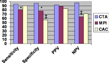
CTA demonstrated significant higher sensitivity than MPI (95% vs. 81%, P < .05). CTA demonstrated significantly higher specificity than both MPI (89% versus 78%, P = .04) and CAC (56%, P = .002). CTA also performed better in a per-vessel analysis (sensitivity 94%, specificity 96%) than both nuclear and CAC. There were no significant differences between the sensitivities and specificities of MPI and CAC.
Conclusion
CTA accurately detects obstructive CAD in symptomatic patients and may be more accurate than MPI or CAC assessment. Larger studies in a more diverse population are needed.
The cost and the risk of invasive angiography have encouraged the development of new diagnostic methods that allow the coronary arteries to be visualized noninvasively. The last decade has seen great strides in the field of cardiac imaging, particularly in the ability of cardiac computed tomographic angiography (CTA) to visualize the coronary lumen with sufficient diagnostic accuracy ( ). Being such a modality, CTA is now being increasingly used in clinical practice. As a result of having high spatial and improved temporal resolution, this imaging modality not only allows branches of the coronary artery to be evaluated, but also allows simultaneous analysis of other cardiac structures, making it extremely useful for other cardiac applications ( ). Moreover, coronary artery calcium (CAC) detected by computed tomography has been shown to be highly specific for atherosclerosis and has prognostic value, yielding valuable information for risk stratification, being both incremental and independent to traditional risk factors ( ).
Other utility has been demonstrated with this modality, including calculation of ejection fraction, as well as assessment of global and regional wall motion evaluation ( ). CTA has been shown to be accurate for determining the presence and severity of coronary artery disease (CAD) ( ).
Get Radiology Tree app to read full this article<
Get Radiology Tree app to read full this article<
Methods
Get Radiology Tree app to read full this article<
Get Radiology Tree app to read full this article<
Angiography Protocol
Get Radiology Tree app to read full this article<
CTA Protocol
Get Radiology Tree app to read full this article<
Get Radiology Tree app to read full this article<
Statistical Methods
Get Radiology Tree app to read full this article<
Results
Get Radiology Tree app to read full this article<
Get Radiology Tree app to read full this article<
Get Radiology Tree app to read full this article<
Get Radiology Tree app to read full this article<
Table 1
Table Showing the Comparison Between Computed Tomography Angiography and Myocardial Perfusion Imaging
Imaging Modality Coronary Artery Calcium Nuclear Myocardial Perfusion Imaging Cardiac Computed Tomography Angiography Sensitivity 90 81 94 Specificity 56 78 96 Positive predictive value 83 89 92 Negative predictive value 71 64 97
Get Radiology Tree app to read full this article<
Discussion
Get Radiology Tree app to read full this article<
Get Radiology Tree app to read full this article<
Get Radiology Tree app to read full this article<
Get Radiology Tree app to read full this article<
Get Radiology Tree app to read full this article<
Get Radiology Tree app to read full this article<
Get Radiology Tree app to read full this article<
Limitations
Get Radiology Tree app to read full this article<
Get Radiology Tree app to read full this article<
Get Radiology Tree app to read full this article<
Get Radiology Tree app to read full this article<
References
1. Leschka S., Alkadhi H., Plass A., et. al.: Accuracy of MDCT coronary angiography with 64-slice technology: first experience. Eur Heart J 2005; 26: pp. 1482-1487.
2. Raff G.L., Gallagher M.J., O’Neill W.W., et. al.: Diagnostic accuracy of noninvasive coronary angiography using 64-slice spiral computed tomography. J Am Coll Cardiol 2005; 46: pp. 552-557.
3. Cademartiri F., Runza G., Belgrano M., et. al.: Introduction to coronary imaging with 64-slice computed tomography. Radiol Med (Torino) 2005; 110: pp. 16-41.
4. Schoenhagen P., Halliburton S.S., Stillman A.E., et. al.: Noninvasive imaging of coronary arteries: current and future role of multi-detector row CT. [review] Radiology 2004; 232: pp. 7-17.
5. Mieres J.H., Shaw L.J., Arai A., et. al.: The role of non-invasive testing in the clinical evaluation of women with suspected coronary artery disease: American Heart Association Consensus Statement. Circulation 2005; 111: pp. 682-696.
6. Taylor A.J., Bindeman J., Feuerstein I., et. al.: Coronary calcium independently predicts incident premature coronary heart disease over measured cardiovascular risk factors. J Am Coll Cardiol 2005; 46: pp. 807-814.
7. Arad Y., Goodman K.J., Roth M., et. al.: Coronary calcification, coronary disease risk factors, C-reactive protein, and atherosclerotic cardiovascular disease events: the St. Francis Heart Study. J Am Coll Cardiol 2005; 46: pp. 158-165.
8. Schroeder S., Kopp A.F., Baumbach A., et. al.: Non-invasive characterisation of coronary lesion morphology by multi-slice computed tomography: a promising new technology for risk stratification of patients with coronary artery disease. Heart 2001; 85: pp. 576-578.
9. Arad Y., Roth M., Newstein D., et. al.: Coronary calcification, coronary risk factors, and atherosclerotic cardiovascular disease events. J Am Coll Cardiol 2005; 46: pp. 158-165.
10. Orakzai S.H., Orakzai R.H., Nasir K., Budoff M.J.: Assessment of Cardiac Function using Multidetector Row Computed Tomography. J Comp Assist Tomogr 2006; 30: pp. 555-563.
11. Leber A.W., Knez A., von Ziegler F., et. al.: Quantification of obstructive and nonobstructive coronary lesions by 64-slice computed tomography: a comparative study with quantitative coronary angiography and intravascular ultrasound. J Am Coll Cardiol 2005; 46: pp. 147-154.
12. Pelzel J.M., Lesser J.R., Flygenring B., et. al.: Multislice CTA determines patient prognosis: six month results in a large ambulatory population. Circulation 2005; 112: pp. II–417.
13. Hacker M., Jakobs T., Matthiesen F., et. al.: Comparison of spiral multidetector CT angiography and myocardial perfusion imaging in the noninvasive detection of functionally relevant coronary artery lesions: first clinical experiences. J Nucl Med 2005; 46: pp. 1294-1300.
14. Iskander S., Iskandrian A.E.: Risk assessment using single-photon emission computed tomographic technetium-99m sestamibi imaging. J Am Coll Cardiol 1998; 32: pp. 57-62.
15. Gibbons R.S.: American Society of Nuclear Cardiology project on myocardial perfusion imaging: measuring outcomes in response to emerging guidelines. J Nucl Cardiol 1996; 3: pp. 436-442.
16. Raiker K., Sinusas A.J., Wackers F.J., et. al.: One-year prognosis of patients with normal planar or single-photon emission computed tomographic technetium 99m-labeled sestamibi exercise imaging. J Nucl Cardiol 1994; 1: pp. 449-456.
17. Brown K.A., Altland E., Rowen M.: Prognostic value of normal technetium-99m sestamibi cardiac imaging. J Nucl Med 1994; 35: pp. 554-557.
18. Pavin D., Delonca J., Siegenthaler M., et. al.: Long-term (10 years) prognostic value of a normal thallium-201 myocardial exercise scintigraphy in patients with coronary artery disease documented by angiography. Eur Heart J 1997; 18: pp. 69-77.
19. Brown K.A., Rowen M.: Prognostic value of a normal exercise myocardial perfusion imaging study in patients with angiographically significant coronary artery disease. Am J Cardiol 1993; 71: pp. 865-867.
20. Budoff M.J., Lu B., Shinbane J.S., et. al.: Methodology for improved detection of coronary stenoses with computed tomographic angiography. Am Heart J 2004; 148: pp. 1085-1090.
21. Lu B., Shavelle D.M., Mao S.S., et. al.: Improved accuracy of noninvasive electron beam coronary angiography. Invest Radiol 2004; 39: pp. 73-79.
22. Mao S., Lu B., Oudiz R.J., et. al.: Coronary artery motion in electron beam tomography. J Comput Assist Tomogr 2000; 24: pp. 253-258.
23. Fleischmann K.E., Hunink M.G., Kuntz K.M., et. al.: Exercise echocardiography or exercise SPECT imaging?. JAMA 1998; 280: pp. 913-920.
24. Shavelle D.M., Budoff M.J., Lamont D.H., et. al.: Exercise testing and electron beam computed tomography in the evaluation of coronary artery disease. J Am Coll Cardiol 2000; 36: pp. 32-38.
25. Schmermund A., Denktas A.E., Rumberger J.A., et. al.: Independent and incremental value of coronary artery calcium for predicting the extent of angiographic coronary artery disease: comparison with cardiac risk factors and radionuclide perfusion imaging. J Am Coll Cardiol 1999; 34: pp. 777-786.
26. Kajinami K., Seki H., Takekoshi N., et. al.: Noninvasive prediction of coronary atherosclerosis by quantification of coronary artery calcification using electron beam computed tomography: comparison with electrocardiographic and thallium exercise stress test results. J Am Coll Cardiol 1995; 26: pp. 1209-1221.
27. Budoff M.J., Jacob B., Rasouli M.L., et. al.: Comparison of electron beam computed tomography and technetium stress testing in differentiating cause of dilated versus ischemic cardiomyopathy. J Comput Assist Tomogr 2005; 29: pp. 699-703.
28. Budoff M.J., Raggi P., Berman D., et. al.: Continuous probabilistic prediction of angiographically significant coronary artery disease using electron beam tomography. Circulation 2002; 105: pp. 1791-1796.
29. Diamond G.A., Rozanski A., Forrester J.S., et. al.: A model for assessing the sensitivity and specificity of tests subject to selection bias. J Chronic Dis 1986; 39: pp. 343-355.
30. Detrano R., Janosi A., Lyons K.P., et. al.: Factors affecting sensitivity and specificity of a diagnostic test: the exercise thallium scintigram. Am J Med 1988; 84: pp. 699-710.
31. Gopal A., Mao S.S., Fischer H., et. al.: Utilization and effectiveness of coronary computed tomographic angiography compared to diagnostic cardiac catheterization for the diagnosis of coronary artery disease. Circulation 2005; 112: II–559
32. Morin R.L., Gerber T.C., McCollough C.H.: Radiation dose in computed tomography of the heart. [review] Circulation 2003; 107: pp. 917-922.
33. Hunold P., Vogt F.M., Schmermund A., et. al.: Radiation exposure during cardiac CT: effective doses at multi-detector row CT and electron-beam CT. Radiology 2003; 226: pp. 145-152.
34. Gerber T.C., Stratmann B.P., Kuzo R.S., et. al.: Effect of acquisition technique on radiation dose and image quality in multidetector row computed tomography coronary angiography with submillimeter collimation. Invest Radiol 2005; 40: pp. 556-563.
35. De Jong H.W., Beekman F.J., Viergever M.A., et. al.: Simultaneous (99m)Tc/(201)Tl dual-isotope SPET with Monte Carlo-based down-scatter correction. Eur J Nucl Med Mol Imaging 2002; 29: pp. 1063-1071.