
Rationale and Objectives
To use a cine balanced steady-state free precession magnetic resonance enterography (cine MRE) pulse sequence to assess the effectiveness of a sublingual (SL) antiperistaltic agent, hyoscyamine sulfate.
Materials and Methods
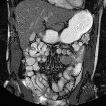
Institutional review board approval was granted with an exemption for informed consent in this Health Insurance Portability and Accountability Act–compliant, retrospective, single-institution study. Of the 288 MRE examinations performed between October 1, 2007 and January 15, 2011, 92 using SL hyoscyamine sulfate for antiperistalsis were included for review, each with cine MRE before and after medication. These 184 cine MRE data sets were randomized, blinded for treatment, and independently reviewed by five attending abdominal radiologists, who rated the degree of whole abdomen bowel motility on each cine MRE data set on a 5-point scale. Pre- and postmedication mean peristalsis ratings, standard deviation, mean difference, and treatment effect sizes were calculated. A repeated measures analysis of variance test was performed using a significance threshold of P = .05. Interobserver reliabilities were also calculated.
Results
Mean peristalsis ratings ranged 2.63–3.34 and 2.36–3.03, before and after medication administration, respectively. The mean differences ranged from 0.22 to 0.46, which are treatment effect sizes of 0.20 to 0.37. The decrease in peristalsis observed by the five reviewing radiologists after SL hyoscyamine sulfate administration was significant (df = 1/182, f = 7.35, P < .01). The interobserver reliabilities were 0.34 for the pretest and 0.33 for the posttest.
Conclusions
Although cine MRE sequences show decreased bowel peristalsis after the use of SL hyoscyamine sulfate, the small size of the observed treatment effect is likely insufficient to justify its use for MRE.
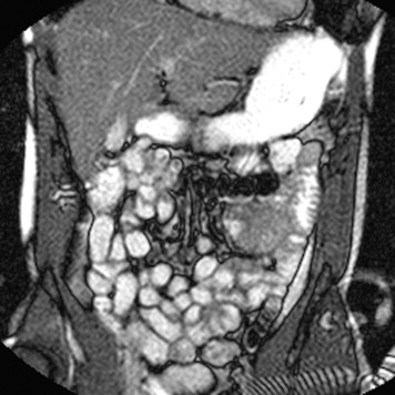
Magnetic resonance enterography (MRE) is a specialized MR examination that is designed to evaluate the small bowel. The most common indication for this procedure is evaluation of inflammatory bowel disease, especially Crohn’s disease. Advantages compared to other imaging modalities include its lack of ionizing radiation, superior soft tissue contrast, a higher safety profile of gadolinium compared to iodinated contrast used for computed tomographic enterography, and the potential for dynamic functional assessment of the bowel . MRE commonly includes repetitive cine images that evaluate intestinal movement, rapid T2-weighted images that highlight fluid and edema, and fat suppressed gradient echo three-dimensional (FatSat-3D) images, which are performed before and after intravenous gadolinium chelate administration to assess bowel enhancement . Previous work has advocated for depressing small bowel peristalsis for the dynamic postcontrast FatSat-3D sequences, which can take up to 25 seconds to acquire, because motion artifact can compromise image quality . Several antimuscarinic agents have been used for this purpose including glucagon (GlucaGen; Novo Nordisk, Bagsvaerd, Denmark) and butylscopolamine (Buscopan; Boehringer Ingelheim Pharma, Ingelheim, Germany), usually via intravenous (IV) administration. IV butylscopolamine has a very favorable side effect profile, but is unavailable for use in the United States. IV glucagon is available in the United States, but is most commonly associated with side effects such as nausea and vomiting, with symptom onset occurring up to 3 hours after administration in some instances . Hyoscyamine sulfate (Levsin; Media Pharmaceuticals, Somerset, NJ), a levo-enantiomer of atropine with anticholinergic and antispasmodic properties, is another antimuscarinic agent used clinically to provide symptom relief to patients with bowel dysmotility syndromes. Hyoscyamine sulfate is available in multiple preparations, but can be administered via the sublingual (SL) route. Previous investigations with the use of this sublingual agent have addressed upper gastrointestinal series, barium enema, and endoscopic retrograde cholangiopancreatography (ERCP) examinations . Prior investigators have noted a favorable side effect profile and a greater cost effectiveness of this agent in comparison to intravenous glucagon; however, the observed efficacy of its antispasmodic activity during investigated procedures has been mixed . Although SL hyoscyamine sulfate use has been investigated in conjunction with other types of imaging studies, there is currently no literature regarding its use as the small bowel antiperistaltic agent for MRE. The aim of this study, therefore, was to assess the effectiveness of SL hyoscyamine sulfate as an antiperistaltic agent for small bowel evaluation at MRE, using a cine MR imaging (MRI) sequence to evaluate the medication’s effect on small bowel peristalsis.
Materials and methods
Participants
This is a single-institution retrospective study performed in compliance with the Health Insurance Portability and Accountability Act. It was institutional review board–approved with a waiver for informed consent. The picture archiving and communication system (PACS) (iSite; Philips, The Netherlands) was retrospectively searched for all MRE studies performed between October 1, 2007 and January 15, 2011 at our institution using the following search criteria: “MR Enterography”, “Small Bowel Enterography”, “MRE”, “Volumen”, “Enterography”, “Levsin”, and “Hyoscyamine”. A total of 288 studies were performed during this time. Of these, 196 studies were excluded for reasons including one or no cine sequences performed, cine sequences permanently labeled in PACS and unable to be randomized, no medication or only one medication tablet given before the study, or improper documentation of medication administration not allowing for verification. Thus, 92 MRE examinations, with 184 individual cine data sets, were included in the study ( Fig 1 ). MRE examinations were performed for a variety of reasons with the most common indication to evaluate Crohn’s disease or other inflammatory bowel disease.
Get Radiology Tree app to read full this article<
MR Scanning
Get Radiology Tree app to read full this article<
Data Analysis
Get Radiology Tree app to read full this article<
Get Radiology Tree app to read full this article<
Statistical Analysis
Get Radiology Tree app to read full this article<
Results
Get Radiology Tree app to read full this article<
Table 1
Cine MR Peristalsis Rating Mean, SD, Mean Difference, t value and Significance Level, and Treatment Effect Size (Cohen’s d) for Each Reader Both before and after SL Hyoscyamine Sulfate Administration, n = 92 for Each Reader/Pre-Post Group
Reader Mean SD Mean Difference_t_ Value and Significance Level Treatment Effect Size (Cohen’s d) 1: pre 3.34 1.22 0.46 3.86 ∗∗∗ 0.37 post 2.88 1.25 2: pre 2.92 1.21 0.33 2.48 ∗ 0.27 post 2.59 1.20 3: pre 3.08 0.92 0.28 2.89 ∗∗ 0.30 post 2.80 0.92 4: pre 2.63 1.14 0.27 2.33 ∗ 0.25 post 2.36 1.05 5: pre 3.25 1.07 0.22 1.82 N.S. 0.20 post 3.03 1.10
MR, magnetic resonance; SD, standard deviation; SL, sublingual.
Statistical significance of mean differences tested by paired t tests are indicated by superscript (* P < .05, ** P < .005, *** P < .0005, N.S., not significant).
Table 2
Repeated Mmeasures ANOVA Table between Subject Effects: Test of Hypothesis That Peristalsis is Lower after SL Hyoscyamine Sulfate Administration
Source df Type III SS Mean Square f Value_P_ Value Pre-post 1 22.23 22.23 7.35 <.01 Error 182 550.44 3.02
ANOVA, analysis of variance; df, degrees of freedom; SL, sublingual; SS, sum of squares.
Get Radiology Tree app to read full this article<
Discussion
Get Radiology Tree app to read full this article<
Get Radiology Tree app to read full this article<
Get Radiology Tree app to read full this article<
Get Radiology Tree app to read full this article<
Get Radiology Tree app to read full this article<
Get Radiology Tree app to read full this article<
Supplementary Data
Get Radiology Tree app to read full this article<
Video 1
Get Radiology Tree app to read full this article<
Video 2
Get Radiology Tree app to read full this article<
Video 3
Get Radiology Tree app to read full this article<
Video 4
Get Radiology Tree app to read full this article<
Get Radiology Tree app to read full this article<
References
1. Fidler J.: MR imaging of the small bowel. Radiol Clin North Am 2007; 45: pp. 317-331.
2. Grand D.J., Beland M., Harris A.: Magnetic resonance enterography. Radiol Clin North Am 2013 1; 51: pp. 99-112.
3. Siddiki H., Fidler J.: MR imaging of the small bowel in Crohn’s disease. Eur J Radiol 2009; 69: pp. 409-417.
4. Chernish S.M., Maglinte D.D.: Glucagon: Common untoward reactions—review and recommendations. Radiology 1990; 177: pp. 145-146.
5. Moeller G., Hughes J.J., Mangano F.A., et. al.: Comparison of L-hyoscyamine, glucagon, and placebo for air-contrast upper gastrointestinal series. Gastrointest Radiol 1992; 17: pp. 195-198.
6. Bova J.G., Jurdi R.A., Bennett W.F.: Antispasmodic drugs to reduce discomfort and colonic spasm during barium enemas: Comparison of oral hyoscyamine, i.v. glucagon, and no drug. AJR Am J Roentgenol 1993; 161: pp. 965-968.
7. Lynch C.R., Khandekar S., Lynch S.M., et. al.: Sublingual L-hyoscyamine for duodenal antimotility during ERCP: A prospective randomized double-blinded study. Gastrointest Endosc 2007 10; 66: pp. 748-752.
8. Janec E., Gandotra V., Chaptini L., et. al.: Initial experience with sublingual hyoscyamine sulfate for suppression of duodenal motility during endoscopic retrograde cholangio-pancreatography (ERCP). Am J Gastroenterol 2003; 98: pp. S306.
9. Complete hyoscyamine sulfate information from drugs.com. Available at: http://www.drugs.com/ppa/hyoscyamine-sulfate.html . Accessed May 1, 2013.
10. Shrout P.E., Fleiss J.L.: Intraclass correlations: uses in assessing rater reliability. Psychol Bull 1979; 86: pp. 420-428.
11. Glucagon official FDA information, side effects and uses. Available at: http://wwwdrugscom/pro/glucagonhtml#fcpfig1a . Accessed May 1, 2013.
12. Hyoscyamine: Drug information. Available at: http://www.uptodate.com/contents/hyoscyamine-drug-information?source=search_result&search=hyoscyamine&selectedTitle=1∼29 . Accessed May 1, 2013.
13. Chaptini L.A., Janec E.M., Seltzer G., et. al.: Sublingual hyoscyamine spray as premedication for colonoscopy: A randomized double-blinded placebo-controlled trial. Am J Surg 2008; 196: pp. 51-55.
14. Marti-Bonmati L., Graells M., Ronchera-Oms C.L.: Reduction of peristaltic artifacts on magnetic resonance imaging of the abdomen: A comparative evaluation of three drugs. Abdom Imaging 1996; 21: pp. 309-313.
15. Froehlich J.M., Patak M.A., von Weymarn C., et. al.: Small bowel motility assessment with magnetic resonance imaging. J Magn Reson Imaging 2005; 21: pp. 370-375.
16. Ajaj W., Lauenstein T., Papanikolaou N., et. al.: Real-time high-resolution MRI for the assessment of gastric motility: Pre- and postpharmacological stimuli. J Magn Reson Imaging 2004; 19: pp. 453-458.
17. Heye T., Stein D., Antolovic D., et. al.: Evaluation of bowel peristalsis by dynamic cine MRI: Detection of relevant functional disturbances—initial experience. J Magn Reson Imaging 2012; 35: pp. 859-867.