
Rationale and Objective
This study aimed to investigate the clinical value of spectral computed tomography (CT) in the detection of cholesterol gallstones from surrounding bile.
Materials and Methods
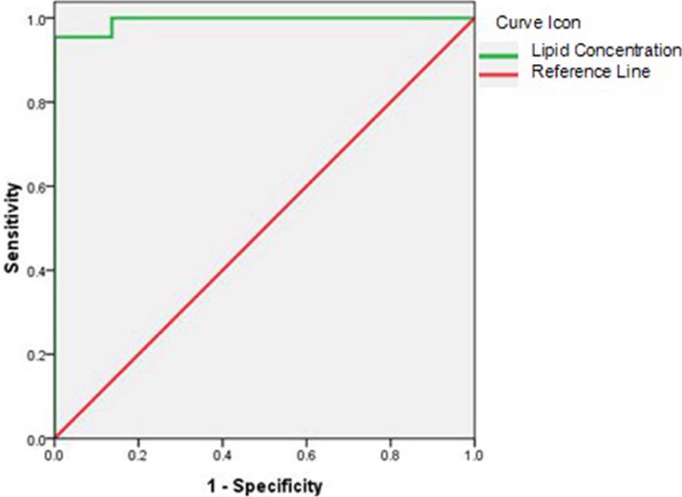
This study was approved by the institutional review board. The unenhanced spectral CT data of 24 patients who had surgically confirmed cholesterol gallstones were analyzed. Lipid concentrations and CT numbers were measured from fat-based material decomposition image and virtual monochromatic image sets (40–140 keV), respectively. The difference in lipid concentration and CT number between cholesterol gallstones and the surrounding bile were statistically analyzed. Receiver operating characteristic analysis was applied to determine the diagnostic accuracy of using lipid concentration to differentiate cholesterol gallstones from bile.
Results
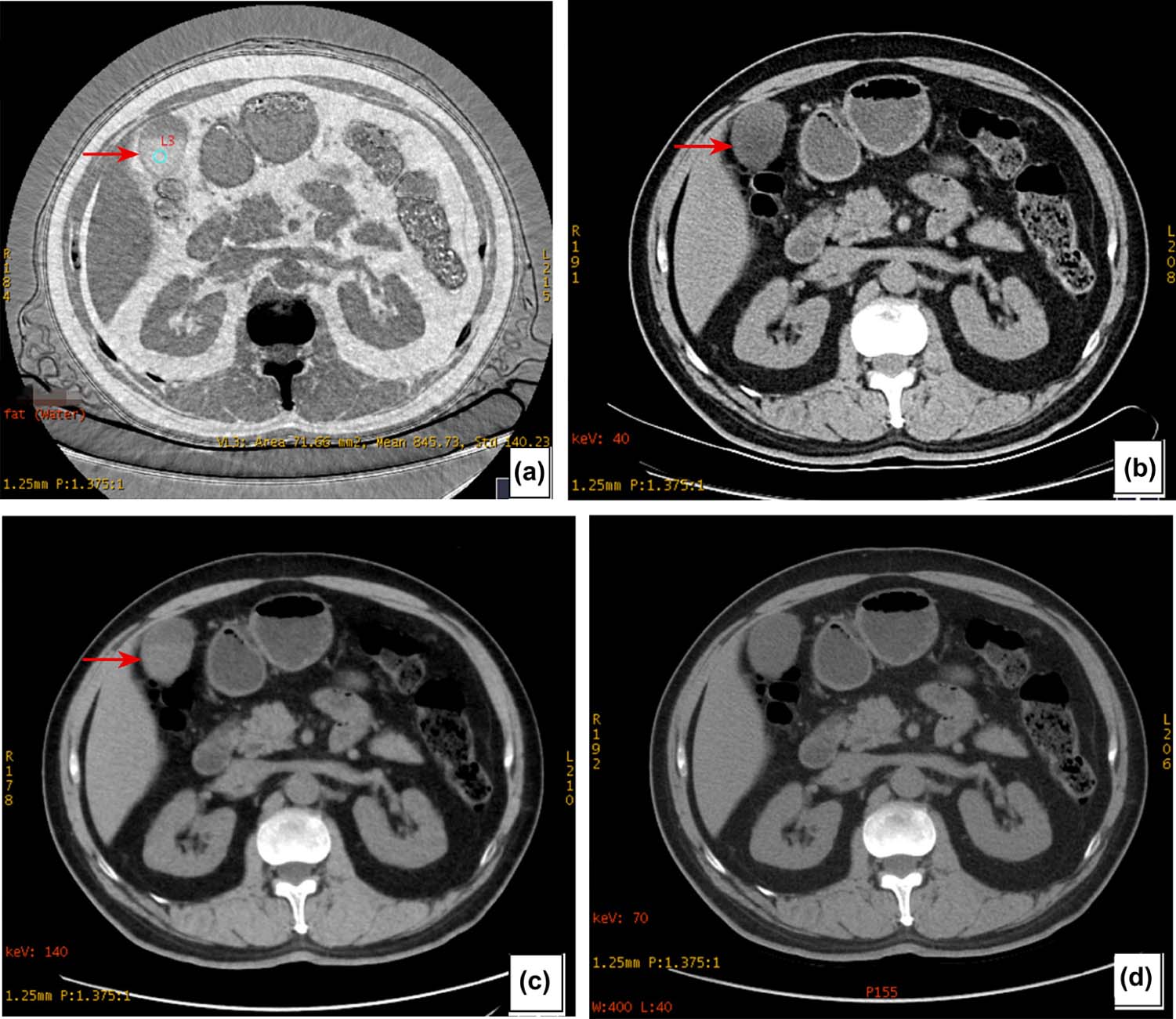
Cholesterol gallstones were bright on fat-based material decomposition images yielding a 92% detection rate (22 of 24). The lipid concentrations (552.65 ± 262.36 mg/mL), CT number at 40 keV (−31.57 ± 16.88 HU) and 140 keV (24.30 ± 5.85 HU) for the cholesterol gallstones were significantly different from those of bile (−13.94 ± 105.12 mg/mL, 12.99 ± 9.39 HU and 6.19 ± 4.97 HU, respectively). Using 182.59 mg/mL as the threshold value for lipid concentration, one could obtain sensitivity of 95.5% and specificity of 100% with accuracy of 0.994 for differentiating cholesterol gallstones from bile.
Conclusions
Virtual monochromatic spectral CT images at 40 keV and 140 keV provide significant CT number differences between cholesterol gallstones and the surrounding bile. Spectral CT provides an excellent detection rate for cholesterol gallstones.
Introduction
Gallstones are a common disease in the gallbladder with an incidence rate of about 10% and occur mostly in middle-aged women. There are three common gallstones: cholesterol, bile pigment, and mixed stone . Cholesterol stone is the main type of gallstone, and is iso- or slightly hypoattenuating relative to bile. The experiment by Brakel et al. proved that the computed tomography (CT) value of the stones negatively correlated with cholesterol content and positively with calcium content. The cholesterol stone has CT attenuation similar to the surrounding bile under the normal X-ray or CT examination and is often difficult to detect. Cholesterol stones are therefore often referred to as negative stones and are easily missed in conventional CT using CT number alone . The recently introduced dual-energy spectral CT imaging uses information from two different energy spectrums to provide additional material density and effective atomic number information for different materials such as different stones , as well as a set of virtual monochromatic images at different photon energy levels. This multiparameter (photon energy-dependent CT number and material density value) approach should improve the separation of materials that have similar CT attenuation value using polychromatic energy beams but different intrinsic material densities such as the cholesterol gallstone and bile. The purpose of this study was to investigate the clinical value of dual-energy spectral CT imaging in the detection of cholesterol gallstones.
Materials and Methods
I. General Information
This retrospective study was approved by the institutional review board. The authors retrospectively analyzed the CT imaging data of 24 patients from July 2013 to June 2014 who underwent unenhanced spectral CT scans for upper abdominal pain in our hospital. Because cholesterol gallstones are easily missed in the conventional CT imaging, the study population was limited to patients who had only this type of stone confirmed by surgery. These patients included 10 men and 14 women with mean age of 48 years (32–67 years), mean body mass index (BMI) of 24.40 ± 4.11 kg/m 2 , no history of biliary surgery, and no fatty diet in the week before the CT examination.
II. CT Scan Technique
Get Radiology Tree app to read full this article<
III. Image Analysis
Get Radiology Tree app to read full this article<
Get Radiology Tree app to read full this article<
Get Radiology Tree app to read full this article<
IV. Statistical Analysis
Get Radiology Tree app to read full this article<
Results
Get Radiology Tree app to read full this article<
Get Radiology Tree app to read full this article<
Get Radiology Tree app to read full this article<
Get Radiology Tree app to read full this article<
Table 1
CT Number Measurements of Bile and Cholesterol Gallstones and CNR Values for Cholesterol Gallstones as Function of Photon Energy ( x¯±s x
¯
±
s ± s)
keV CT Number (HU) Measurement_Z_ Value_P_ Value Contrast-to-Noise Ratio for Gallstones Bile Gallstone Gallstone 40 12.99 ± 9.39 −31.57±16.88 −5.68 <.001 1.38 ± 0.38 50 10.50 ± 6.01 −10.62±9.35 −5.34 <.001 0.87 ± 0.42 60 7.85 ± 4.32 7.67 ± 4.04 −0.42 .673 0.01 ± 0.03 70 8.57 ± 2.51 8.58 ± 2.58 −0.12 .907 0.00 ± 0.01 80 8.91 ± 2.05 9.28 ± 2.16 −0.81 .418 0.03 ± 0.06 90 6.84 ± 4.30 15.18 ± 4.18 −5.09 <.001 0.73 ± 0.48 100 6.52 ± 4.58 19.37 ± 4.94 −5.63 <.001 1.22 ± 0.62 110 6.39 ± 4.71 21.34 ± 5.30 −5.68 <.001 1.49 ± 0.68 120 6.30 ± 4.80 22.51 ± 5.49 −5.68 <.001 1.69 ± 0.72 130 6.25 ± 4.90 23.25 ± 5.58 −5.68 <.001 1.82 ± 0.75 140 6.19 ± 4.97 24.30 ± 5.85 −5.68 <.001 2.00 ± 0.75
CNR, contrast-to-noise ratio; CT, computed tomography; HU; Hounsfield unit.
Get Radiology Tree app to read full this article<
Get Radiology Tree app to read full this article<
Get Radiology Tree app to read full this article<
Discussions
Get Radiology Tree app to read full this article<
Get Radiology Tree app to read full this article<
Get Radiology Tree app to read full this article<
Get Radiology Tree app to read full this article<
Get Radiology Tree app to read full this article<
Conclusions
Get Radiology Tree app to read full this article<
Acknowledgments
Get Radiology Tree app to read full this article<
References
1. Kim I.S., Myung S.J., Lee S.S., et. al.: Classification and nomenclature of gallstones revisited. Yonsei Med J 2003; 44: pp. 561. 12950109
2. Brakel K., Lameris J.S., Nijs S.J., et. al.: Predicting gallstone composition with CT: in vivo and in vitro analysis. Radiology 1990; 174: pp. 337-341. 2296642
3. Anderson S.W., Lucey B.C., Varghese J.C., et. al.: Accuracy of MDCT in the diagnosis of choledocholithiasis. AJR Am J Roentgenol 2006; 187: pp. 174. 16794173
4. Chan W.C., Joe B.N., Coakley F.V., et. al.: Gallstone detection at CT in vitro: effect of peak voltage setting. Radiology 2006; 241: pp. 546-553. 17057073
5. Potretzke T.A., Brace C.L., Lubner M.G., et. al.: Early small-bowel ischemia: dual-energy CT improves conspicuity compared with conventional CT in a swine model. Radiology 2015; 275: pp. 119-126. 25426772 November 26:140875. [Epub ahead of print]
6. Bova J.G., Schwesinger W.H., Kurtin W.E.: In vivo analysis of gallstone composition by computed tomography. Gastrointest Radiol 1992; 17: pp. 253-256. 1612311
7. Patel N.B., Oto A., Thomas S.: Multidetector CT of emergent biliary pathologic conditions. Radiographics 2013; 33: pp. 1867-1888. 24224584
8. Jeffrey R.B., Federle M.P., Laing F.C., et. al.: Computed tomography of choledocholithiasis. AJR Am J Roentgenol 1983; 140: pp. 1179-1183. 6602489
9. Baron R.L.: Diagnosing choledocholithiasis: how far can we push helical CT?. Radiology 1997; 203: pp. 601-603. 9169674
10. Johnson T.R., Krauss B., Sedlmair M., et. al.: Material differentiation by dual energy CT: initial experience. Eur Radiol 2007; 17: pp. 1510-1517. 17151859
11. Wang X.L., Meier D., Taguchi K., et. al.: Material separation in X-ray CT with energy resolved photon-counting detectors. Med Phys 2011; 38: pp. 1534-1546. 21520865
12. Graser A., Johnson T.R., Bader M., et. al.: Dual energy CT characterization of urinary calculi: initial in vitro and clinical experience. Invest Radiol 2008; 43: pp. 112-119. 18197063
13. Hidas G., Eliahou R., Duvdevani M., et. al.: Determination of renal stone composition with dual-energy CT: in vivo analysis and comparison with X-ray diffraction. Radiology 2010; 257: pp. 394-401. 20807846
14. Brown C.L., Hartman B.P., Dzyubak O.P., et. al.: Dual-energy CT iodine overlay technique for characterization of renal masses as cyst or solid: a phantom feasibility study. Eur Radiol 2009; 19: pp. 1289-1295. 19153744
15. Zhang D., Li X., Wang J.: Objective characterization of GE Discovery CT750 HD scanner: gemstone spectral imaging mode. Med Phys 2011; 38: pp. 1178. 21520830