
Three-dimensional (3D) printing refers to a number of manufacturing technologies that create physical models from digital information. Radiology is poised to advance the application of 3D printing in health care because our specialty has an established history of acquiring and managing the digital information needed to create such models. The 3D Printing Task Force of the Radiology Research Alliance presents a review of the clinical applications of this burgeoning technology, with a focus on the opportunities for radiology. Topics include uses for treatment planning, medical education, and procedural simulation, as well as patient education. Challenges for creating custom implantable devices including financial and regulatory processes for clinical application are reviewed. Precedent procedures that may translate to this new technology are discussed. The task force identifies research opportunities needed to document the value of 3D printing as it relates to patient care.
Introduction
What began as a largely industrial tool to facilitate concept-to-prototype development, three-dimensional (3D) printing has evolved into a widely used technology, affecting many aspects of modern society. The term “3D printing” grew out of the research and development laboratories of the automotive and aerospace industries . The technology was developed throughout the 1980s and 1990s , and medical applications were initially reported in the early 2000s . Initially, these reports focused on custom prostheses , but as the technology improved, reports of using anatomic models for preoperative planning began appearing . Today, 3D printing continues to find new applications: customized eyeglasses can be printed to exact specifications , an increasing number of foods can be printed on demand , and there are plans to manufacture cars entirely using 3D printing .
Recent rapid growth of 3D printing in medicine has been staggering. A search of Pubmed.gov using the term “3D printing” yielded only six publications in the year 2000, 61 publications in 2010, and more than 1100 publications in 2016. To encourage continued growth of this technology, the National Additive Manufacturing Innovation Institute was launched in 2012 . Many professional societies have also advocated the use of this technology in medicine. For example, the Society for Manufacturing Engineers has a dedicated medical 3D printing workgroup . In 2016, the Radiological Society of North America formed the 3D Printing Special Interest Group. The 3D Printing Special Interest Group has already sponsored many educational sessions at the annual meeting and is committed to building evidence for clinical utility of 3D printing .
Undoubtedly, this topic has gained popularity because of the tremendous potential it offers to radiologists, our colleagues, and patients. If implemented correctly, 3D printing promises to improve patient care and enhance the relative contribution to that care by radiologists. Specifically, 3D printing can deliver personalized medicine based on the anatomic data radiologists acquire and interpret every day. Providing such a service offers a new way to interact with referring clinicians and a potential way radiology can demonstrate value in patient care.
Radiologists have witnessed the evolution of medical imaging that allows for 3D printing. Multiplanar imaging with computed tomography (CT) and magnetic resonance imaging gave rise to 3D reconstructions that improved the evaluation of complex anatomy . At its most basic level, 3D printing takes imaging data from the two dimensions of a computer screen to the three dimensions of the real world .
3D printing has been used in a wide range of healthcare settings including Cardiology , Cardiothoracic Surgery , Critical Care , Gastroenterology , General Surgery , Interventional Radiology , Neurosurgery , Ophthalmology , Oral and Maxillofacial Surgery , Orthopedic Surgery , Otolaryngology , Plastic Surgery , Podiatry , Pulmonology , Radiation Oncology , Transplant Surgery , Urology , and Vascular Surgery .
Get Radiology Tree app to read full this article<
Get Radiology Tree app to read full this article<
Get Radiology Tree app to read full this article<
Transforming Clinical Care
Treatment Planning
Get Radiology Tree app to read full this article<
Get Radiology Tree app to read full this article<
Get Radiology Tree app to read full this article<
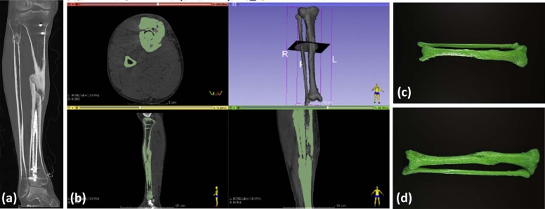
Fracture fixation
Get Radiology Tree app to read full this article<
Get Radiology Tree app to read full this article<
Get Radiology Tree app to read full this article<
Get Radiology Tree app to read full this article<
Resection of renal tumors
Get Radiology Tree app to read full this article<
Cardiovascular applications
Get Radiology Tree app to read full this article<
Customized Surgical Tools and Prostheses
Get Radiology Tree app to read full this article<
Surgical Tools
Get Radiology Tree app to read full this article<
Prostheses
Get Radiology Tree app to read full this article<
Get Radiology Tree app to read full this article<
Get Radiology Tree app to read full this article<
Maxillofacial prostheses
Get Radiology Tree app to read full this article<
Joint prostheses
Get Radiology Tree app to read full this article<
Get Radiology Tree app to read full this article<
Get Radiology Tree app to read full this article<
Patient Education
Get Radiology Tree app to read full this article<
Get Radiology Tree app to read full this article<
Transforming Medical Education
Get Radiology Tree app to read full this article<
Anatomy Teaching
Get Radiology Tree app to read full this article<
Get Radiology Tree app to read full this article<
Get Radiology Tree app to read full this article<
Get Radiology Tree app to read full this article<
Operative Rehearsal
Get Radiology Tree app to read full this article<
Get Radiology Tree app to read full this article<
Get Radiology Tree app to read full this article<
Get Radiology Tree app to read full this article<
Get Radiology Tree app to read full this article<
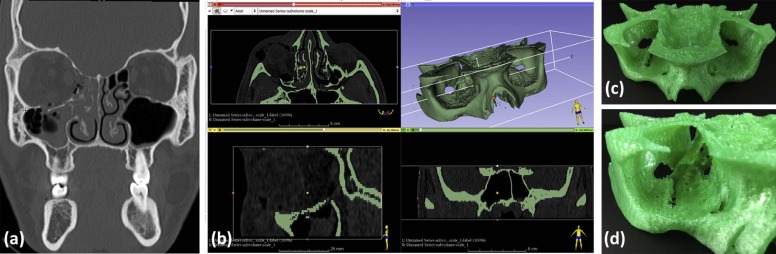
Applications in Radiology
Get Radiology Tree app to read full this article<
Get Radiology Tree app to read full this article<
Get Radiology Tree app to read full this article<
Get Radiology Tree app to read full this article<
Research Directions
Bioprinting Revolution
Get Radiology Tree app to read full this article<
Get Radiology Tree app to read full this article<
Get Radiology Tree app to read full this article<
Get Radiology Tree app to read full this article<
Get Radiology Tree app to read full this article<
Get Radiology Tree app to read full this article<
Get Radiology Tree app to read full this article<
Get Radiology Tree app to read full this article<
Get Radiology Tree app to read full this article<
Get Radiology Tree app to read full this article<
Research Opportunities for Radiology
Get Radiology Tree app to read full this article<
Get Radiology Tree app to read full this article<
Get Radiology Tree app to read full this article<
Challenges to Clinical Application of 3D Printing
Get Radiology Tree app to read full this article<
Financial Challenges
Get Radiology Tree app to read full this article<
Get Radiology Tree app to read full this article<
Get Radiology Tree app to read full this article<
Get Radiology Tree app to read full this article<
Get Radiology Tree app to read full this article<
Get Radiology Tree app to read full this article<
Get Radiology Tree app to read full this article<
Get Radiology Tree app to read full this article<
Regulatory Challenges
Get Radiology Tree app to read full this article<
Get Radiology Tree app to read full this article<
Get Radiology Tree app to read full this article<
Get Radiology Tree app to read full this article<
Get Radiology Tree app to read full this article<
Get Radiology Tree app to read full this article<
Get Radiology Tree app to read full this article<
Get Radiology Tree app to read full this article<
Medicolegal Challenges
Get Radiology Tree app to read full this article<
Get Radiology Tree app to read full this article<
Conclusion
Get Radiology Tree app to read full this article<
Get Radiology Tree app to read full this article<
Get Radiology Tree app to read full this article<
Get Radiology Tree app to read full this article<
Acknowledgment
Get Radiology Tree app to read full this article<
References
1. Sniderman D.: American Society Of Mechanical Engineers 3D printing takes off. American Society Of Mechanical Engineers website; Available at: http://bit.ly/2vAauKy
2. Hull C.W.: Apparatus for production of three-dimensional objects by stereolithography. Uvp, Inc., inventor; Uvp, Inc., assignee; United States patent US 4,575,330; Available at: http://www.google.com/patents/us4575330
3. Sachs E., Cima M., Cornie J.: Three-dimensional printing: rapid tooling and prototypes directly from a CAD model. CIRP Annals—Manuf Tech 1990; 39: pp. 201-204.
4. Sachs E.M., Haggerty J.S., Cima M.J., et. al.: Three-dimensional printing techniques. United States patent US 5,204,055; Available at: http://www.google.com/patents/US5204055
5. Gross B.C., Erkal J.L., Lockwood S.Y., et. al.: Evaluation of 3D printing and its potential impact on biotechnology and the chemical sciences. Anal Chem 2014; 86: pp. 3240-3253.
6. Curodeau A., Sachs E., Caldarise S.: Design and fabrication of cast orthopedic implants with freeform surface textures from 3-D printed ceramic shell. J Biomed Mater Res 2000; 53: pp. 525-535.
7. Hong S.B., Eliaz N., Leisk G.G., et. al.: A new Ti-5Ag alloy for customized prostheses by three-dimensional printing (3DP). J Dent Res 2001; 80: pp. 860-863.
8. Melican M.C., Zimmerman M.C., Dhillon M.S., et. al.: Three-dimensional printing and porous metallic surfaces: a new orthopedic application. J Biomed Mater Res 2001; 55: pp. 194-202.
9. Rowe C.W., Katstra W.E., Palazzolo R.D., et. al.: Multimechanism oral dosage forms fabricated by three dimensional printing. J Control Release 2000; 66: pp. 11-17.
10. Sodian R., Schmauss D., Markert M., et. al.: Three-dimensional printing creates models for surgical planning of aortic valve replacement after previous coronary bypass grafting. Ann Thorac Surg 2008; 85: pp. 2105-2108.
11. Sodian R., Weber S., Markert M., et. al.: Pediatric cardiac transplantation: three-dimensional printing of anatomic models for surgical planning of heart transplantation in patients with univentricular heart. J Thorac Cardiovasc Surg 2008; 136: pp. 1098-1099.
12. Wilasrusmee C., Suvikrom J., Suthakorn J., et. al.: Three-dimensional aortic aneurysm model and endovascular repair: an educational tool for surgical trainees. Int J Angiol 2008; 17: pp. 129-133.
13. Bruyère F., Leroux C., Brunereau L., et. al.: Rapid prototyping model for percutaneous nephrolithotomy training. J Endourol 2008; 22: pp. 91-96.
14. Protos Eyewear website. Available at: http://www.protoseyewear.com/
15. Natural Machines website. Available at: http://www.naturalmachines.com/
16. Kessler A.M.: A 3-D Printed Car, Ready for the Road. The New York Times. Available at: http://nyti.ms/2u6eiUj
17. Davies C.T., Baird L.M., Jacobson M.D., et. al.: Reed Smith white paper: 3D printing of medical devices: when a novel technology meets traditional legal principles. Reed Smith website; Available at: http://bit.ly/2oH5PY4
18. SME Medical Additive Manufacturing/3D Printing Workgroup : Society for manufacturing engineers website. Available at: http://www.sme.org/medical-am3dp-workgroup/
19. RSNA 3D Printing Special Interest Group: Radiological society of North America website. Available at: http://www.rsna.org/3D-Printing-SIG/
20. Kijima S., Sasaki T., Nagata K., et. al.: Preoperative evaluation of colorectal cancer using CT colonography, MRI, and PET/CT. World J Gastroenterol 2014; 20: pp. 16964-16975.
21. Sakamoto T.: Roles of universal three-dimensional image analysis devices that assist surgical operations. J Hepatobiliary Pancreat Sci 2014; 21: pp. 230-234.
22. Fayad L.M., Patra A., Fishman E.K.: Value of 3D CT in defining skeletal complications of orthopedic hardware in the postoperative patient. AJR Am J Roentgenol 2009; 193: pp. 1155-1163.
23. Farooqi K.M., Saeed O., Zaidi A., et. al.: 3D printing to guide ventricular assist device placement in adults with Congenital Heart Disease and heart failure. JACC Heart Fail 2016; 4: pp. 301-311.
24. Schmauss D., Haeberle S., Hagl C., et. al.: Three-dimensional printing in cardiac surgery and interventional cardiology: a single-centre experience. Eur J Cardiothorac Surg 2015; 47: pp. 1044-1052.
25. Costello J.P., Olivieri L.J., Su L., et. al.: Incorporating three-dimensional printing into a simulation-based congenital heart disease and critical care training curriculum for resident physicians. Congenit Heart Dis 2015; 10: pp. 185-190.
26. Dhir V., Itoi T., Fockens P., et. al.: Novel ex vivo model for hands-on teaching of and training in EUS-guided biliary drainage: creation of “Mumbai EUS” stereolithography/3D printing bile duct prototype (with videos). Gastrointest Endosc 2015; 81: pp. 440-446.
27. Malik H.H., Darwood A.R.J., Shaunak S., et. al.: Three-dimensional printing in surgery: a review of current surgical applications. J Surg Res 2015; 199: pp. 512-522.
28. Itagaki M.W.: Using 3D printed models for planning and guidance during endovascular intervention: a technical advance. Diagn Interv Radiol 2015; 21: pp. 338-341.
29. Anderson J.R., Thompson W.L., Alkattan A.K., et. al.: Three-dimensional printing of anatomically accurate, patient specific intracranial aneurysm models. J Neurointerv Surg 2016; 8: pp. 517-520.
30. Weinstock P., Prabhu S.P., Flynn K., et. al.: Optimizing cerebrovascular surgical and endovascular procedures in children via personalized 3D printing. J Neurosurg Pediatr 2015; 16: pp. 584-589.
31. Huang W., Zhang X.: 3D Printing: print the future of ophthalmology. Invest Ophthalmol Vis Sci 2014; 55: pp. 5380-5381.
32. Obregon F., Vaquette C., Ivanovski S., et. al.: Three-dimensional bioprinting for regenerative dentistry and craniofacial tissue engineering. J Dent Res 2015; 94: pp. 143-152.
33. Groth C., Kravitz N.D., Jones P.E., et. al.: Three-dimensional printing technology. J Clin Orthod 2014; 48: pp. 475S-485S.
34. Eltorai A.E.M., Nguyen E., Daniels A.H.: Three-dimensional printing in orthopedic surgery. Lindeque BGP, ed Orthopedics 2015; 38: pp. 684-687.
35. VanKoevering K.K., Hollister S.J., Green G.E.: Advances in 3-dimensional printing in otolaryngology: a review. JAMA Otolaryngol Head Neck Surg 2017; 143: pp. 178-183.
36. Zopf D.A., Hollister S.J., Nelson M.E., et. al.: Bioresorbable airway splint created with a three-dimensional printer. N Engl J Med 2013; 368: pp. 2043-2045.
37. Chae M.P., Rozen W.M., McMenamin P.G., et. al.: Emerging applications of bedside 3D printing in plastic surgery. Front Surg 2015; 2: pp. 25.
38. Jastifer J.R., Gustafson P.A.: Three-dimensional printing and surgical simulation for preoperative planning of deformity correction in foot and ankle surgery. J Foot Ankle Surg 2017; 56: pp. 191-195.
39. Bustamante S., Shravan Cheruku M.D.: 3D printing for simulation in thoracic anesthesia. J Cardiothorac Vasc Anesth 2016; 30: pp. 61-63.
40. Burleson S., Baker J., Hsia A.T., et. al.: Use of 3D printers to create a patient-specific 3D bolus for external beam therapy. J Appl Clin Med Phys 2015; 16: pp. 5247.
41. Zein N.N., Hanouneh I.A., Bishop P.D., et. al.: Three-dimensional print of a liver for preoperative planning in living donor liver transplantation. Liver Transpl 2013; 19: pp. 1304-1310.
42. Youssef R.F., Spradling K., Yoon R., et. al.: Applications of three-dimensional printing technology in urological practice. BJU Int 2015; 116: pp. 697-702.
43. Hoch E., Tovar G.E.M., Borchers K.: Bioprinting of artificial blood vessels: current approaches towards a demanding goal. Eur J Cardiothorac Surg 2014; 46: pp. 767-778.
44. Matsumoto J.S., Morris J.M., Rose P.S.: 3-Dimensional printed anatomic models as planning aids in complex oncology surgery. JAMA Oncol 2016; 2: pp. 1121-1122.
45. Tack P., Victor J., Gemmel P., et. al.: 3D-printing techniques in a medical setting: a systematic literature review. Biomed Eng Online 2016; 15: pp. 115.
46. Mao Y., Xu C., Xu J., et. al.: The use of customized cages in revision total hip arthroplasty for Paprosky type III acetabular bone defects. Int Orthop 2015; 39: pp. 2023-2030.
47. Chung K.J., Hong D.Y., Kim Y.T., et. al.: Preshaping plates for minimally invasive fixation of calcaneal fractures using a real-size 3D-printed model as a preoperative and intraoperative tool. Foot Ankle Int 2014; 35: pp. 1231-1236.
48. Huang H., Hsieh M.-F., Zhang G., et. al.: Improved accuracy of 3D-printed navigational template during complicated tibial plateau fracture surgery. Australas Phys Eng Sci Med 2015; 38: pp. 109-117.
49. Pacione D., Tanweer O., Berman P., et. al.: The utility of a multimaterial 3D printed model for surgical planning of complex deformity of the skull base and craniovertebral junction. J Neurosurg 2016; 125: pp. 1194-1197.
50. Zeng C., Xing W., Wu Z., et. al.: A combination of three-dimensional printing and computer-assisted virtual surgical procedure for preoperative planning of acetabular fracture reduction. Injury 2016; 47: pp. 2223-2227.
51. Silberstein J.L., Maddox M.M., Dorsey P., et. al.: Physical models of renal malignancies using standard cross-sectional imaging and 3-dimensional printers: a pilot study. Urology 2014; 84: pp. 268-272.
52. Wake N., Rude T., Kang S.K., et. al.: 3D printed renal cancer models derived from MRI data: application in pre-surgical planning. Abdom Radiol 2017; 42: pp. 1501-1509.
53. Pepper J., Petrou M., Rega F., et. al.: Implantation of an individually computer-designed and manufactured external support for the Marfan aortic root. Multimed Man Cardiothorac Surg 2013; 2013: mmt004
54. Hossien A., Gesomino S., Maessen J., et. al.: The interactive use of multi-dimensional modeling and 3D printing in preplanning of type A aortic dissection. J Card Surg 2016; 31: pp. 441-445.
55. Wang Z., Luo H., Gao C., et. al.: Three-dimensional printing model for the postoperative follow-up of atrial septal defect. Int J Cardiol 2016; 222: pp. 891-892.
56. Weisman J.A., Nicholson J.C., Tappa K., et. al.: Antibiotic and chemotherapeutic enhanced three-dimensional printer filaments and constructs for biomedical applications. Int J Nanomedicine 2015; 10: pp. 357-370.
57. Weisman J.A., Jammalamadaka U., Tappa K., et. al.: 3D printing antibiotic and chemotherapeutic eluting catheters and constructs. J Vasc Interv Radiol 2015; 26: pp. S12.
58. Ballard D.H., Weisman J.A., Jammalamadaka U., et. al.: Three-dimensional printing of bioactive hernia meshes: in vitro proof of principle. Surgery 2017; 161: pp. 1479-1481.
59. Tappa K., Jammalamadaka U., Ballard D.H., et. al.: Medication eluting devices for the field of OBGYN (MEDOBGYN): 3D printed biodegradable hormone eluting constructs, a proof of concept study. PLoS ONE 2017; 12: e0182929
60. Rankin T.M., Giovinco N.A., Cucher D.J., et. al.: Three-dimensional printing surgical instruments: are we there yet?. J Surg Res 2014; 189: pp. 193-197.
61. Fuller S.M., Butz D.R., Vevang C.B., et. al.: Application of 3-dimensional printing in hand surgery for production of a novel bone reduction clamp. J Hand Surg Am 2014; 39: pp. 1840-1845.
62. Coletta E.M.: Care of the elderly patient with lower extremity amputation. J Am Board Fam Pract 2000; 13: pp. 23-34.
63. Dillingham T.R., Pezzin L.E., MacKenzie E.J., et. al.: Use and satisfaction with prosthetic devices among persons with trauma-related amputations: a long-term outcome study. Am J Phys Med Rehabil 2001; 80: pp. 563-571.
64. Cunha J.A.M., Mellis K., Sethi R., et. al.: Evaluation of PC-ISO for customized, 3D printed, gynecologic 192-Ir HDR brachytherapy applicators. J Appl Clin Med Phys 2015; 16: pp. 5168.
65. Ricotti R., Vavassori A., Bazani A., et. al.: 3D-printed applicators for high dose rate brachytherapy: dosimetric assessment at different infill percentage. Phys Med 2016; 32: pp. 1698-1706.
66. Sethi R., Cunha A., Mellis K., et. al.: Clinical applications of custom-made vaginal cylinders constructed using three-dimensional printing technology. J Contemp Brachytherapy 2016; 8: pp. 208-214.
67. Ventola C.L.: Medical applications for 3D printing: current and projected uses. P T 2014; 39: pp. 704-711.
68. Decker S., Ford J., Ching J.: Patient-specific jaw splint for edentulous and partially edentulous patients presenting with jaw fractures. Int J Comput Assist Radiol Surg 2014; 9: pp. S258-S259.
69. Mitsouras D., Liacouras P., Imanzadeh A., et. al.: Medical 3D printing for the radiologist. Radiographics 2015; 35: pp. 1965-1988.
70. Jin Y., Plott J., Chen R., et. al.: Additive manufacturing of custom orthoses and prostheses—a review. Procedia CIRP 2015; 36: pp. 199-204.
71. Tanaka K.S., Lightdale-Miric N.: Advances in 3D-printed pediatric prostheses for upper extremity differences. J Bone Joint Surg Am 2016; 98: pp. 1320-1326.
72. Christensen A., Rybicki F.J.: Maintaining safety and efficacy for 3D printing in medicine. 3D Printing in Medicine 2017; 3:
73. Roundtable on Health Literacy , Board on Population Health and Public Health Practice , Health and Medicine Division , et. al.: Relevance of health literacy to precision medicine: proceedings of a workshop. Washington (DC): National Academies Press (US); Available at: http://www.ncbi.nlm.nih.gov/books/NBK396049/
74. Michalski M.H., Ross J.S.: The shape of things to come: 3D printing in medicine. JAMA 2014; 312: pp. 2213-2214.
75. Bernhard J.C., Isotani S., Matsugasumi T., et. al.: Personalized 3D printed model of the kidney and tumor anatomy: a useful tool for patient education. World J Urol 2016; 34: pp. 337-345.
76. Tominaga T., Takagi K., Takeshita H., et. al.: Usefulness of three-dimensional printing models for patients with stoma construction. Case Rep Gastroenterol 2016; 10: pp. 57-62.
77. Andolfi C., Plana A., Kania P., et. al.: Usefulness of three-dimensional modeling in surgical planning, resident training, and patient education. J Laparoendosc Adv Surg Tech A 2017; 27: pp. 512-515.
78. Trace A.P., Ortiz D., Deal A., et. al.: Radiology’s emerging role in 3-D printing applications in health care. J Am Coll Radiol 2016; 13: pp. 856-862. e4
79. McMenamin P.G., Quayle M.R., McHenry C.R., et. al.: The production of anatomical teaching resources using three-dimensional (3D) printing technology. Anat Sci Educ 2014; 7: pp. 479-486.
80. Lim K.H.A., Loo Z.Y., Goldie S.J., et. al.: Use of 3D printed models in medical education: a randomized control trial comparing 3D prints versus cadaveric materials for learning external cardiac anatomy: use of 3D prints in medical education. Anat Sci Educ 2016; 9: pp. 213-221.
81. Adams J.W., Paxton L., Dawes K., et. al.: 3D printed reproductions of orbital dissections: a novel mode of visualising anatomy for trainees in ophthalmology or optometry. Br J Ophthalmol 2015; 99: pp. 1162-1167.
82. Wang K., Wu C., Qian Z., et. al.: Dual-material 3D printed metamaterials with tunable mechanical properties for patient-specific tissue-mimicking phantoms. Addit Manuf 2016; 12: pp. 31-37.
83. Yoo S.-J., Spray T., Austin E.H., et. al.: Hands-on surgical training of congenital heart surgery using 3-dimensional print models. J Thorac Cardiovasc Surg 2017; 153: pp. 1530-1540.
84. Cabalag M.S., Chae M.P., Miller G.S., et. al.: Use of three-dimensional printed “haptic” models for preoperative planning in an Australian plastic surgery unit. ANZ J Surg 2015;
85. Xu Y., Fan F., Kang N., et. al.: Tissue engineering of human nasal alar cartilage precisely by using three-dimensional printing. Plast Reconstr Surg 2015; 135: pp. 451-458.
86. Nishimoto S., Sotsuka Y., Kawai K., et. al.: Three-dimensional mock-up model for chondral framework in auricular reconstruction, built with a personal three-dimensional printer. Plast Reconstr Surg 2014; 134: pp. 180-181.
87. Bos E.J., Scholten T., Song Y., et. al.: Developing a parametric ear model for auricular reconstruction: a new step towards patient-specific implants. J Craniomaxillofac Surg 2015; 43: pp. 390-395.
88. Chae M.P., Lin F., Spychal R.T., et. al.: 3D-printed haptic “reverse” models for preoperative planning in soft tissue reconstruction: a case report. Microsurgery 2015; 35: pp. 148-153.
89. Chae M.P., Hunter-Smith D.J., Spychal R.T., et. al.: 3D volumetric analysis for planning breast reconstructive surgery. Breast Cancer Res Treat 2014; 146: pp. 457-460.
90. Hermsen J.L., Burke T.M., Seslar S.P., et. al.: Scan, plan, print, practice, perform: development and use of a patient-specific 3-dimensional printed model in adult cardiac surgery. J Thorac Cardiovasc Surg 2017; 153: pp. 132-140.
91. Wang H., Liu J., Zheng X., et. al.: Three-dimensional virtual surgery models for percutaneous coronary intervention (PCI) optimization strategies. Sci Rep 2015; 5: pp. 10945.
92. Wurm G., Tomancok B., Pogady P., et. al.: Cerebrovascular stereolithographic biomodeling for aneurysm surgery. J Neurosurg 2004; 100: pp. 139-145. Technical note
93. Abdel-Sayed P., Kalejs M., von Segesser L.K.: A new training set-up for trans-apical aortic valve replacement. Interact Cardiovasc Thorac Surg 2009; 8: pp. 599-601.
94. Bustamante S., Bose S., Bishop P., et. al.: Novel application of rapid prototyping for simulation of bronchoscopic anatomy. J Cardiothorac Vasc Anesth 2014; 28: pp. 1122-1125.
95. Bieniosek M.F., Lee B.J., Levin C.S.: Technical note: characterization of custom 3D printed multimodality imaging phantoms. Med Phys 2015; 42: pp. 5913-5918.
96. Baron K.B., Choi A.D., Chen M.Y.: Low radiation dose calcium scoring: evidence and techniques. Curr Cardiovasc Imaging Rep 2016; 9: pp. 12.
97. Mitsouras D., Lee T.C., Liacouras P., et. al.: Three-dimensional printing of MRI-visible phantoms and MR image-guided therapy simulation. Magn Reson Med 2017; 77: pp. 613-622.
98. Ebert L.C., Thali M.J., Ross S.: Getting in touch—3D printing in forensic imaging. Forensic Sci Int 2011; 211: pp. e1-e6.
99. Woźniak K., Rzepecka-Woźniak E., Moskała A., et. al.: Weapon identification using antemortem computed tomography with virtual 3D and rapid prototype modeling—a report in a case of blunt force head injury. Forensic Sci Int 2012; 222: pp. e29-e32.
100. Kettner M., Schmidt P., Potente S., et. al.: Reverse engineering—rapid prototyping of the skull in forensic trauma analysis. J Forensic Sci 2011; 56: pp. 1015-1017.
101. Laronda M.M., Rutz A.L., Xiao S., et. al.: A bioprosthetic ovary created using 3D printed microporous scaffolds restores ovarian function in sterilized mice. Nat Commun 2017; 8: pp. 15261.
102. Kang H.-W., Lee S.J., Ko I.K., et. al.: A 3D bioprinting system to produce human-scale tissue constructs with structural integrity. Nat Biotechnol 2016; 34: pp. 312-319.
103. United States Food and Drug Administration : Highlights of prescribing information—spritam. Available at: http://www.accessdata.fda.gov/drugsatfda_docs/label/2015/207958s000lbl.pdf
104. Goyanes A., Det-Amornrat U., Wang J., et. al.: 3D scanning and 3D printing as innovative technologies for fabricating personalized topical drug delivery systems. J Control Release 2016; 234: pp. 41-48.
105. Homan K.A., Kolesky D.B., Skylar-Scott M.A., et. al.: Bioprinting of 3D convoluted renal proximal tubules on perfusable chips. Sci Rep 2016; 6: pp. 34845.
106. Pourchet L.J., Thepot A., Albouy M., et. al.: Human skin 3D bioprinting using scaffold-free approach. Adv Healthc Mater 2017; 6:
107. Borovjagin A.V., Ogle B.M., Berry J.L., et. al.: From microscale devices to 3D printing: advances in fabrication of 3D cardiovascular tissues. Circ Res 2017; 120: pp. 150-165.
108. Sawkins M.J., Mistry P., Brown B.N., et. al.: Cell and protein compatible 3D bioprinting of mechanically strong constructs for bone repair. Biofabrication 2015; 7: pp. 035004.
109. Hockaday L.A., Kang K.H., Colangelo N.W., et. al.: Rapid 3D printing of anatomically accurate and mechanically heterogeneous aortic valve hydrogel scaffolds. Biofabrication 2012; 4: pp. 035005.
110. Zhang K., Fu Q., Yoo J., et. al.: 3D bioprinting of urethra with PCL/PLCL blend and dual autologous cells in fibrin hydrogel: an in vitro evaluation of biomimetic mechanical property and cell growth environment. Acta Biomater 2017; 50: pp. 154-164.
111. Mannoor M.S., Jiang Z., James T., et. al.: 3D printed bionic ears. Nano Lett 2013; 13: pp. 2634-2639.
112. Zuniga J., Katsavelis D., Peck J., et. al.: Cyborg beast: a low-cost 3D-printed prosthetic hand for children with upper-limb differences. BMC Res Notes 2015; 8: pp. 10.
113. Ripley B., Levin D., Kelil T., et. al.: 3D printing from MRI data: harnessing strengths and minimizing weaknesses. J Magn Reson Imaging 2017; 45: pp. 635-645.
114. Matsumoto J.S., Morris J.M., Foley T.A., et. al.: Three-dimensional physical modeling: applications and experience at Mayo Clinic. Radiographics 2015; 35: pp. 1989-2006.
115. Soejima Y., Shimada M., Suehiro T., et. al.: Outcome analysis in adult-to-adult living donor liver transplantation using the left lobe. Liver Transpl 2003; 9: pp. 581-586.
116. Atala A., Bauer S.B., Soker S., et. al.: Tissue-engineered autologous bladders for patients needing cystoplasty. Lancet 2006; 367: pp. 1241-1246.
117. Raya-Rivera A., Esquiliano D.R., Yoo J.J., et. al.: Tissue-engineered autologous urethras for patients who need reconstruction: an observational study. Lancet 2011; 377: pp. 1175-1182.
118. Chepelev L., Giannopoulos A., Tang A., et. al.: Medical 3D printing: methods to standardize terminology and report trends. 3D Printing in Med 2017; 3: pp. 4.
119. Javan R., Herrin D., Tangestanipoor A.: Understanding spatially complex segmental and branch anatomy using 3D printing. Acad Radiol 2016; 23: pp. 1183-1189.
120. Leng S., McGee K., Morris J., et. al.: Anatomic modeling using 3D printing: quality assurance and optimization. 3D Printing in Med 2017; 3: pp. 6.
121. Di Prima M., Coburn J., Hwang D., et. al.: Additively manufactured medical products—the FDA perspective. 3D Printing in Med 2015; 2: pp. 1.
122. Morrison R.J., Hollister S.J., Niedner M.F., et. al.: Mitigation of tracheobronchomalacia with 3D-printed personalized medical devices in pediatric patients. Sci Transl Med 2015; 7: 285ra64
123. Yu A.W., Khan M.: On-demand three-dimensional printing of surgical supplies in conflict zones. J Trauma Acute Care Surg 2015; 78: pp. 201-203.
124. Cheng G.Z., San Jose Estepar R., Folch E., et. al.: Three-dimensional printing and 3D slicer: powerful tools in understanding and treating structural lung disease. Chest 2016; 149: pp. 1136-1142.
125. DICOM approves new working group to address 3D printing: Medical Imaging and Technology Alliance website. Available at: http://bit.ly/2wIFTzm
126. Process Validation: General Principles and Practices. United States Food and Drug Administration, Center for Drug Evaluation and Research, Center for Biologics Evaluation Research, Center for Veterinary Medicine; Available at: http://www.fda.gov/downloads/drugs/guidances/ucm070336.pdf
127. Preece D., Williams S.B., Lam R., et. al.: “Let’s get physical”: advantages of a physical model over 3D computer models and textbooks in learning imaging anatomy. Anat Sci Educ 2013; 6: pp. 216-224.
128. Rogers-Vizena C.R., Sporn S.F., Daniels K.M., et. al.: Cost-benefit analysis of three-dimensional craniofacial models for midfacial distraction: a pilot study. Cleft Palate Craniofac J 2016; 54: pp. 612-617.
129. Biglino G., Capelli C., Wray J., et. al.: 3D-manufactured patient-specific models of congenital heart defects for communication in clinical practice: feasibility and acceptability. BMJ Open 2015; 5: pp. e007165.
130. NIH 3D Print Exchange. National Institutes of Health; Available at: http://3dprint.nih.gov