
Rationale and Objectives
To evaluate the relationship between measurements of lung volume (LV) on inspiratory/expiratory computed tomography (CT) scans, pulmonary function tests (PFT), and CT measurements of emphysema in individuals with chronic obstructive pulmonary disease.
Materials and Methods
Forty-six smokers (20 females and 26 males; age range 46–81 years), enrolled in the Lung Tissue Research Consortium, underwent PFT and chest CT at full inspiration and expiration. Inspiratory and expiratory LV values were automatically measured by open-source software, and the expiratory/inspiratory (E/I) ratio of LV was calculated. Mean lung density (MLD) and low attenuation area percent (<−950 HU) were also measured. Correlations of LV measurements with lung function and other CT indices were evaluated by the Spearman rank correlation test.
Results
LV E/I ratio significantly correlated with the following: the percentage of predicted value of forced expiratory volume in the first second (FEV 1 ), the ratio of FEV 1 to forced vital capacity (FVC), and the ratio of residual volume (RV) to total lung capacity (TLC) (FEV 1 %P, R = −0.56, P < .0001; FEV 1 /FVC, r = −0.59, P < .0001; RV/TLC, r = 0.57, P < .0001, respectively). A higher correlation coefficient was observed between expiratory LV and expiratory MLD ( r = −0.73, P < .0001) than between inspiratory LV and inspiratory MLD ( r = −0.46, P < .01). LV E/I ratio showed a very strong correlation to MLD E/I ratio ( r = 0.95, P < .0001).
Conclusions
LV E/I ratio can be considered to be equivalent to MLD E/I ratio and to reflect airflow limitation and air-trapping. Higher collapsibility of lung volume, observed by inspiratory/expiratory CT, indicates less severe conditions in chronic obstructive pulmonary disease.
With the development of imaging analysis using computed tomography (CT) in individuals with chronic obstructive pulmonary disease (COPD), several quantitative CT indices have been advocated and proved to be significant for predicting lung function. The more commonly used indices are the percentage of low attenuation area (LAA%) and mean lung density (MLD). Further, these indices on expiratory CT scans have been often reported to be stronger predictors for lung function than those on inspiratory scans . Regarding MLD, it is also known that the expiratory/inspiratory (E/I) ratio of MLD demonstrates significant correlations with pulmonary function tests (PFTs) .
In contrast, lung volume (LV) measured by CT has not been as rigorously assessed in subjects with COPD. Although inspiratory/expiratory LV and plethysmographic measures of total lung capacity (TLC) and residual volume (RV) , the relationship between CT-based LV measurements, including LV E/I ratio, and airflow limitation or air-trapping is still undefined.
Get Radiology Tree app to read full this article<
Get Radiology Tree app to read full this article<
Materials and methods
Get Radiology Tree app to read full this article<
Subjects
Get Radiology Tree app to read full this article<
Table 1
Clinical Characteristics of 46 LTRC Subjects
Mean ± SD Range Age (y) 67.7 ± 7.9 46–81 Smoking index (pack-years) 50.8 ± 38.4 5–180 FEV 1 (%predicted) 57.9 ± 24.6 15–114 FEV 1 /FVC 0.55 ± 0.14 0.25–0.81 RV/TLC 0.50 ± 0.12 0.32–0.73 DL co (%predicted) 61.6 ± 22.2 22–103
DLco, diffusing capacity for carbon monoxide; FEV 1 , forced expiratory volume in the first second; FVC, forced vital capacity; LTRC, Lung Tissue Research Consortium; TLC, total lung capacity; RV, ratio of residual volume.
Get Radiology Tree app to read full this article<
Pulmonary Function Tests
Get Radiology Tree app to read full this article<
Get Radiology Tree app to read full this article<
Thin-section CT
Get Radiology Tree app to read full this article<
Measurements of Lung Volume and other CT Indices
Get Radiology Tree app to read full this article<
![Figure 1, A 69-year-old man with chronic obstructive pulmonary disease (Global Initiative for Chronic Obstructive Lung Disease [GOLD] stage 2) Reconstructed coronal computed tomography (CT) images, which are made by the software, at full inspiration (a) and full expiration (b) are shown. Lung volume (LV) expiratory/inspiratory (E/I) ratio is 0.70, and mean lung density (MLD) E/I ratio is 0.95. Note that LV is calculated based on all axial images, not on the coronal images.](https://storage.googleapis.com/dl.dentistrykey.com/clinical/CollapsibilityofLungVolumebyPairedInspiratoryandExpiratoryCTScans/0_1s20S1076633209006291.jpg)
Get Radiology Tree app to read full this article<
Statistical Analysis
Get Radiology Tree app to read full this article<
Results
CT Measurements and Correlations with Lung Function
Get Radiology Tree app to read full this article<
Table 2
CT Measurements and Correlations with Lung Function
CT Measurements Correlations with Pulmonary Function Tests Mean ± SD Range FEV 1 %P FEV 1 /FVC RV/TLC DLco%P Insp-LAA% (%) 14.1 ± 11.9 1.2 to 49.7 −0.625 ∗ −0.713 ∗ 0.532 † −0.606 ∗ Exp-LAA% (%) 9.3 ± 11.2 0.6 to 46.8 −0.637 ∗ −0.729 ∗ 0.574 ∗ −0.633 ∗ Insp-MLD (HU) −848.8 ± 35.7 −906.3 to −755.3 0.494 † 0.562 ∗ −0.397 ‡ 0.284 Exp-MLD (HU) −796.1 ± 60.7 −893.4 to −660.5 0.661 ∗ 0.743 ∗ −0.607 ∗ 0.411 ‡ MLD E/I ratio 0.94 ± 0.05 0.78 to 0.99 −0.583 ∗ −0.648 ∗ 0.537 † 0.366 § Insp-LV (L) 5.12 ± 1.27 3.08 to 9.09 −0.010 −0.198 −0.168 0.149 Exp-LV (L) 3.74 ± 1.07 1.76 to 6.31 −0.406 ‡ −0.588 ∗ 0.252 −0.160 LV E/I ratio 0.73 ± 0.14 0.39 to 0.95 −0.563 ∗ −0.594 ∗ 0.571 ∗ −0.358 §
FEV 1 , forced expiratory volume in the first second; FVC, forced vital capacity; RV, ratio of residual volume; TLC, total lung capacity; insp, inspiratory; exp, expiratory; LAA%, low attenuation area percent (<−950 HU); MLD, mean lung density; E/I, expiratory/inspiratory; LV, lung volume.
Get Radiology Tree app to read full this article<
Get Radiology Tree app to read full this article<
Get Radiology Tree app to read full this article<
Get Radiology Tree app to read full this article<
Get Radiology Tree app to read full this article<
Get Radiology Tree app to read full this article<
Get Radiology Tree app to read full this article<

Get Radiology Tree app to read full this article<
Correlations of LV Measurements with MLD and LAA%
Get Radiology Tree app to read full this article<
Table 3
Correlations between CT Lung Volume and Other Indices
Insp-LAA% Exp-LAA% Insp-MLD Exp-MLD MLD E/I ratio Insp-LV 0.175 0.096 −0.463 ‡ −0.233 0.030 Exp-LV 0.449 ‡ 0.482 † −0.565 ∗ −0.725 ∗ 0.628 ∗ LV E/I ratio 0.411 ‡ 0.554 ∗ −0.256 −0.767 ∗ 0.952 ∗
insp, inspiratory; exp, expiratory; LAA%, low attenuation area percent (<−950 HU); MLD, mean lung density; E/I, expiratory/inspiratory; LV, lung volume.
Get Radiology Tree app to read full this article<
Get Radiology Tree app to read full this article<
Get Radiology Tree app to read full this article<
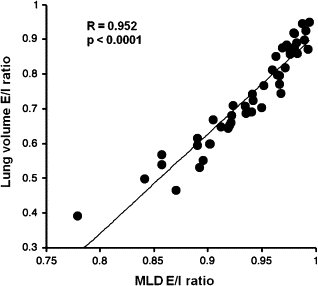
Table 4
Multivariate Analysis Using LV and LAA% to Predict MLD
LV LAA% R 2 Std B_P_ Std B_P_ Inspiratory phase 0.721 −0.336 <.001 −0.723 <.0001 Expiratory phase 0.760 −0.458 <.0001 −0.553 <.0001
MLD, mean lung density; LV, lung volume; LAA%, low attenuation area percent (<−950 HU); Std B, standardized coefficient B.
Get Radiology Tree app to read full this article<
Discussion
Get Radiology Tree app to read full this article<
Get Radiology Tree app to read full this article<
Get Radiology Tree app to read full this article<
Get Radiology Tree app to read full this article<
Get Radiology Tree app to read full this article<
Get Radiology Tree app to read full this article<
Get Radiology Tree app to read full this article<
Get Radiology Tree app to read full this article<
Get Radiology Tree app to read full this article<
Get Radiology Tree app to read full this article<
Get Radiology Tree app to read full this article<
Get Radiology Tree app to read full this article<
Get Radiology Tree app to read full this article<
Acknowledgment
Get Radiology Tree app to read full this article<
Get Radiology Tree app to read full this article<
Get Radiology Tree app to read full this article<
Get Radiology Tree app to read full this article<
References
1. Kauczor H.U., Heussel C.P., Fischer B., et. al.: Assessment of lung volumes using helical CT at inspiration and expiration: comparison with pulmonary function tests. Am J Roentgenol 1998; 171: pp. 1091-1095.
2. Zaporozhan J., Ley S., Eberhardt R., et. al.: Paired inspiratory/expiratory volumetric thin-section CT scan for emphysema analysis: comparison of different quantitative evaluations and pulmonary function test. Chest 2005; 128: pp. 3212-3220.
3. Akira M., Toyokawa K., Inoue Y., et. al.: Quantitative CT in chronic obstructive pulmonary disease: inspiratory and expiratory assessment. Am J Roentgenol 2009; 192: pp. 267-272.
4. Lee Y.K., Oh Y.M., Lee J.H., et. al.: Quantitative assessment of emphysema, air trapping, and airway thickening on computed tomography. Lung 2008; 186: pp. 157-165.
5. O’Donnell R.A., Peebles C., Ward J.A., et. al.: Relationship between peripheral airway dysfunction, airflow obstruction, and neutrophilic inflammation in COPD. Thorax 2004; 59: pp. 837-842.
6. Arakawa A., Yamashita Y., Nakayama Y., et. al.: Assessment of lung volumes in pulmonary emphysema using multidetector helical CT: correlation with pulmonary function tests. Comput Med Imaging Graph 2001; 25: pp. 399-404.
7. Kauczor H.U., Hast J., Heussel C.P., et. al.: CT attenuation of HRCT scans obtained at full inspiratory and expiratory position: comparison with pulmonary function tests. Eur Radiol 2002; 12: pp. 2757-2763.
8. Mergo P.J., Williams W.F., Gonzalez-Rothi R., et. al.: Three-dimensional volumetric assessment of abnormally low attenuation of the lung from routine helical CT: inspiratory and expiratory quantification. Am J Roentgenol 1998; 170: pp. 1355-1360.
9. Matsuoka S., Kurihara Y., Yagihashi K., et. al.: Quantitative assessment of air trapping in chronic obstructive pulmonary disease using inspiratory and expiratory volumetric MDCT. Am J Roentgenol 2008; 190: pp. 762-769.
10. Matsuoka S., Kurihara Y., Yagihashi K., et. al.: Quantitative assessment of peripheral airway obstruction on paired expiratory/inspiratory thin-section computed tomography in chronic obstructive pulmonary disease with emphysema. J Comput Assist Tomogr 2007; 31: pp. 384-389.
11. Nishino M., Roberts D.H., Sitek A., et. al.: Loss of anteroposterior intralobar attenuation gradient of the lung: correlation with pulmonary function. Acad Radiol 2006; 13: pp. 589-597.
12. Kubo K., Eda S., Yamamoto H., et. al.: Expiratory and inspiratory chest computed tomography and pulmonary function tests in cigarette smokers. Eur Respir J 1999; 13: pp. 52-256.
13. Iwano S., Okada T., Satake H., et. al.: 3D-CT volumetry of the lung using multidetector row CT: comparison with pulmonary function tests. Acad Radiol 2009; 16: pp. 250-256.
14. Camiciottoli G., Cavigli E., Grassi L., et. al.: Prevalence and correlates of pulmonary emphysema in smokers and former smokers: a densitometric study of participants in the ITALUNG trial. Eur Radiol 2009; 19: pp. 58-66.
15. Miller M.R., Hankinson J., Brusasco V., et. al.: ATS/ERS task force: standardisation of spirometry. Eur Respir J 2005; 26: pp. 319-338.
16. Stocks J., Quanjer P.H.: Reference values for residual volume, functional residual capacity and total lung capacity: ATS workshop on lung volume measurements/official statement of the European Respiratory Society. Eur Respir J 1995; 8: pp. 492-506.
17. Rabe K.F., Hurd S., Anzueto A., et. al.: Global strategy for the diagnosis, management, and prevention of chronic obstructive pulmonary disease: GOLD executive summary. Am J Respir Crit Care Med 2007; 176: pp. 532-555.
18. Dransfield M.T., Washko G.R., Foreman M.G., et. al.: Gender differences in the severity of CT emphysema in COPD. Chest 2007; 132: pp. 464-470.
19. Washko G.R., Dransfield M.T., San Jose Estepar R., et. al.: Airway wall attenuation: a biomarker of airway disease in subjects with COPD. J Appl Physiol 2009; 107: pp. 185-191.
20. Becker M.D., Berkmen Y.M., Austin J.H., et. al.: Lung volumes before and after lung volume reduction surgery: quantitative CT analysis. Am J Respir Crit Care Med 1998; 157: pp. 1593-1599.
21. Bae K.T., Slone R.M., Gierada D.S., et. al.: Patients with emphysema: quantitative CT analysis before and after lung volume reduction surgery. Radiology 1997; 203: pp. 705-714.
22. Coxson H.O., Nasute Fauerbach P.V., Storness-Bliss C., et. al.: Computed tomography assessment of lung volume changes after bronchial valve treatment. Eur Respir J 2008; 32: pp. 1443-1450.
23. Cohen J., Douma W.R., van Ooijen P.M., et. al.: Localization and quantification of regional and segmental air trapping in asthma. J Comput Assist Tomogr 2008; 32: pp. 562-569.
24. Stoel B.C., Putter H., Bakker M.E., et. al.: Volume correction in computed tomography densitometry for follow-up studies on pulmonary emphysema. Proc Am Thorac Soc 2008; 5: pp. 919-924.
25. Boedeker K.L., McNitt-Gray M.F., Rogers S.R., et. al.: Emphysema: effect of reconstruction algorithm on CT imaging measures. Radiology 2004; 232: pp. 295-301.
26. Ley-Zaporozhan J., Ley S., Weinheimer O., et. al.: Quantitative analysis of emphysema in 3D using MDCT: influence of different reconstruction algorithms. Eur J Radiol 2008; 65: pp. 228-234.
27. Matsuoka S., Kurihara Y., Yagihashi K., et. al.: Airway dimensions at inspiratory and expiratory multisection CT in chronic obstructive pulmonary disease: correlation with airflow limitation. Radiology 2008; 248: pp. 1042-1049.
28. D’Andrilli A., Vismara L., Rolla M., et. al.: Computed tomography with volume rendering for the evaluation of parenchymal hyperinflation after bronchoscopic lung volume reduction. Eur J Cardiothorac Surg 2009; 35: pp. 403-407.
29. O’Donnell C.R., Loring S.H.: Comparison of plethysmographic, helium dilution and CT-derive total lung capacity. [abstract] Am J Respir Crit Care Med 2005; 171: pp. A293.
30. Garfield J.L., Marchetti N., Gaughan J.P., et. al.: Lung volume by plethysmography and CT in advanced COPD. [abstract] Am J Respir Crit Care Med 2009; 179: pp. A2902.