
Rationale and Objectives
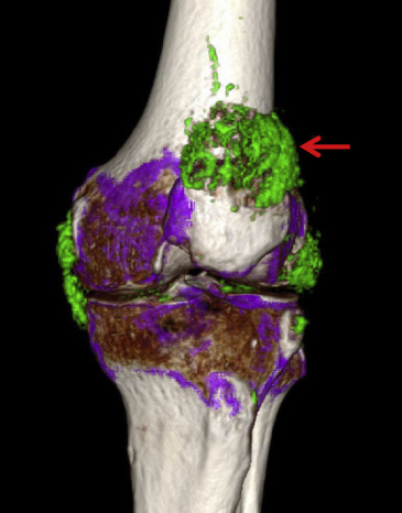
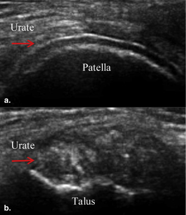
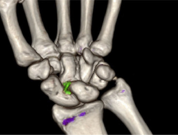
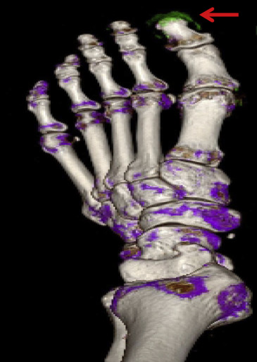
Dual-energy computed tomography (DECT) and ultrasound are both used to assess gouty arthritis. The present study was designed to compare the diagnostic accuracy of DECT and ultrasound in detecting monosodium urate (MSU) crystal deposition in various joints.
Materials and Methods
This study enrolled 40 patients diagnosed with acute gouty arthritis. All affected and contralateral joints were scanned (128 in total) using both DECT and ultrasound to determine the MSU deposition in upper limbs (wrist and elbow) and lower limbs (the first metatarsophalangeal joints, ankles, and knee). The MSU crystal accumulation detected by each method was compared for various joints.
Results
The 128 scanned joints included 52 of the upper limbs and 76 of the lower limbs. For the upper limbs, the percentage of MSU crystal accumulation detected by DECT (22/52, 42.3%) was significantly higher than that by ultrasound (10/52, 19.2%; P = .0027). The detection rates of the two methods for the lower limbs were similar ( P = .3173).
Conclusions
For detection of MSU crystal deposition in the upper limb joints, DECT was superior to ultrasound, whereas there was no difference between the two methods for the lower limbs. Therefore, ultrasound can be used for primary screening, and DECT afterward. Although the modalities are similar in making the initial diagnosis, DECT is far superior at displaying the anatomic extent of the disease.
Gouty arthritis (gout) is a form of inflammatory arthritis, most prevalent among middle-aged to elderly men and postmenopausal women. It is characterized by abnormal purine metabolism, hyperuricemia, and subsequent abnormal deposition of monosodium urate (MSU) crystals . The incidence of gout is increasing; the number of cases increased by approximately two per 1000 during the 10-year period between 1990 and 1999 . The disease affects about 3.9% of the population in the United States and 1.4% of the population in the United Kingdom and Germany . Although the prevalence is relatively low in China (reportedly 1.14%) , the country still suffers a significant disease burden because of its large population.
The pathogenesis of gout remains to be elucidated. Long-term deposition of MSU crystals in the joints leads to damage to articular cartilage and bone and may result in organ dysfunction, especially renal impairment and cardiac diseases . Clinical outcomes can be severe, limiting daily activity and impairing the quality of life of gout patients.
Get Radiology Tree app to read full this article<
Get Radiology Tree app to read full this article<
Get Radiology Tree app to read full this article<
Get Radiology Tree app to read full this article<
Materials and methods
Patients
Get Radiology Tree app to read full this article<
Get Radiology Tree app to read full this article<
Get Radiology Tree app to read full this article<
Dual-energy computed tomography
Get Radiology Tree app to read full this article<
Get Radiology Tree app to read full this article<
Ultrasound
Get Radiology Tree app to read full this article<
Get Radiology Tree app to read full this article<
Statistical analysis
Get Radiology Tree app to read full this article<
Results
Get Radiology Tree app to read full this article<
Table 1
Demographic and Clinical Characteristics of Forty patients ∗
Age, y 58.5; 53–68 Men, n (%) 38 (95.0) Women, n (%) 2 (5.0) Disease duration, y 6.5; 4.5–8.5 Monosodium urate crystal deposition in synovial fluid, n (%) 6 (15) Macroscopic tophi, n (%) 13 (32.5) Acute attack duration, d 4.0; 2.5–6.5 d Screening serum uric acid, mmol/L 530; 485–570
Get Radiology Tree app to read full this article<
Get Radiology Tree app to read full this article<
Get Radiology Tree app to read full this article<
Get Radiology Tree app to read full this article<
Get Radiology Tree app to read full this article<
Table 2
Comparison of DECT to Ultrasound for Detecting MSU in Scanned Joints
Scanned Joints Ultrasound DECT + − Total All + 47 17 64 − 3 61 64 Total 50 78 128 Upper limbs ∗ + 8 14 22 − 2 28 30 Total 10 42 52 Lower limbs † + 39 3 42 − 1 33 34 Total 40 36 76
DECT, dual-energy computed tomography; MSU, monosodium urate.
+ (−): Positive (negative) for MSU crystal deposition.
Get Radiology Tree app to read full this article<
Get Radiology Tree app to read full this article<
Table 3
Comparison of DECT to Ultrasound for Detecting MSU in the Contralateral Joints
Ultrasound DECT + − Total + 22 4 26 − 0 56 56 Total 22 60 82
DECT, dual-energy computed tomography; MSU, monosodium urate.
+ (−): Positive (negative) for MSU crystal deposition.
Get Radiology Tree app to read full this article<
Get Radiology Tree app to read full this article<
Discussion
Get Radiology Tree app to read full this article<
Get Radiology Tree app to read full this article<
Get Radiology Tree app to read full this article<
Get Radiology Tree app to read full this article<
Get Radiology Tree app to read full this article<
Get Radiology Tree app to read full this article<
Get Radiology Tree app to read full this article<
Get Radiology Tree app to read full this article<
Get Radiology Tree app to read full this article<
Get Radiology Tree app to read full this article<
Conclusions
Get Radiology Tree app to read full this article<
Get Radiology Tree app to read full this article<
Get Radiology Tree app to read full this article<
References
1. Singh J.A.: Racial and gender disparities among patients with gout. Curr Rheumatol Rep 2013; 15: pp. 307.
2. Brixner D.I., Ho M.J.: Clinical, humanistic, and economic outcomes of gout. Am J Manag Care 2005; 11: pp. S459-S464. quiz S65–8
3. Annemans L., Spaepen E., Gaskin M., et. al.: Gout in the UK and Germany: prevalence, comorbidities and management in general practice 2000-2005. Ann Rheum Dis 2008; 67: pp. 960-966.
4. Gruber M., Bodner G., Rath E., et. al.: Dual-energy computed tomography compared with ultrasound in the diagnosis of gout. Rheumatology (Oxford) 2014; 53: pp. 173-179.
5. Miao Z., Li C., Chen Y., et. al.: Dietary and lifestyle changes associated with high prevalence of hyperuricemia and gout in the Shandong coastal cities of Eastern China. J Rheumatol 2008; 35: pp. 1859-1864.
6. Chilappa C.S., Aronow W.S., Shapiro D., et. al.: Gout and hyperuricemia. Compr Ther 2010; 36: pp. 3-13.
7. Gonzalez-Rozas M., Prieto de Paula J.M., Franco Hidalgo S., et. al.: Chronic tophaceous gout. Semergen 2013; 39: pp. e29-34.
8. Roddy E., Zhang W., Doherty M.: The changing epidemiology of gout. Nat Clin Pract Rheumatol 2007; 3: pp. 443-449.
9. Ruilope L.M., Pontremoli R.: Serum uric acid and cardio-renal diseases. Curr Med Res Opin 2013; 29: pp. 25-31.
10. Girish G., Glazebrook K.N., Jacobson J.A.: Advanced imaging in gout. AJR Am J Roentgenol 2013; 201: pp. 515-525.
11. McQueen F.M., Reeves Q., Dalbeth N.: New insights into an old disease: advanced imaging in the diagnosis and management of gout. Postgrad Med J 2013; 89: pp. 87-93.
12. Carter J.D., Kedar R.P., Anderson S.R., et. al.: An analysis of MRI and ultrasound imaging in patients with gout who have normal plain radiographs. Rheumatology (Oxford) 2009; 48: pp. 1442-1446.
13. De Miguel E., Puig J.G., Castillo C., et. al.: Diagnosis of gout in patients with asymptomatic hyperuricaemia: a pilot ultrasound study. Ann Rheum Dis 2012; 71: pp. 157-158.
14. Filippucci E., Riveros M.G., Georgescu D., et. al.: Hyaline cartilage involvement in patients with gout and calcium pyrophosphate deposition disease. An ultrasound study. Osteoarthritis Cartilage 2009; 17: pp. 178-181.
15. Zhang W., Doherty M., Bardin T., et. al.: EULAR evidence based recommendations for gout. Part II: management. Report of a task force of the EULAR Standing Committee for International Clinical Studies Including Therapeutics (ESCISIT). Ann Rheum Dis 2006; 65: pp. 1312-1324.
16. Zhang W., Doherty M., Pascual E., et. al.: EULAR evidence based recommendations for gout. Part I: diagnosis. Report of a task force of the Standing Committee for International Clinical Studies Including Therapeutics (ESCISIT). Ann Rheum Dis 2006; 65: pp. 1301-1311.
17. Lyburn I.D., Torreggiani W.C., Harris A.C., et. al.: Tophaceous podagra: ultrasound diagnosis. Hosp Med 2002; 63: pp. 48-49.
18. Thiele R.G.: Role of ultrasound and other advanced imaging in the diagnosis and management of gout. Curr Rheumatol Rep 2011; 13: pp. 146-153.
19. Desai M.A., Peterson J.J., Garner H.W., et. al.: Clinical utility of dual-energy CT for evaluation of tophaceous gout. Radiographics 2011; 31: pp. 1365-1375. discussion 76–7
20. Nicolaou S., Yong-Hing C.J., Galea-Soler S., et. al.: Dual-energy CT as a potential new diagnostic tool in the management of gout in the acute setting. AJR Am J Roentgenol 2010; 194: pp. 1072-1078.
21. Choi H.K., Al-Arfaj A.M., Eftekhari A., et. al.: Dual energy computed tomography in tophaceous gout. Ann Rheum Dis 2009; 68: pp. 1609-1612.
22. Huppertz A., Hermann K.G., Diekhoff T., et. al.: Systemic staging for urate crystal deposits with dual-energy CT and ultrasound in patients with suspected gout. Rheumatol Int 2014; 34: pp. 763-771.
23. Ogdie A., Taylor W.J., Weatherall M., et. al.: Imaging modalities for the classification of gout: systematic literature review and meta-analysis. Ann Rheum Dis 2014; 74: pp. 1868-1874.
24. Wallace S.L., Robinson H., Masi A.T., et. al.: Preliminary criteria for the classification of the acute arthritis of primary gout. Arthritis Rheum 1977; 20: pp. 895-900.
25. Glazebrook K.N., Guimaraes L.S., Murthy N.S., et. al.: Identification of intraarticular and periarticular uric acid crystals with dual-energy CT: initial evaluation. Radiology 2011; 261: pp. 516-524.
26. Meenagh G., Filippucci E., Delle Sedie A., et. al.: Ultrasound imaging for the rheumatologist XXX. Sonographic assessment of the painful knee. Clin Exp Rheumatol 2010; 28: pp. 803-805.
27. Ottaviani S., Allard A., Bardin T., et. al.: An exploratory ultrasound study of early gout. Clin Exp Rheumatol 2011; 29: pp. 816-821.
28. Thiele R.G., Schlesinger N.: Diagnosis of gout by ultrasound. Rheumatology (Oxford) 2007; 46: pp. 1116-1121.