
Rationale and Objectives
Patients with known Crohn’s disease (CD) and an acute onset of severe abdominal pain attending an emergency room frequently undergo contrast-enhanced emergency computed tomography (CT) for complication assessment. To assess small bowel changes, an additional dedicated imaging procedure such as magnetic resonance enterography (MRE) is regularly performed. Therefore, these patients undergo two imaging procedures, although the clinical and diagnostic value of such an approach is not known. In a retrospective study, we compared the diagnostic value of a conventional abdominal CT with a dedicated small bowel MRE to assess bowel wall changes as well as typical complications in patients with advanced CD.
Materials and Methods
We retrospectively evaluated 53 patients with CD having a conventional abdominal multidetector-CT (MD-CT) and MRE within 2 days. Image quality and bowel inflammation was analyzed for each bowel segment. Lymph nodes, abscesses, and fistulas were evaluated.
Results
For small bowel and colon assessment, there was no significant difference for image quality between CT and MRE. Inflammation diagnosis was not significantly different between CT (69.4%) and MRE (71.4%). Colonic inflammation was diagnosed in 30.2% based on CT and 14.3% based on MRE. The difference for the detection of lymph nodes was significant (CT 49; MRE 27), whereas the differences between fistula (CT 25, MRE 27) or abscesses (CT and MRE 32) detection were not significant.
Conclusions
In patients with known advanced CD with acute abdominal pain conventional abdominal MD-CT, which is frequently performed as an emergency imaging procedure, is sufficient for bowel wall assessment. Based on our data, additional dedicated small bowel imaging such as MRE seems not to be necessary.
Crohn’s disease (CD) is an incurable chronic condition that can affect the entire gastrointestinal system. Histologically, it typically affects the whole bowel wall in contrast to ulcerative colitis, which is restricted to the mucosal intestinal layer. Because of these particular spreading features, a subset of patients develops major complications during the diseases course, such as fistulas and consecutive abscesses. Frequently, patients with advanced CD are undergoing emergency imaging because of acute abdominal pain for the evaluation of complications such as abscesses with consecutive radiological interventional therapy by computed tomography (CT)-guided abscess drainage.
Most of these patients are examined by a routine CT of the abdomen applying positive contrast orally and rectally with an additional intravenous contrast. After establishing the diagnosis of CD or therapy of the complications, such as abscess drainage, in some of these patients, a further diagnostic workup for the small bowel is performed. In addition to the conventional examinations such as small bowel enteroclysis or small bowel follow-through, sectional imaging methods such as MR- and CT-enteroclysis or magnetic resonance enterography (MRE) and CT-enterography are now often used to assess the small bowel . For CT-enterography and MRE, intraluminal contrast medium is given orally . These studies are also used to assess extra-enteric lesions .
Get Radiology Tree app to read full this article<
Material and methods
Patients
Get Radiology Tree app to read full this article<
MD-CT
Get Radiology Tree app to read full this article<
MRE
Get Radiology Tree app to read full this article<
Data Analysis
Get Radiology Tree app to read full this article<
Get Radiology Tree app to read full this article<
Get Radiology Tree app to read full this article<
Get Radiology Tree app to read full this article<
Get Radiology Tree app to read full this article<
Get Radiology Tree app to read full this article<
Results
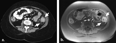
Pathologic Findings of the Small Bowel
Get Radiology Tree app to read full this article<
Table 1
Results of Image Quality and Inflamed Bowel Segments in 53 Patients with Advanced Crohn’s Disease and Abdominal Pain
Jejunum Ileum Terminal Ileum Cecum and Ascending Colon Transverse Colon Descending Colon Sigmoid Colon/ Rectum Diagnostic quality CT 32/53 (60%) 46/53 (87%) 49/53 (92%) 49/53 (92%) 52/53 (98%) 51/53 (96%) 50/53 (94%) MRE 32/53 (60%) 47/53 (89%) 49/53 (92%) 45/53 (92%) 48/53 (91%) 48/53 (91%) 47/53 (89%) Suboptimal quality CT 21/53 (40%) 7/53 (13%) 4/53 (8%) 4/53 (8%) 1/53 (2%) 2/53 (4%) 3/53 (6%) MRE 21/53 (40%) 6/53 (11%) 4/53 (8%) 8/53 (8%) 5/53 (9%) 5/53 (9%) 6/53 (11%) Not inflamed CT 25/32 (78%) 29/46 (63%) 15/49 (31%) 34/49 (69%) 40/52 (77%) 36/51 (71%) 31/50 (62%) MRE 23/32 (72%) 31/47 (66%) 14/49 (29%) 31/45 (69%) 35/48 (73%) 37/48 (77%) 31/47 (66%) Moderately inflamed CT 5/32 (16%) 9/46 (20%) 13/49 (27%) 6/49 (12%) 2/52 (4%) 7/51 (14%) 11/50 (22%) MRE 7/32 (22%) 8/47 (17%) 12/49 (24%) 6/45 (13%) 6/48 (13%) 4/48 (8%) 11/47 (23%) Inflamed CT 2/32 (6%) 11/46 (24%) 21/49 (43%) 9/49 (18%) 10/52 (19%) 8/51 (16%) 8/50 (16%) MRE 2/32 (6%) 10/47 (21%) 23/49 (47%) 8/45 (18%) 7/48 (14%) 7/48 (15%) 5/47 (11%)
CT: computed tomography; MRE: magnetic resonance enterography.
Image quality as well as inflammation grading (not inflamed, moderately inflamed, inflamed) are assessed in seven different bowel segments by routine abdominal multidetector-CT (CT) and MR-enterography (MRE).
Patients n = 53, bowel segments n = 371.
Get Radiology Tree app to read full this article<
Get Radiology Tree app to read full this article<
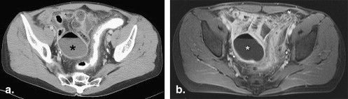
Pathologic Findings of the Colon
Get Radiology Tree app to read full this article<
Get Radiology Tree app to read full this article<
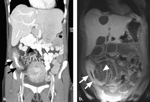
Evaluation of Extraintestinal Pathologies
Get Radiology Tree app to read full this article<
Table 2
Additional Diagnostic Features such as Presence of a Fistula, Abscess, or Local Enlarged Lymph Nodes >1cm in 53 Patients with Acute Abdominal Pain and Known Active Crohn’s Disease
Fistula Abscess Enlarged lymph Nodes Computed tomography 25 32 49 Magnetic resonance enterography 27 32 37
Get Radiology Tree app to read full this article<
Get Radiology Tree app to read full this article<
Get Radiology Tree app to read full this article<
Get Radiology Tree app to read full this article<
Discussion
Get Radiology Tree app to read full this article<
Get Radiology Tree app to read full this article<
Get Radiology Tree app to read full this article<
Get Radiology Tree app to read full this article<
Get Radiology Tree app to read full this article<
Get Radiology Tree app to read full this article<
Get Radiology Tree app to read full this article<
Get Radiology Tree app to read full this article<
Get Radiology Tree app to read full this article<
Get Radiology Tree app to read full this article<
Get Radiology Tree app to read full this article<
Get Radiology Tree app to read full this article<
Conclusion
Get Radiology Tree app to read full this article<
References
1. Schreyer A.G., Geissler A., Albrich H., et. al.: Abdominal MRI after enteroclysis or with oral contrast in patients with suspected or proven Crohn’s disease. Clin Gastroenterol Hepatol 2004; 2: pp. 491-497.
2. Rieber A., Wruk D., Potthast S., et. al.: Diagnostic imaging in Crohn’s disease: comparison of magnetic resonance imaging and conventional imaging methods. Int J Colorectal Dis 2000; 15: pp. 176-181.
3. Hara A.K., Alam S., Heigh R.I., et. al.: Using CT enterography to monitor Crohn’s disease activity: a preliminary study. AJR Am J Roentgenol 2008; 190: pp. 1512-1516.
4. Paulsen S.R., Huprich J.E., Fletcher J.G., et. al.: CT enterography as a diagnostic tool in evaluating small bowel disorders: review of clinical experience with over 700 cases. Radiographics 2006; 26: pp. 641-657. discussion 657–662
5. Masselli G., Casciani E., Polettini E., et. al.: Comparison of MR enteroclysis with MR enterography and conventional enteroclysis in patients with Crohn’s disease. Eur Radiol 2008; 18: pp. 438-447.
6. Fletcher J.G.: CT enterography technique: theme and variations. Abdom Imaging 2009; 34: pp. 283-288.
7. Herfarth H.H., Grunert M., Klebl F., et. al.: Frequency and nature of incidental extra-enteric lesions found on magnetic resonance enterography (MR-E) in patients with inflammatory bowel diseases (IBD). PLoS ONE 2009; 4: pp. e4863.
8. Schreyer A.G., Golder S., Scheibl K., et. al.: Dark lumen magnetic resonance enteroclysis in combination with MRI colonography for whole bowel assessment in patients with Crohn’s disease: first clinical experience. Inflamm Bowel Dis 2005; 11: pp. 388-394.
9. Schreyer A.G., Herfarth H., Kikinis R., et. al.: 3D modeling and virtual endoscopy of the small bowel based on magnetic resonance imaging in patients with inflammatory bowel disease. Invest Radiol 2002; 37: pp. 528-533.
10. Paolantonio P., Ferrari R., Vecchietti F., et. al.: Current status of MR imaging in the evaluation of IBD in a pediatric population of patients. Eur J Radiol 2009; 69: pp. 418-424.
11. Martin D.R., Lauenstein T., Sitaraman S.V.: Utility of magnetic resonance imaging in small bowel Crohn’s disease. Gastroenterology 2007; 133: pp. 385-390.
12. Chiorean M.V., Sandrasegaran K., Saxena R., et. al.: Correlation of CT enteroclysis with surgical pathology in Crohn’s disease. Am J Gastroenterol 2007; 102: pp. 2541-2550.
13. Lee S.S., Kim A.Y., Yang S.K., et. al.: Crohn disease of the small bowel: comparison of CT enterography, MR enterography, and small-bowel follow-through as diagnostic techniques. Radiology 2009; 251: pp. 751-761.
14. Hoffmann J.C., Preiss J.C., Autschbach F., et. al.: Clinical practice guideline on diagnosis and treatment of Crohn’s disease. Z Gastroenterol 2008; 46: pp. 1094-1146.
15. Hanauer S.B., Sandborn W.J.: European evidence-based consensus on the diagnosis and management of Crohn’s disease. Gut 2007; 56: pp. 161-163.
16. Desmond A.N., O’Regan K., Curran C., et. al.: Crohn’s disease: factors associated with exposure to high levels of diagnostic radiation. Gut 2008; 57: pp. 1524-1529.
17. Peloquin J.M., Pardi D.S., Sandborn W.J., et. al.: Diagnostic ionizing radiation exposure in a population-based cohort of patients with inflammatory bowel disease. Am J Gastroenterol 2008; 103: pp. 2015-2022.
18. Gaca A.M., Jaffe T.A., Delaney S., et. al.: Radiation doses from small-bowel follow-through and abdomen/pelvis MDCT in pediatric Crohn disease. Pediatr Radiol 2008; 38: pp. 285-291.
19. Jaffe T.A., Gaca A.M., Delaney S., et. al.: Radiation doses from small-bowel follow-through and abdominopelvic MDCT in Crohn’s disease. AJR Am J Roentgenol 2007; 189: pp. 1015-1022.
20. Muirhead C.R., O’Hagan J.A., Haylock R.G., et. al.: Mortality and cancer incidence following occupational radiation exposure: third analysis of the National Registry for Radiation Workers. Br J Cancer 2009; 100: pp. 206-212.
21. Sodickson A., Baeyens P.F., Andriole K.P., et. al.: Recurrent CT, cumulative radiation exposure, and associated radiation-induced cancer risks from CT of adults. Radiology 2009; 251: pp. 175-184.
22. Schreyer A.G., Rath H.C., Kikinis R., et. al.: Comparison of magnetic resonance imaging colonography with conventional colonoscopy for the assessment of intestinal inflammation in patients with inflammatory bowel disease: a feasibility study. Gut 2005; 54: pp. 250-256.
23. Schmidt S., Lepori D., Meuwly J.Y., et. al.: Prospective comparison of MR enteroclysis with multidetector spiral-CT enteroclysis: interobserver agreement and sensitivity by means of “sign-by-sign” correlation. Eur Radiol 2003; 13: pp. 1303-1311.
24. Siddiki H.A., Fidler J.L., Fletcher J.G., et. al.: Prospective comparison of state-of-the-art MR enterography and CT enterography in small-bowel Crohn’s disease. AJR Am J Roentgenol 2009; 193: pp. 113-121.
25. Horsthuis K., Bipat S., Bennink R.J., Stoker J.: Inflammatory bowel disease diagnosed with US, MR, scintigraphy, and CT: meta-analysis of prospective studies. Radiology 2008; 247: pp. 64-79.