
Rationale and Objectives
To compare differences in diffusion tensor imaging (DTI) and dynamic susceptibility-weighted contrast-enhanced (DSC) magnetic resonance (MR) perfusion imaging characteristics of recurrent neoplasm and radiation necrosis in patients with brain tumors previously treated with radiotherapy with or without surgery and chemotherapy.
Materials and Methods
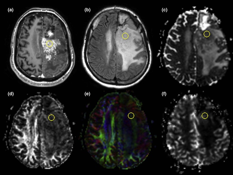
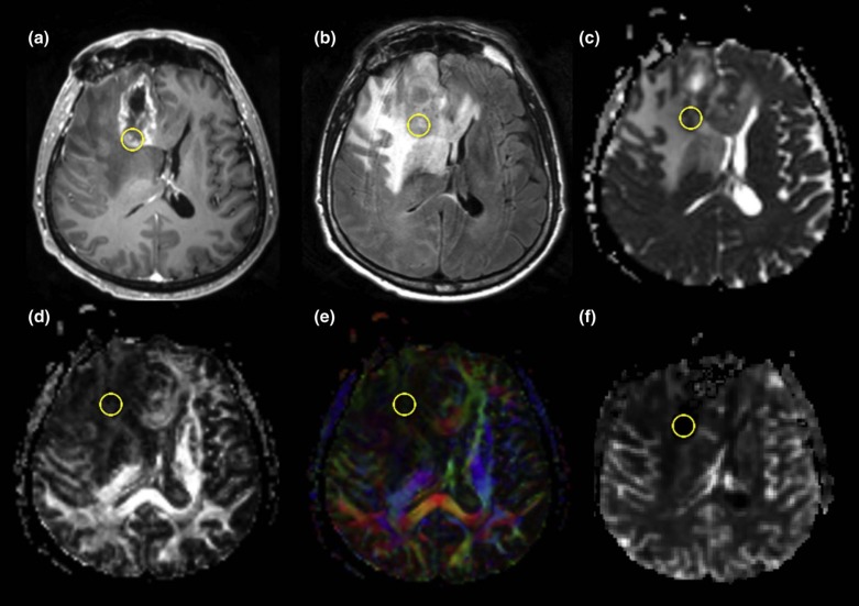
Patients with a history of brain neoplasm previously treated with radiotherapy with or without chemotherapy and surgery who developed a new enhancing lesion on posttreatment surveillance MRI were enrolled. DSC perfusion MRI and DTI were performed. Region of interest cursors were manually drawn in the contrast-enhancing lesions, in the perilesional white matter edema, and in the contralateral normal-appearing frontal lobe white matter. DTI and DSC perfusion MR indices were compared in recurrent tumor versus radiation necrosis.
Results
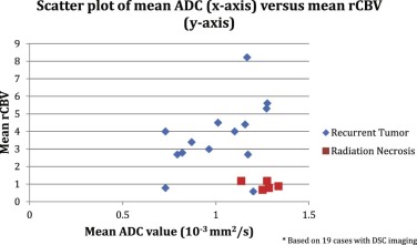
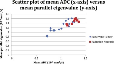
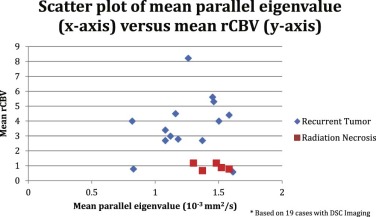
Twenty-two patients with 24 lesions were included. Sixteen (67%) lesions were placed into the recurrent neoplasm group and eight (33%) lesions were placed into the radiation necrosis group using biopsy results as the gold standard in all but three patients. Mean apparent diffusion coefficient values, mean parallel eigenvalues, and mean perpendicular eigenvalues in the contrast-enhancing lesion were significantly lower, and relative cerebral blood volume was significantly higher for the recurrent neoplasm group compared to the radiation necrosis group ( P < 0.01, P = 0.03, P < 0.01, and P < 0.01, respectively).
Conclusions
The combined assessment of DTI and DSC MR perfusion properties of new contrast-enhancing lesions is helpful in distinguishing recurrent neoplasm from radiation necrosis in patients with a history of brain neoplasm previously treated with radiotherapy with or without surgery and chemotherapy.
Introduction
Conventional magnetic resonance imaging (MRI) is not reliable in distinguishing radiation necrosis from recurrent brain neoplasm in patients with brain tumors previously treated with radiation therapy and surgery . Stereotactic biopsy and resection remain the most reliable methods for the classification of enhancing lesions that develop in the posttreatment period . In recent years, dynamic susceptibility-weighted contrast-enhanced (DSC) MR perfusion imaging and diffusion tensor imaging (DTI) have been used to evaluate posttreatment brain tumor patients. Multiple studies have shown significantly higher relative cerebral blood volume (rCBV) in the contrast-enhancing lesions of patients with recurrent tumor compared to those of patients with radiation necrosis . Whereas some studies have shown enhancing lesion apparent diffusion coefficient (ADC) values or ratios to be lower in recurrent neoplasms as opposed to radiation necrosis , other studies have shown contradictory findings . Few studies have published findings specifically examining the DTI characteristics (fractional anisotropy [FA], eigenvalues) of these lesions .
Our study prospectively analyzes both DSC MR perfusion and DTI characteristics of new enhancing lesions in patients with brain tumors previously treated with radiation therapy with or without surgery and chemotherapy.
Materials and Methods
Get Radiology Tree app to read full this article<
Subjects
Get Radiology Tree app to read full this article<
Table 1
Patient Demographics
Patient No. Sex Age Primary Tumor Diagnosis Time to Lesion Detection 1 Male 18 Diffuse astrocytoma Tumor 42 2 Female 57 Metastatic breast cancer Tumor 51 3 Male 41 Mixed glioma Tumor 61 4 Male 42 GBM Necrosis 14 5 Male 20 GBM Tumor 10 6 Male \* 28 Anaplastic glioma Necrosis/Tumor 14, 28 7 Male 48 Anaplastic astrocytoma Necrosis 11 8 Male 43 Metastatic lung cancer Necrosis 41 9 Male 63 GBM Tumor 12 10 Male 51 GBM Tumor 18 11 Male 54 GBM Necrosis 4 12 Male 69 Metastatic lung cancer Tumor 8 13 Female 62 GBM Tumor 17 14 Male \* 62 Anaplastic oligodendroglioma Necrosis/Tumor 173, 210 15 Male 68 Anaplastic oligodendroglioma Tumor 395 16 Male 56 GBM Tumor 23 17 Female 56 GBM Necrosis 15 18 Female 51 Anaplastic astrocytoma Tumor 68 19 Female 60 Mixed glioma Tumor 8 20 Female 66 Oligodendroglioma Tumor 146 21 Female 34 Anaplastic oligodendroglioma Tumor 37 22 Female 78 GBM Necrosis 50
GBM, glioblastoma multiforme; Necrosis, radiation necrosis; Tumor, recurrent tumor.
Get Radiology Tree app to read full this article<
Get Radiology Tree app to read full this article<
Get Radiology Tree app to read full this article<
MRI Technique
Get Radiology Tree app to read full this article<
Data Acquisition and Postprocessing
Get Radiology Tree app to read full this article<
Get Radiology Tree app to read full this article<
Histologic Analysis
Get Radiology Tree app to read full this article<
Data Analysis
Get Radiology Tree app to read full this article<
Get Radiology Tree app to read full this article<
Results
Get Radiology Tree app to read full this article<
Get Radiology Tree app to read full this article<
Table 2
Results
ADC FA λ1 λ⊥ rCBV Group 1 (contrast-enhancing lesion) 1.01 ± 0.19 0.23 ± 0.10 1.25 × ±0.24 0.89 × ±0.19 3.76 ± 1.95 Group 2 (contrast-enhancing lesion) 1.26 ± 0.08 0.16 ± 0.06 1.46 × ±0.11 1.15 × ±0.09 0.99 ± 0.25P value (contrast-enhancing lesion) <0.01 0.07 0.03 <0.01 <0.01 Group 1 (perilesional edema) 1.24 ± 0.44 0.27 ± 0.15 1.55 ± 0.42 1.08 ± 0.47 1.48 ± 1.29 Group 2 (perilesional edema) 1.28 ± 0.29 0.21 ± 0.10 1.56 ± 0.33 1.15 ± 0.29 0.62 ± 0.52P value (perilesional edema) 0.79 0.32 0.95 0.72 0.17 Group 1 (contralateral white matter) 0.80 ± 0.08 0.56 ± 0.08 1.35 ± 0.19 0.52 ± 0.08 n/a Group 2 contralateral white matter) 0.75 ± 0.05 0.50 ± 0.12 1.21 ± 0.16 0.53 ± 0.9 n/a_P_ value (contralateral white matter) 0.18 0.23 0.09 0.93 n/a
ADC, apparent diffusion coefficient; FA, fractional anisotropy; rCBV, relative cerebral blood volume.
ADC values and eigenvalues are in units of 10 −3 mm 2 /s. FA and rCBV values are unit-less.
Group 1, recurrent neoplasm; Group 2, radiation necrosis.
Get Radiology Tree app to read full this article<
Discussion
Get Radiology Tree app to read full this article<
Get Radiology Tree app to read full this article<
Get Radiology Tree app to read full this article<
Get Radiology Tree app to read full this article<
Get Radiology Tree app to read full this article<
Get Radiology Tree app to read full this article<
Get Radiology Tree app to read full this article<
Get Radiology Tree app to read full this article<
Get Radiology Tree app to read full this article<
Get Radiology Tree app to read full this article<
Get Radiology Tree app to read full this article<
Acknowledgments
Get Radiology Tree app to read full this article<
References
1. Jain R., Narang J., Sundgren P., et. al.: Treatment induced necrosis versus recurrent/progressing brain tumor: going beyond the boundaries of conventional morphologic imaging. J Neurooncol 2010; 100: pp. 17-29.
2. Verma N., Cowperthwaite M.C., Burnett M.G., et. al.: Differentiating tumor recurrence from treatment necrosis: a review of neuro-oncologic imaging strategies. Neuro Oncol 2013; 15: pp. 515-534.
3. Alexiou G.A., Tsioris S., Kyritsis A.P., et. al.: Glioma recurrence versus radiation necrosis: accuracy of current imaging modalities. J Neurooncol 2009; 95: pp. 1-11.
4. Fatterpekar G.M., Galheigo D., Narayana A., et. al.: Treatment-related change versus tumor recurrence in high-grade gliomas: a diagnostic conundrum—use of dynamic susceptibility contrast-enhanced (DSC) perfusion MRI. AJR 2012; 198: pp. 19-26.
5. Barajas R.F., Chang J.S., Segal M.R., et. al.: Differentiation of recurrent glioblastoma multiforme from radiation necrosis after external beam radiation therapy with dynamic susceptibility-weighted contrast-enhanced perfusion MR imaging. Radiology 2009; 253: pp. 486-496.
6. Hu L.S., Baxter L.C., Smith K.A., et. al.: Relative cerebral blood volume values to differentiate high-grade glioma recurrence from posttreatment radiation effect: direct correlation between image-guided tissue histopathology and localized dynamic susceptibility-weighted contrast-enhanced perfusion MR imaging measurements. AJNR 2009; 30: pp. 552-558.
7. Alexiou G., Zikou A., Tsiouris S., et. al.: Comparison of diffusion tensor, dynamic susceptibility contrast MRI and 99m Tc-Tetrofosmin brain SPECT for the detection of recurrent high-grade glioma. Magn Reson Imaging 2014; 32: pp. 854-859.
8. Sugahara T., Korogi Y., Tomiguchi S., et. al.: Posttherapeutic intraaxial brain tumor: the value of perfusion-sensitive contrast-enhanced MR imaging for differentiating tumor recurrence from nonneoplastic contrast-enhancing tissue. AJNR 2000; 21: pp. 901-909.
9. Kim H.S., Goh M.J., Kim N., et. al.: Which combination of MR imaging modalities is best for predicting recurrent glioblastoma? Study of diagnostic accuracy and reproducibility. Radiology 2014; 373: pp. 831-843.
10. Fink J.R., Carr R.B., Matsusue E.: Comparison of 3 tesla proton MR spectroscopy, MR perfusion, and MR diffusion for distinguishing glioma recurrence from posttreatment effects. JMRI 2012; 35: pp. 56-63.
11. Asao C., Korogi Y., Kitajima M., et. al.: Diffusion-weighted imaging of radiation-induced brain injury for differentiation from tumor recurrence. AJNR 2004; 26: pp. 1455-1460.
12. Hein P., Eskey C., Dunn J., et. al.: Diffusion-weighted imaging in the follow-up of treated high-grade gliomas: tumor recurrence versus radiation injury. AJNR 2004; 25: pp. 201-209.
13. Xu J., Li Y., Lian J., et. al.: Distinction between postoperative recurrent glioma and radiation injury using MR diffusion tensor imaging. Neuroradiology 2010; 52: pp. 1193-1199.
14. Sundgren P., Fan X., Weybright P., et. al.: Differentiation of recurrent brain tumor versus radiation injury using diffusion tensor imaging in patients with new contrast-enhancing lesions. Magn Reson Imaging 2006; 24: pp. 1131-1142.
15. Wang S., Chen Y., Lal B., et. al.: Evaluation of radiation necrosis and malignant glioma in rat models using diffusion tensor MR imaging. J Neurooncol 2012; 107: pp. 51-60.
16. Rahmathulla G., Marko N.F., Weil R.J.: Cerebral radiation necrosis: a review of the pathobiology, diagnosis and management considerations. J Clin Neurosci 2013; 20: pp. 485-502.
17. Miyatake S., Nonoguchi N., Furuse M., et. al.: Pathophysiology, diagnosis, and treatment of radiation necrosis in the brain. Neurol Med Chir 2015; 55: pp. 50-59.
18. Bauer A.H., Erly W., Moser F.G., et. al.: Differentiation of solitary brain metastasis from glioblastoma multiforme: a predictive multiparametric approach using combined MR diffusion and perfusion. Neuroradiology 2015; 57: pp. 697-703.
19. Weybright P., Sundgren P.C., Maly P., et. al.: Differentiation between brain tumor recurrence and radiation injury using MR spectroscopy. AJR 2005; 185: pp. 1471-1476.
20. Smith E.A., Carlos R.C., Junck L.R., et. al.: Developing a clinical decision model: use of MR spectroscopy to differentiate between recurrent tumor and radiation change in patients with new contrast enhancing lesions. AJR 2009; 192: pp. 45-52.
21. Saindane A.M.: Recent advances in brain and spine imaging. Radiol Clin North Am 2015; 53: pp. 477-496.
22. Verhaak R.G., Hoadley K.A., Purdom E., et. al.: Integrated genomics analysis identifies clinically relevant subtypes of glioblastoma characterized by abnormalities in PDGFRA, IDH1, EGFR, and NF1. Cancer Cell 2010; 17: pp. 98-110.
23. Dunn G.P., Rinne M.L., Wykosky J., et. al.: Emerging insights into the molecular and cellular basis of glioblastoma. Genes Dev 2012; 26: pp. 756-784.
24. Kim Y.H., Oh S.W., Lim Y.J., et. al.: Differentiating radiation necrosis from tumor recurrence in high-grade gliomas: assessing the efficacy of 18F-FDG PET, 11C-methionine PET and perfusion MRI. Clin Neurol Nerosurg 2010; 112: pp. 758-765.
25. Ozsunar Y., Mullins M.E., Kwong K., et. al.: Glioma recurrence versus radiation necrosis? A pilot comparison of arterial spin-labeled, dynamic susceptibility contrast enhanced MRI, and FDG-PET imaging. Acad Radiol 2010; 17: pp. 282-290.
26. Wang S., Kim S.J., Poptani H., et. al.: Diagnostic utility of diffusion tensor imaging in differentiating glioblastomas from brain metastases. AJNR 2014; 35: pp. 928-934.
27. Yamasaki F., Kurisu K., Satoh K., et. al.: Apparent diffusion coefficient of human brain tumors at MR imaging. Radiology 2005; 235: pp. 985-991.