
Rationale and Objectives
To compare diffusion-weighted (DW) with standard T2-weighted imaging for quantitative evaluation of small hepatocellular carcinoma (HCC) in cirrhosis.
Materials and Methods
Fourteen patients (all men; mean age, 58.6 years; age range, 45-69 years) with 22 small HCCs (<3 cm and >1 cm in diameter) in cirrhosis were included in the study. DW imaging with breath-hold single-shot echo planar imaging (b = 0, 800 seconds/mm 2 ) and T2-weighted imaging with respiratory triggering fat-suppressed fast spin-echo sequence were performed on a 3-T magnetic resonance unit using an eight-channel torso phased-array coil. The signal intensity (SI) of HCC and liver were measured at workstation. Contrast-to-noise ratio (CNR), contrast ratio (CR, SI lesion /SI liver ), and apparent diffusion coefficient (ADC) values were calculated. CNRs and CRs obtained with DW and T2-weighted images, and ADCs of HCC and liver were compared using nonparametric tests.
Results
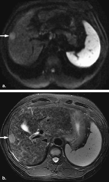
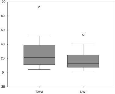
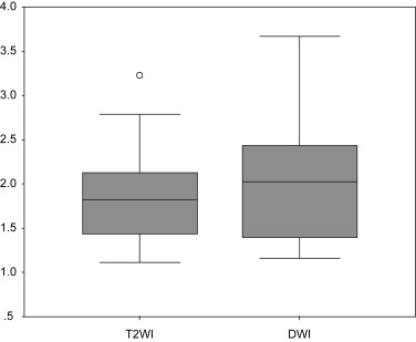
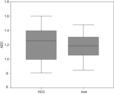
Two lesions were excluded because of artifacts on DW images. Thus 20 lesions were analyzed. The CNRs obtained with T2-weighted images (27.12 ± 21.12) were significantly higher ( P = .02) than those with DW images (17.52 ± 13.50). There were no significant difference between the CRs obtained with T2-weighted images (1.83 ± 0.56) and DW images (2.01 ± 0.67). There were no significant difference between the mean ADCs of HCC (1.22 × 10 −3 mm 2 /second ± 0.24) and the cirrhotic liver (1.17 × 10 −3 mm 2 /second ± 0.17), either.
Conclusion
DW imaging with high b value was not superior to standard T2-weighted imaging in terms of lesion conspicuity of small HCC in cirrhosis.
Hepatocellular carcinoma (HCC) is the most common primary malignant tumor of the liver and develops predominantly in patients who have underlying chronic hepatitis or cirrhosis, with highest incidences occurring in Africa, Southeast Asia, and China . Therefore, it is clinically important to detect HCC at an early stage for prompt surgical resection, transplantation, or local therapy to ensure a better chance of survival.
Diffusion-weighted (DW) imaging is a noninvasive method used to show microscopic motion of water in tissue. Some studies have been performed on the use of DW imaging for focal liver lesion (FLL) detection and characterization. A limited number of studies involved the direct comparison of DW and T2-weighted imaging in terms of FLL detection. Results showed improved assessment with DW imaging by using low b values compared with that with T2-weighted imaging.
Get Radiology Tree app to read full this article<
Materials and methods
Patients
Get Radiology Tree app to read full this article<
Get Radiology Tree app to read full this article<
MR Imaging
Get Radiology Tree app to read full this article<
DW imaging
Get Radiology Tree app to read full this article<
T2-weighted imaging
Get Radiology Tree app to read full this article<
Image Analysis
Get Radiology Tree app to read full this article<
Get Radiology Tree app to read full this article<
Statistical Analysis
Get Radiology Tree app to read full this article<
Results
Get Radiology Tree app to read full this article<
Get Radiology Tree app to read full this article<
Discussion
Get Radiology Tree app to read full this article<
Get Radiology Tree app to read full this article<
Get Radiology Tree app to read full this article<
Get Radiology Tree app to read full this article<
Get Radiology Tree app to read full this article<
Get Radiology Tree app to read full this article<
References
1. Unoura M., Kaneko S., Matsushita E., et. al.: High-risk groups and screening strategies for early detection of hepatocellular carcinoma in patients with chronic liver disease. Hepatogastroenterology 1993; 40: pp. 305-310.
2. Parkin D.M.: Global cancer statistics in the year 2000. Lancet Oncol 2001; 2: pp. 533-543.
3. Bosch F.X., Ribes J., Borras J.: Epidemiology of primary liver cancer. Semin Liver Dis 1999; 19: pp. 271-285.
4. Namimoto T., Yamashita Y., Sumi S., et. al.: Focal liver masses: characterization with diffusion-weighted echo-planar MR imaging. Radiology 1997; 204: pp. 739-744.
5. Ichikawa T., Haradome H., Hachiya J., et. al.: Diffusion-weighted MR imaging with a single-shot echoplanar sequence: detection and characterization of focal hepatic lesions. AJR Am J Roentgenol 1998; 170: pp. 397-402.
6. Kim T., Murakami T., Takahashi S., et. al.: Diffusion-weighted single-shot echoplanar MR imaging for liver disease. AJR Am J Roentgenol 1999; 173: pp. 393-398.
7. Taouli B., Vilgrain V., Dumont E., et. al.: Evaluation of liver diffusion isotropy and characterization of focal hepatic lesions with two single-shot echoplanar MR imaging sequences: prospective study in 66 patients. Radiology 2003; 226: pp. 71-78.
8. Yoshikawa T., Kawamitsu H., Mitchell D.G., et. al.: ADC measurement of abdominal organs and lesions using parallel imaging technique. AJR Am J Roentgenol 2006; 187: pp. 1521-1530.
9. Moteki T., Horikoshi H.: Evaluation of hepatic lesions and hepatic parenchyma using diffusion-weighted echo-planar MR with three values of gradient b-factor. J Magn Reson Imaging 2006; 24: pp. 637-645.
10. Okada Y., Ohtomo K., Kiryu S., et. al.: Breath-hold T2-weighted MRI of hepatic tumors: value of echo planar imaging with diffusion-sensitizing gradient. J Comput Assist Tomogr 1998; 22: pp. 364-371.
11. Nasu K., Kuroki Y., Nawano S., et. al.: Hepatic metastases: diffusion-weighted sensitivity-encoding versus SPIO-enhanced MR imaging. Radiology 2006; 239: pp. 122-130.
12. Hussain S.M., De Becker J., Hop W.C., et. al.: Can a singleshot black-blood T2-weighted spin-echo echo-planar imaging sequence with sensitivity encoding replace the respiratory-triggered turbo spin-echo sequence for the liver? An optimization and feasibility study. J Magn Reson Imaging 2005; 21: pp. 219-229.
13. Coenegrachts K., Delanote J., Ter Beek L., et. al.: Improved focal liver lesion detection: comparison of single-shot diffusion-weighted echoplanar and single-shot T2 weighted turbo spin echo techniques. Br J Radiol 2007; 80: pp. 524-531.
14. Parikh T., Drew S.J., Lee V.S., et. al.: Focal liver lesion detection and characterization with diffusion-weighted MR imaging: comparison with standard breath-hold T2-weighted imaging. Radiology 2008; 246: pp. 812-822.
15. Zech C.J., Herrmann K.A., Dietrich O., et. al.: Black-blood diffusion-weighted EPI acquisition of the liver with parallel imaging: comparison with a standard T2-weighted sequence for detection of focal liver lesions. Invest Radiol 2008; 43: pp. 261-266.
16. Bruegel M., Gaa J., Waldt S., et. al.: Diagnosis of hepatic metastasis: comparison of respiration-triggered diffusion-weighted echo-planar MRI and five t2-weighted turbo spin-echo sequences. AJR Am J Roentgenol 2008; 191: pp. 1421-1429.
17. Kadoya M., Matsui O., Takashima T., et. al.: Hepatocellular carcinoma: correlation of MR imaging and histopathologic findings. Radiology 1992; 183: pp. 819-825.
18. Earls J.P., Theise N.D., Weinreb J.C., et. al.: Dysplastic nodules and HCC: thin-section MR imaging of explanted cirrhotic livers with pathologic correlation. Radiology 1996; 201: pp. 207-214.
19. Outwater E.K., Mitchell D.G., Vinitski S.: Abdominal MR imaging: evaluation of a fast spin-echo sequence. Radiology 1994; 190: pp. 425-429.
20. Coates G.G., Borrello J.A., McFarland E.G., et. al.: Hepatic T2-weighted MRI: a prospective comparison of sequences, including breath-hold, half-Fourier turbo spin echo (HASTE). J Magn Reson Imaging 1998; 8: pp. 642-649.
21. Marti-Bonmati L., Talens A., del Olmo J., et. al.: Chronic hepatitis and cirrhosis: evaluation by means of MR imaging with histologic correlation. Radiology 1993; 188: pp. 37-43.
22. Semelka R.C., Chung J.J., Hussain S.M., et. al.: Chronic hepatitis: correlation of early patchy and late linear enhancement patterns on gadolinium-enhanced MR images with histopathology initial experience. J Magn Reson Imaging 2001; 13: pp. 385-391.
23. Hussain H.K., Syed I., Nghiem H.V., et. al.: T2-weighted MR imaging in the assessment of cirrhotic liver. Radiology 2004; 230: pp. 637-644.
24. Hecht E.M., Holland A.E., Israel G.M., et. al.: Hepatocellular carcinoma in the cirrhotic liver: gadolinium-enhanced 3D T1-weighted MR imaging as a stand-alone sequence for diagnosis. Radiology 2006; 239: pp. 438-447.
25. van den Bos I.C., Hussain S.M., Krestin G.P., et. al.: Liver imaging at 3.0 T: diffusion-induced black-blood echo-planar imaging with large anatomic volumetric coverage as an alternative for specific absorption rate-intensive echo-train spin-echo sequences: feasibility study. Radiology 2008; 248: pp. 264-271.
26. Le Bihan D., Breton E., Lallemand D., et. al.: Separation of diffusion and perfusion in intravoxel incoherent motion MR imaging. Radiology 1988; 168: pp. 497-505.
27. Schindera S.T., Merkle E.M., Dale B.M., et. al.: Abdominal magnetic resonance imaging at 3.0 T: what is the ultimate gain in signal-to-noise ratio?. Acad Radiol 2006; 13: pp. 1236-1243.
28. Blaimer M., Breuer F., Mueller M., et. al.: SMASH, SENSE, PILS, GRAPPA: how to choose the optimal method. Top Magn Reson Imaging 2004; 15: pp. 223-236.
29. Oner A.Y., Celik H., Oktar S.O., et. al.: Single breath-hold diffusion-weighted MRI of the liver with parallel imaging: initial experience. Clin Radiol 2006; 61: pp. 959-965.
30. Taouli B., Tolia A.J., Losada M., et. al.: Diffusion-weighted MRI for quantification of liver fibrosis: preliminary experience. AJR Am J Roentgenol 2007; 189: pp. 799-806.
31. Taouli B., Chouli M., Martin A.J., et. al.: Chronic hepatitis: role of diffusion-weighted imaging and diffusion tensor imaging for the diagnosis of liver fibrosis and inflammation. J Magn Reson Imaging 2008; 28: pp. 89-95.
32. Girometti R., Furlan A., Esposito G., et. al.: Relevance of b-values in evaluating liver fibrosis: a study in healthy and cirrhotic subjects using two single-shot spin-echo echo-planar diffusion-weighted sequences. J Magn Reson Imaging 2008; 28: pp. 411-419.