
Rationale and Objectives
Gadofosveset trisodium is a blood-pool contrast agent (BPA) that shows a less pronounced r1 relaxivity advantage over gadobenate dimeglumine at 3T than at 1.5T. However, there are few data on image quality during first-pass imaging of the thoracic vasculature with gadofosveset trisodium at 3 T. Therefore, it was the aim of this study to compare first-pass imaging characteristics of gadofosveset trisodium to gadobenate dimeglumine during time-resolved contrast-enhanced three-dimensional magnetic resonance angiography (CE MRA) at 3 T.
Materials and Methods
Twenty volunteers underwent time-resolved CE MRA on a 3 T magnetic resonance (MR) system with a standard eight-channel phased-array surface coil, receiving either gadofosveset trisodium (blood pool agent [BPA], n = 10) or gadobenate dimeglumine (standard contrast agent, [SCA], n = 10). Image quality was assessed by two independent readers using a Likert scale ranging from 0 = poor quality to 3 = excellent quality, and relative signal-to-noise and contrast-to-noise ratios were calculated.
Results
Equally good to excellent first-pass image quality was confirmed for time-resolved CE MRA using BPA and SCA (arteries, 2.8 ± 0.2 and 2.6 ± 0.4; veins, 2.5 ± 0.3 and 2.2 ± 0.4; artifacts, 2.4 ± 0.2 and 2.3 ± 0.1). Signal-to-noise and contrast-to-noise ratios showed nonsignificant differences, except for left subclavian artery values. There was an overall nonsignificant superiority in signal-to-noise and contrast-to-noise ratios for standard contrast agent in arterial values and BPA regarding venous values.
Conclusions
Despite a markedly decreased r 1/ r 2 relaxivity ratio, first-pass imaging characteristics of gadofosveset trisodium and gadobenate dimeglumine are equally well suited for first pass time-resolved CE MRA at 3 T.
The most widely used gadolinium-based agents for contrast-enhanced magnetic resonance angiography (CE-MRA) have small molecular sizes, resulting in short blood-pool residence times. Gadofosveset trisodium is a gadolinium-based blood-pool contrast agent (BPA) based on a higher albumin-binding capacity and thus a considerably prolonged intravascular residence time. In addition, the multiple paramagnetic ions attached to each macromolecule result in a relatively high T1 relaxivity, as shown by Rohrer et al (at 1.5 T, gadofosveset trisodium r 1 = 19 vs gadobenate dimeglumine r 1 = 6.3 in plasma at 37°C). However, at 3 T, contrast agents show individual field-strength dependencies that result in a less pronounced differences between relaxivities (at 3 T, gadofosveset trisodium r 1 = 9.9 vs gadobenate dimeglumine r 1 = 5.5 in plasma at 37°C) and hence altered r 1/ r 2 ratios .
Previous studies have focused on the benefits of the longer acquisition window of BPAs that can be exploited for time-intensive high-resolution assessment of arteries and veins at low concentrations at 1.5 T . Preclinical tests of gadofosveset trisodium were presented as early as 1996 . Phase II and III studies were performed for peripheral arteries in which the prolonged decrease of the blood T1 was used for improved image quality . Early applications reported in the literature included evaluations of aortoiliac occlusive disease and the carotid arteries . Such early clinical reports demonstrated the feasibility of steady-state and dynamic (ie, time-resolved) three-dimensional CE MRA compared to two-dimensional time-of-flight MRA as a reference standard. A recently reported study showed significantly increased image quality with gadofosveset trisodium compared to the low-albumin-binding contrast agent gadopentetate dimeglumine for nondynamic CE MRA at 1.5 T as part of a whole-body imaging approach .
Get Radiology Tree app to read full this article<
Materials and methods
MR Imaging
Get Radiology Tree app to read full this article<
Get Radiology Tree app to read full this article<
Get Radiology Tree app to read full this article<
Get Radiology Tree app to read full this article<
Qualitative Data Analysis
Get Radiology Tree app to read full this article<
Get Radiology Tree app to read full this article<
Quantitative Data Analysis
Get Radiology Tree app to read full this article<
Get Radiology Tree app to read full this article<
Get Radiology Tree app to read full this article<
SNR=mean(ROIavg)SD(ROIsub), SNR
=
mean
(
ROI
avg
)
SD
(
ROI
sub
)
,
where mean denotes the average signal intensity and SD the standard deviation of all pixels within the ROI. The relative CNR (CNR rel ) was calculated for all vascular ROIs referenced to the surrounding fatty and muscular tissue as
CNRrel=SNRvasc−SNRrefSNRref, CNR
rel
=
SNR
vasc
−
SNR
ref
SNR
ref
,
where SNR vasc is the SNR of the vessel of interest, and SNR ref is calculated for either fat or muscle.
Get Radiology Tree app to read full this article<
Statistical Analysis
Get Radiology Tree app to read full this article<
Results
Get Radiology Tree app to read full this article<
Qualitative Data Analysis
Get Radiology Tree app to read full this article<
Get Radiology Tree app to read full this article<
Get Radiology Tree app to read full this article<
Table 1
Summary of Qualitative Image Grading
Arteries Aorta First branch Second branch Total SCA 2.7 ± 0.3 (2.5) 3.0 ± 0.0 (3.0) 2.8 ± 0.3 (2.8) 2.8 ± 0.3 (2.8) BPA 2.6 ± 0.4 (2.5) 2.6 ± 0.4 (3.0) 2.5 ± 0.5 (2.5) 2.6 ± 0.4 (2.7) Veins Vena cava First branch Second branch Total SCA 2.9 ± 0.2 (2.5) 2.9 ± 0.2 (3.0) 2.2 ± 0.5 (2.0) 2.6 ± 0.4 (2.5) BPA 2.4 ± 0.5 (2.3) 2.4 ± 0.5 (2.5) 2.1 ± 0.5 (2.0) 2.2 ± 0.5 (2.3) Artifacts Blurring Ghosting Fold-over Total SCA 2.1 ± 0.2 (3.0) 2.9 ± 0.2 (3.0) 2.2 ± 0.4 (2.0) 2.4 ± 0.2 (2.3) BPA 2.0 ± 0.2 (3.0) 3.0 ± 0.0 (3.0) 2.1 ± 0.2 (2.0) 2.3 ± 0.1 (2.3)
BPA, blood-pool contrast agent (gadofosveset trisodium); SCA, standard contrast agent (gadobenate dimeglumine).
Data are expressed as mean ± standard deviation (median). No significant differences in image quality between SCA and BPA were observed.
Get Radiology Tree app to read full this article<
Quantitative Data Analysis
Get Radiology Tree app to read full this article<
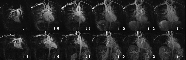
![Figure 2, Maximum intensity projection (MIP) shortly after maximum arterial contrast enhancement and arterial and venous regions for signal-to-noise ratio (SNR) and contrast-to-noise ratio (CNR) analysis. For each region, mean relative (rel) CNR fat and CNR muscle are shown for gadobenate dimeglumine (standard contrast agent [SCA]; middle) and gadofosveset trisodium (blood-pool agent [BPA] below). AAo, ascending aorta; AnonV, anonymous vein; BrachTrunc, brachiocephalic trunk; BrachV, brachiocephalic vein, DAo, descending aorta; L subcl A, left subclavian artery; PulmArt, pulmonary artery; R subcl A, right subclavian artery; SVC, superior vena cava. P < .05.](https://storage.googleapis.com/dl.dentistrykey.com/clinical/ComparisonofGadofosvesetTrisodiumandGadobenateDimeglumineDuringTimeResolvedThoracicMRAngiographyat3T/1_1s20S1076633210003132.jpg)
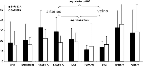
![Figure 4, Mean signal-to-noise ratio (SNR) and contrast-to-noise ratio (CNR) averaged for all evaluated arteries and veins. Significant difference between gadobenate dimeglumine (standard contrast agent [SCA]) and gadofosveset trisodium (blood-pool contrast agent [BPA]) values ( P < .05).](https://storage.googleapis.com/dl.dentistrykey.com/clinical/ComparisonofGadofosvesetTrisodiumandGadobenateDimeglumineDuringTimeResolvedThoracicMRAngiographyat3T/3_1s20S1076633210003132.jpg)
Get Radiology Tree app to read full this article<
Discussion
Get Radiology Tree app to read full this article<
Get Radiology Tree app to read full this article<
Get Radiology Tree app to read full this article<
Get Radiology Tree app to read full this article<
Get Radiology Tree app to read full this article<
Get Radiology Tree app to read full this article<
Get Radiology Tree app to read full this article<
Get Radiology Tree app to read full this article<
Get Radiology Tree app to read full this article<
Get Radiology Tree app to read full this article<
Conclusions
Get Radiology Tree app to read full this article<
Get Radiology Tree app to read full this article<
References
1. Rohrer M., Bauer H., Mintorovitch J., et. al.: Comparison of magnetic properties of MRI contrast media solutions at different magnetic field strengths. Invest Radiol 2005; 40: pp. 715-724.
2. Brasch R.C.: Rationale and applications for macromolecular Gd-based contrast agents. Magn Reson Med 1991; 22: pp. 282-287.
3. Lauffer R.B., Parmelee D.J., Ouellet H.S., et. al.: MS-325: a small-molecule vascular imaging agent for magnetic resonance imaging. Acad Radiol 1996; 3: pp. S356-S358.
4. Perreault P., Edelman M.A., Baum R.A., et. al.: MR angiography with gadofosveset trisodium for peripheral vascular disease: phase II trial. Radiology 2003; 229: pp. 811-820.
5. Rapp J.H., Wolff S.D., Quinn S.F., et. al.: Aortoiliac occlusive disease in patients with known or suspected peripheral vascular disease: safety and efficacy of gadofosveset-enhanced MR angiography—multicenter comparative phase III study. Radiology 2005; 236: pp. 71-78.
6. Bluemke D.A., Stillman A.E., Bis K.G., et. al.: Carotid MR angiography: phase II study of safety and efficacy for MS-325. Radiology 2001; 219: pp. 114-122.
7. Klessen C., Hein P.A., Huppertz A., et. al.: First-pass whole-body magnetic resonance angiography (MRA) using the blood-pool contrast medium gadofosveset trisodium: comparison to gadopentetate dimeglumine. Invest Radiol 2007; 42: pp. 659-664.
8. Korosec F.R., Frayne R., Grist T.M., et. al.: Time-resolved contrast-enhanced 3D MR angiography. Magn Reson Med 1996; 36: pp. 345-351.
9. Lim R.P., Shapiro M., Wang E.Y., et. al.: 3D time-resolved MR angiography (MRA) of the carotid arteries with time-resolved imaging with stochastic trajectories: comparison with 3D contrast-enhanced bolus-chase MRA and 3D time-of-flight MRA. AJNR Am J Neuroradiol 2008; 29: pp. 1847-1854.
10. Willinek W.A., Hadizadeh D.R., von Falkenhausen M., et. al.: 4D time-resolved MR angiography with keyhole (4D-TRAK): more than 60 times accelerated MRA using a combination of CENTRA, keyhole, and SENSE at 3.0T. J Magn Reson Imaging 2008; 27: pp. 1455-1460.
11. Nielsen Y.W., Eiberg J.P., Logager V.B., et. al.: Whole-body MR angiography with body coil acquisition at 3 T in patients with peripheral arterial disease using the contrast agent gadofosveset trisodium. Acad Radiol 2009; 16: pp. 654-661.
12. Naehle C.P., Muller A., Willinek W.A., et. al.: First-pass and steady-state magnetic resonance angiography of the thoracic vasculature using gadofosveset trisodium. J Magn Reson Imaging 2009; 30: pp. 809-816.
13. Nissen J.C., Attenberger U.I., Fink C., et. al.: Thoracic and abdominal MRA with gadofosveset: influence of injection rate on vessel signal and image quality. Eur Radiol 2009; 19: pp. 1932-1938.
14. Maki J.H., Wang M., Wilson G.J., et. al.: Highly accelerated first-pass contrast-enhanced magnetic resonance angiography of the peripheral vasculature: comparison of gadofosveset trisodium with gadopentetate dimeglumine contrast agents. J Magn Reson Imaging 2009; 30: pp. 1085-1092.
15. Iezzi R., Soulez G., Thurnher S., et. al.: Contrast-enhanced MRA of the renal and aorto-iliac-femoral arteries: comparison of gadobenate dimeglumine and gadofosveset trisodium. Eur J Radiol 2009; [Epub ahead of print]
16. Griswold M.A., Jakob P.M., Heidemann R.M., et. al.: Generalized autocalibrating partially parallel acquisitions (GRAPPA). Magn Reson Med 2002; 47: pp. 1202-1210.
17. Pruessmann K.P.: Parallel imaging at high field strength: synergies and joint potential. Top Magn Reson Imaging 2004; 15: pp. 237-244.
18. Reeder S.B., Wintersperger B.J., Dietrich O., et. al.: Practical approaches to the evaluation of signal-to-noise ratio performance with parallel imaging: application with cardiac imaging and a 32-channel cardiac coil. Magn Reson Med 2005; 54: pp. 748-754.
19. Laurent S., Elst L.V., Muller R.N.: Comparative study of the physicochemical properties of six clinical low molecular weight gadolinium contrast agents. Contrast Media Mol Imaging 2006; 1: pp. 128-137.
20. Cavagna F.M., Marzola P., Dapra M., et. al.: Binding of gadobenate dimeglumine to proteins extravasated into interstitial space enhances conspicuity of reperfused infarcts. Invest Radiol 1994; 29: pp. S50-S53.
21. Stalder A.F., Elverfeldt D.V., Paul D., et. al.: Variable echo time imaging: signal characteristics of 1-M gadobutrol contrast agent at 1.5 and 3T. Magn Reson Med 2008; 59: pp. 113-123.