
Rationale and Objectives
To compare the image quality of dedicated coronary computed tomography angiography (cCTA) to that of triple rule-out (TRO) CTA designed to evaluate the coronary arteries, thoracic aorta, and pulmonary arteries.
Materials and Methods
Consecutive cCTA examinations performed by a single radiologist over 1 year were reviewed. Biphasic injection protocols were employed: 70 mL of optiray-350 followed by 40 mL of saline injected at 5.5 mL/second for dedicated cCTA; 70 mL of optiray-350 followed by 25 mL of the contrast diluted with 25 mL of saline injected at 5.0 mL/second for TRO-CTA. Two independent cardiovascular radiologists reviewed the coronary vessels in each case and rated diagnostic image quality on a 5 point scale (1, suboptimal; 3, adequate; 5, excellent). Vascular enhancement was measured in the coronary arteries, aorta, and pulmonary arteries.
Results
There was excellent interobserver agreement between the cardiovascular radiologists (kappa = 0.91). Coronary image quality score were similar among 260 dedicated cCTA studies and 168 TRO-CTA studies (mean: 3.8–3.9. P > .18). At least one coronary segment demonstrated suboptimal image quality in 8% of examinations, including 18 dedicated cCTA studies and 16 TRO studies ( P = .94). Enhancement was greater in the distal thoracic aorta of TRO patients (336 vs. 311 Hounsfield units; P = .01); no other significant differences in enhancement were identified in the aorta and coronary arteries of dedicated cCTA and TRO studies. Vascular enhancement was adequate for diagnostic evaluation of the pulmonary arteries in all TRO studies.
Conclusions
A TRO-CTA protocol using 95 mL of contrast can provide comparable coronary image quality and coronary vascular enhancement as compared to dedicated cCTA with 70 mL of contrast.
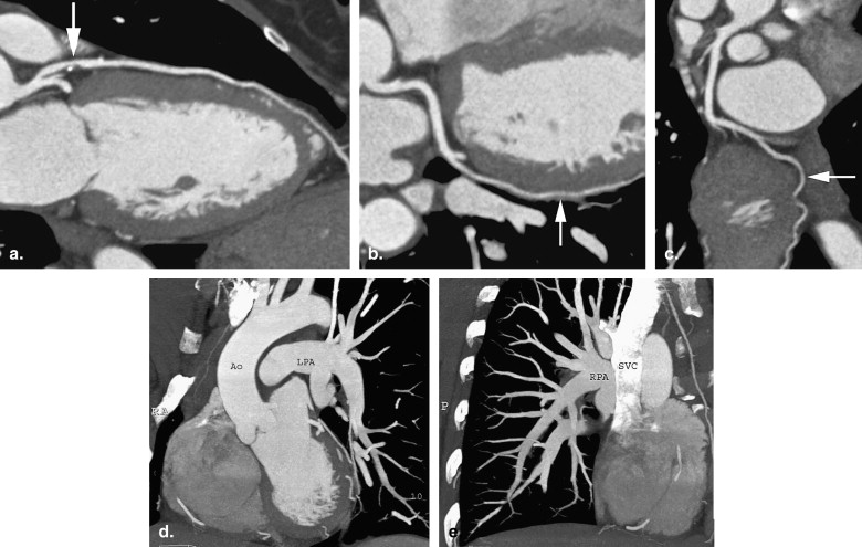
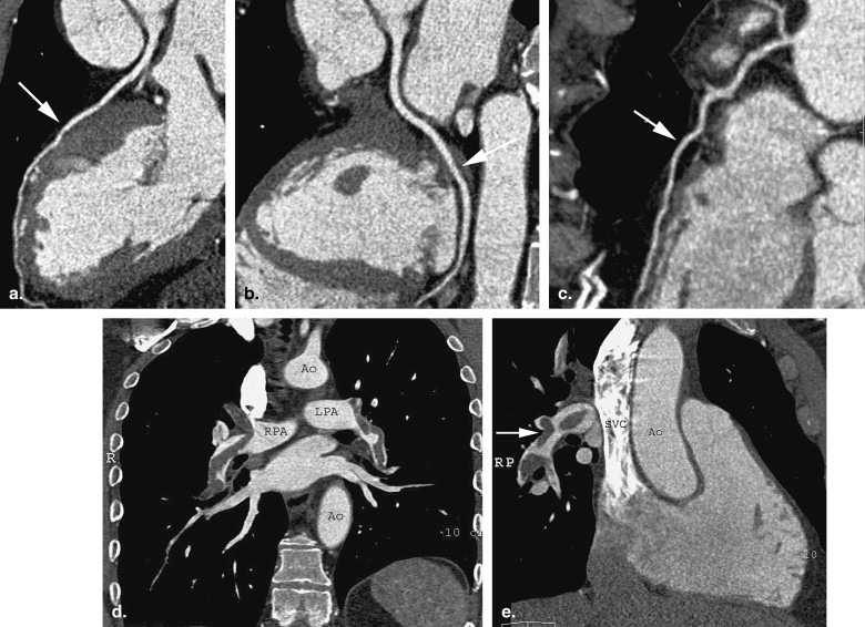
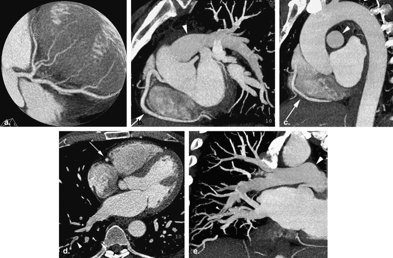
Although much has been written about the application of dedicated coronary computed tomography angiography (cCTA) to the evaluation of coronary disease, fewer studies have examined the use of computed tomography (CT) to simultaneously evaluate the thoracic aorta, and pulmonary arteries . The “triple rule-out” (TRO) CTA study is a tailored examination that is designed to specifically evaluate these three major vascular beds in the thorax. A recent survey of radiology practices found that 33% were involved with CT in the emergency department workup of chest pain, and that 18% were using the TRO study . Physicians may be reluctant to perform TRO studies because of an impression that the TRO is too technically challenging or that the quality of the coronary artery study is compromised in the TRO examination.
Imaging results and clinical outcomes for the emergency department patients in this study have been previously reported . TRO studies demonstrated the presence of moderate to severe coronary disease (>50% luminal diameter stenosis) in 11% of patients, demonstrated a process other than coronary atherosclerosis that explained the clinical presentation in another 11% of patients, and allowed for disposition of the patient without further diagnostic testing in 76% of patients. The current study is designed to compare the image quality of TRO CTA to that of dedicated cCTA to determine whether there is a difference in coronary artery image quality between dedicated cCTA and TRO studies.
Methods
Get Radiology Tree app to read full this article<
Get Radiology Tree app to read full this article<
Coronary CTA Scan Protocol
Get Radiology Tree app to read full this article<
Get Radiology Tree app to read full this article<
Get Radiology Tree app to read full this article<
Get Radiology Tree app to read full this article<
Vascular Enhancement and Image Quality Analysis
Get Radiology Tree app to read full this article<
Get Radiology Tree app to read full this article<
Get Radiology Tree app to read full this article<
Get Radiology Tree app to read full this article<
Statistical Analysis
Get Radiology Tree app to read full this article<
Get Radiology Tree app to read full this article<
Results
Get Radiology Tree app to read full this article<
Table 1
Demographics for Patients in Dedicated cCTA and TRO Groups
Dedicated cCTA TRO_P_ Value Age 58 ± 12 years 50 ± 12 y <.001 Sex (male:female ratio) 57 : 43 45 : 55 .05 Height 1.7 ± 0.09 m 1.7 ± 0.10 m .17 Weight 89 ± 25 kg 85 ± 21 kg .24 Body mass index 30.3 ± 7.6 29.6 ± 6.7 .55
cCTA: coronary computed tomography angiography; TRO: triple rule-out.
Table 2
Study Parameters for Patients in Dedicated cCTA and TRO Groups
Dedicated cCTA TRO_P_ Value Beta-blocker dose 12.2 ± 0.8 mg 11.7 ± 1.0 mg .84 Heart rate 61.5 ± 0.8 beats/min 63.0 ± 1.0 bpm .24 Tube current modulation 43% 48% .52 Scan length 18.4 ± 2.9 cm 21.0 ± 2.0 <.001
cCTA: coronary computed tomography angiography; TRO: triple rule-out.
Get Radiology Tree app to read full this article<
Get Radiology Tree app to read full this article<
Table 3
Subjective Coronary Artery Image Quality in Dedicated cCTA and TRO Patients
Subjective Image Quality Score 1 2 3 4 5 cCTA 3 15 51 111 80 TRO 5 11 37 71 44
cCTA: coronary computed tomography angiography; TRO: triple rule-out.
Get Radiology Tree app to read full this article<
Get Radiology Tree app to read full this article<
Get Radiology Tree app to read full this article<
Table 4
Post-hoc Power Analysis
Difference in Image Quality Scores Power to Detect this Difference 0.4 0.99 0.3 0.88 0.27 0.81 0.26 0.78 0.25 0.75
Get Radiology Tree app to read full this article<
Get Radiology Tree app to read full this article<
Get Radiology Tree app to read full this article<
Table 5
Enhancement of the Aorta and Left Ventricle in Dedicated cCTA and TRO Patients (Expressed in Hounsfield Units)
Dedicated cCTA TRO_P_ Value Aortic root 341 ± 83 329 ± 84 .25 Upper aorta 322 ± 79 327 ± 85 .60 Mid aorta 316 ± 83 304 ± 85 .24 Lower aorta 337 ± 95 311 ± 88 .03 Upper LV 324 ± 85 310 ± 78 .16 Lower LV 326 ± 93 320 ± 80 .59 Left main CA 308 ± 74 313 ± 78 .59 Right CA 315 ± 77 316 ± 85 .93
cCTA: coronary computed tomography angiography; TRO: triple rule-out; LV: left ventricle; CA: coronary artery.
Get Radiology Tree app to read full this article<
Get Radiology Tree app to read full this article<
Table 6
Pulmonary Artery Opacification in Triple Rule-out Studies
Hounsfield Units Main pulmonary artery 341 ± 101 Right pulmonary artery 325 ± 95 Left pulmonary artery 334 ± 97 Right upper lobe pulmonary artery 331 ± 108 Right lower lobe pulmonary artery 318 ± 99 Left upper lobe pulmonary artery 327 ± 96 Left lower lobe pulmonary artery 331 ± 101
Get Radiology Tree app to read full this article<
Discussion
Get Radiology Tree app to read full this article<
Get Radiology Tree app to read full this article<
Get Radiology Tree app to read full this article<
Get Radiology Tree app to read full this article<
Get Radiology Tree app to read full this article<
Get Radiology Tree app to read full this article<
Get Radiology Tree app to read full this article<
Radiation Dose
Get Radiology Tree app to read full this article<
Study Limitations
Get Radiology Tree app to read full this article<
Get Radiology Tree app to read full this article<
Get Radiology Tree app to read full this article<
Get Radiology Tree app to read full this article<
Conclusion
Get Radiology Tree app to read full this article<
References
1. Gallagher M.J., Raff G.L.: Use of multislice CT for the evaluation of emergency room patients with chest pain: the so-called “triple rule-out”. Catheter Cardiovasc Interv 2008; 71: pp. 92-99.
2. Thomas J., Rideau A.M., Paulson E.K., et. al.: Emergency department imaging: current practice. J Am Coll Radiol 2008; 5: pp. 811-816e2.
3. Takakuwa K.M., Halpern E.J.: Evaluation of a “triple rule-out” coronary CT angiography protocol using 64-slice multidetector computed tomography in low-to-moderate risk emergency department patients with suspected acute coronary syndrome. Radiology 2008; 248: pp. 438-446.
4. Halpern EJ. Triple rule-out CT Angiography for evaluation of acute chest pain and suspected acute coronary syndrome. Radiology. In press.
5. Meijer A.B., O Y.L., Geleijns J., et. al.: Meta-analysis of 40- and 64-MDCT angiography for assessing coronary artery stenosis. AJR Am J Roentgenol 2008; 191: pp. 1667-1675.
6. Limkakeng A.T., Halpern E.J., Takakuwa K.: Sixty-four-slice multidetector computed tomography: the future of ED cardiac care. Am J Emerg Med 2007; 25: pp. 450-458.
7. White C.S., Kuo D., Kelemen M., et. al.: Chest pain evaluation in the emergency department: can MDCT provide a comprehensive evaluation?. AJR Am J Roentgenol 2005; 185: pp. 533-540.
8. Sato Y., Matsumoto N., Ichikawa M., Kunimasa T., et. al.: Efficacy of multislice computed tomography for the detection of acute coronary syndrome in the emergency department. Circ J 2005; 69: pp. 1047-1051.
9. Savino G., Herzog C., Costello P., et. al.: 64 slice cardiovascular CT in the emergency department: concepts and first experiences. Radiol Med (Torino) 2006; 111: pp. 481-496.
10. Schussler J.M., Smith E.R.: Sixty-four-slice computed tomographic coronary angiography: will the “triple rule out” change chest pain evaluation in the ED?. Am J Emerg Med 2007; 25: pp. 367-375.
11. Jeudy J., White C.S.: Evaluation of acute chest pain in the emergency department: utility of multidetector computed tomography. Semin Ultrasound CT MR 2007; 28: pp. 109-114.
12. Dodd J.D., Kalva S., Pena A., et. al.: Emergency cardiac CT for suspected acute coronary syndrome: qualitative and quantitative assessment of coronary, pulmonary, and aortic image quality. AJR Am J Roentgenol 2008; 191: pp. 870-877.
13. Fleischmann D., Rubin G.D., Bankier A.A., et. al.: Improved uniformity of aortic enhancement with customized contrast medium injection protocols at CT angiography. Radiology 2000; 214: pp. 363-371.
14. Jones S.E., Wittram C.: The indeterminate CT pulmonary angiogram: imaging characteristics and patient clinical outcome. Radiology 2005; 237: pp. 329-337.
15. Bédard J.P., Blais C., Patenaude Y.G., et. al.: Pulmonary embolism: prospective comparison of iso-osmolar and low-osmolarity nonionic contrast agents for contrast enhancement at CT angiography. Radiology 2005; 234: pp. 929-933.
16. Schoellnast H., Deutschmann H.A., Berghold A., et. al.: MDCT angiography of the pulmonary arteries: influence of body weight, body mass index, and scan length on arterial enhancement at different iodine flow rates. AJR Am J Roentgenol 2006; 187: pp. 1074-1078.
17. Haidary A., Bis K., Vrachliotis T., et. al.: Enhancement performance of a 64-slice triple rule-out protocol vs 16-slice and 10-slice multidetector CT-angiography protocols for evaluation of aortic and pulmonary vasculature. J Comput Assist Tomogr 2007; 31: pp. 917-923.
18. Roggenland D., Peters S.A., Lemburg S.P., et. al.: CT angiography in suspected pulmonary embolism: impact of patient characteristics and different venous lines on vessel enhancement and image quality. AJR Am J Roentgenol 2008; 190: pp. W351-W359.
19. Vrachliotis T.G., Bis K.G., Haidary A., et. al.: Atypical chest pain: coronary, aortic, and pulmonary vasculature enhancement at biphasic single-injection 64-section CT angiography. Radiology 2007; 243: pp. 368-376.
20. Litmanovich D., Zamboni G.A., Hauser T.H., et. al.: ECG-gated chest CT angiography with 64-MDCT and tri-phasic IV contrast administration regimen in patients with acute non-specific chest pain. Eur Radiol 2008; 18: pp. 308-317.
21. Takakuwa K.M., Halpern E.J., Gingold E.L., et. al.: Radiation dose in a “triple rule-out” coronary CT angiography protocol of emergency department patients using 64-slice multidetector computed tomography: impact of scan parameters, prospective ECG-based tube current modulation, age, gender and body mass index. AJR Am J Roentgenol 2009; 192: pp. 866-872.
22. Menzel H.G.Schibilla H.Teunen D.European guidelines on quality criteria for computed tomography. Publication No. EUR 16262 EN.2000.European CommissionLuxembourg:
23. Stillman A.E., Oudkerk M., Ackerman M., et. al.: Use of multidetector computed tomography for the assessment of acute chest pain: a consensus statement of the North American Society of Cardiac Imaging and the European Society of Cardiac Radiology. Eur Radiol 2007; 17: pp. 2196-2207.