
Rationale and Objectives
Obstructive pulmonary disease phenotypes are related to variable combinations of emphysema and small-airway disease, the latter manifested as air trapping (AT) on imaging. The investigators propose a method to extract AT information quantitatively from thoracic multi–detector row high-resolution computed tomography (HRCT), validated by pulmonary function testing (PFT) correlation.
Materials and Methods
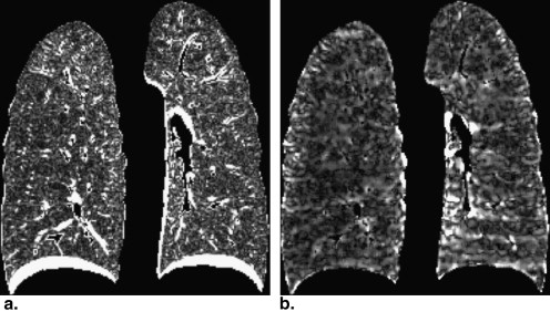
Seventeen patients with obstructive pulmonary disease who underwent HRCT and PFT within a 3-day interval were retrospectively identified. Thin-section volumetric HRCT in inspiration and expiration was registered and analyzed using custom-made software. Nonaerated regions of lung were segmented through exclusion of voxels > −50 Hounsfield units (HU); emphysematous areas were segmented as voxels < −950 HU on inspiratory images. Small-airway AT volume (ATV) was segmented as regions of lung voxels whose attenuation values increased by less than a specified change threshold (set from 5 to 300 HU in 25-HU increments) between inspiration and expiration. Inspiratory and expiratory total segmented lung volumes, emphysema volume (EV), and ATV for each threshold were subsequently calculated and correlated with PFT parameters.
Results
A strong positive correlation was obtained between total segmented lung volume in inspiration and total lung capacity ( r = 0.83). A strong negative correlation ( r = −0.80) was obtained between EV and the ratio between forced expiratory volume in 1 second and forced vital capacity. Stronger negative correlation with forced expiratory volume in 1 second/forced vital capacity ( r = −0.85) was demonstrated when ATV (threshold, 50 HU) was added to EV, indicating improved quantification of total AT to predict obstructive disease severity. A moderately strong positive correlation between ATV and residual volume was observed, with a maximum r value of 0.72 (threshold, 25 HU), greater than that between EV and residual volume ( r = 0.58). The benefit of ATV quantification was greater in a subgroup of patients with negligible emphysema compared to patients with moderate to severe emphysema.
Conclusions
Small-airway AT segmentation in conjunction with emphysema segmentation through computer-assisted methodologies may provide better correlations with key PFT parameters, suggesting that the quantification of emphysema-related and small airway–related components of AT from thoracic HRCT has great potential to elucidate phenotypic differences in patients with chronic obstructive pulmonary disease.
Obstructive pulmonary disease is most often due to chronic obstructive pulmonary disease (COPD), which is a major global public health problem. It is the fourth leading cause of chronic morbidity and mortality in the United States and is anticipated to rank fifth in 2020 in burden of disease caused worldwide . Moreover, among the four major causes of mortality, namely, cardiovascular disease, malignant neoplasm, cerebrovascular disease, and COPD, the last is the only one that has been steadily rising in prevalence .
COPD is defined as chronic, progressive airflow limitation that is not fully reversible, associated with a range of pathologic changes in the lungs with significant extrapulmonary effects, caused by chronic inflammation and structural changes . The chronic airflow limitation is caused by a mixture of small-airway disease (obstructive bronchiolitis) and parenchymal destruction (emphysema). The relative contributions of these two components vary substantially from patient to patient. The presence and extent of each component has the potential to affect clinical presentation, disease severity, prognosis, and therapeutic response .
Get Radiology Tree app to read full this article<
Get Radiology Tree app to read full this article<
Get Radiology Tree app to read full this article<
Get Radiology Tree app to read full this article<
Materials and methods
Get Radiology Tree app to read full this article<
Patient Selection
Get Radiology Tree app to read full this article<
Image Acquisition and Reconstruction
Get Radiology Tree app to read full this article<
Image Analysis
Get Radiology Tree app to read full this article<
Get Radiology Tree app to read full this article<
Get Radiology Tree app to read full this article<
Get Radiology Tree app to read full this article<
Get Radiology Tree app to read full this article<
Get Radiology Tree app to read full this article<
Get Radiology Tree app to read full this article<
Statistical Analysis
Get Radiology Tree app to read full this article<
Results
Get Radiology Tree app to read full this article<
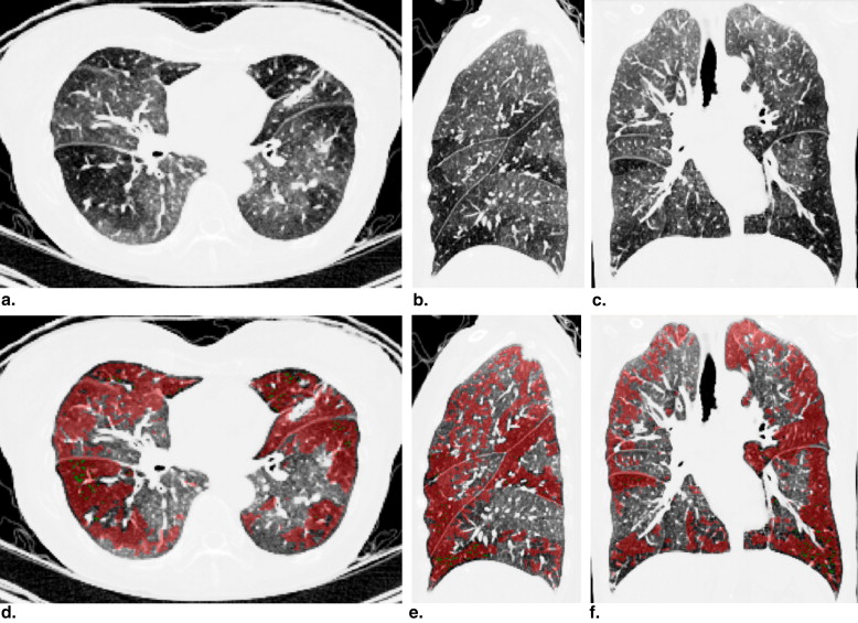
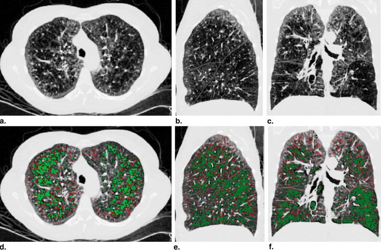
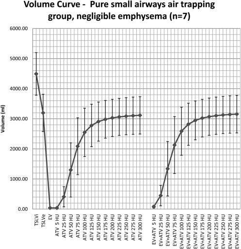
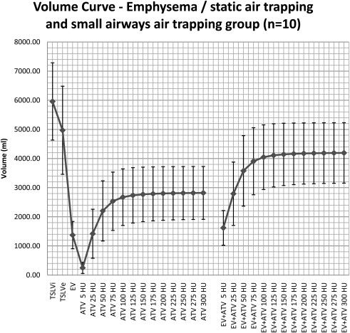
Table 1
Calculated Volumes and Sample Standard Deviations for All Patients ( n = 17), Subgroup with Negligible Emphysema ( n = 7), and Subgroup with Moderate or Severe Emphysema ( n = 10)
Group TSLVi (mL) TSLVe (mL) EV (mL) EV (%) ∗ TSLVi − TSLVe (%) † Volume SD Volume SD Volume SD Volume SD Volume SD All patients 5355.43 1315.38 4240.41 1496.48 824.07 762.14 14.10 12.45 22.35 10.77 Negligible EV 4492.36 706.90 3196.84 619.85 40.10 32.13 0.88 0.68 28.59 ‡ 10.06 Moderate or severe EV 5959.58 1325.86 4970.90 1511.68 1372.85 468.30 23.35 6.66 17.98 ‡ 9.35
EV, emphysema volume; SD, standard deviation; TSLVe, total segmented lung volume in expiration; TSLVi, total segmented lung volume in inspiration.
Get Radiology Tree app to read full this article<
Get Radiology Tree app to read full this article<
Get Radiology Tree app to read full this article<
Get Radiology Tree app to read full this article<
Get Radiology Tree app to read full this article<
Table 2
Pearson’s Linear Correlation Coefficients and Respective P Values Between Key PFT Parameters and Calculated EV and ATV
Volume FVC (%P) FEV 1 (%P) FEV 1 /FVC (%P) FEF 25%–75% (%P) ERV (%P) RV (%P) ∗ TLC (%P) ∗ r__P__r__P__r__P__r__P__r__P__r__P__r__P EV −0.037 .889 −0.550 .022 −0.802 <.001 −0.460 .063 0.582 .029 0.579 .030 0.631 .016 ATV 5 HU −0.140 .591 −0.528 .029 −0.709 .001 −0.423 .091 0.653 .011 0.725 .003 0.860 <.001 25 HU −0.027 .917 −0.548 .023 −0.770 <.001 −0.511 .036 0.771 .001 0.723 .003 0.933 <.001 50 HU 0.033 .901 −0.521 .032 −0.701 .002 −0.604 .010 0.675 .008 0.649 .012 0.881 <.001 75 HU 0.139 .595 −0.386 .126 −0.574 .016 −0.557 .020 0.574 .032 0.545 .044 0.791 .001 100 HU 0.271 .292 −0.193 .457 −0.422 .092 −0.395 .117 0.562 .036 0.418 .137 0.711 .004 125 HU 0.366 .149 −0.050 .848 −0.313 .221 −0.262 .309 0.568 .034 0.321 .262 0.656 .011 150 HU 0.433 .082 0.050 .848 −0.242 .349 −0.170 .515 0.575 .032 0.251 .386 0.623 .017 175 HU 0.474 .055 0.113 .666 −0.198 .446 −0.112 .668 0.577 .031 0.205 .481 0.602 .023 200 HU 0.500 .041 0.155 .553 −0.168 .520 −0.073 .781 0.578 .030 0.174 .553 0.586 .028 225 HU 0.518 .033 0.184 .479 −0.146 .575 −0.045 .864 0.578 .030 0.150 .608 0.575 .032 250 HU 0.530 .029 0.206 .428 −0.131 .617 −0.024 .926 0.577 .031 0.133 .651 0.566 .035 275 HU 0.539 .026 0.221 .393 −0.119 .649 −0.009 .973 0.577 .031 0.120 .683 0.559 .038 300 HU 0.546 .023 0.233 .368 −0.110 .674 0.003 .991 0.577 .031 0.110 .708 0.553 .040 EV + ATV 5 HU −0.061 .816 −0.565 .018 −0.810 <.001 −0.469 .058 0.631 .016 0.646 .013 0.722 .004 25 HU −0.034 .897 −0.593 .012 −0.848 <.001 −0.529 .029 0.749 .002 0.716 .004 0.876 <.001 50 HU 0.007 .979 −0.613 .009 −0.852 <.001 −0.632 .007 0.720 .004 0.700 .005 0.893 <.001 75 HU 0.085 .747 −0.561 .019 −0.826 <.001 −0.643 .005 0.682 .007 0.658 .011 0.871 <.001 100 HU 0.185 .476 −0.459 .064 −0.776 <.001 −0.561 .019 0.711 .004 0.595 .025 0.851 <.001 125 HU 0.262 .311 −0.376 .137 −0.734 .001 −0.485 .048 0.743 .002 0.544 .044 0.837 <.001 150 HU 0.318 .213 −0.313 .222 −0.705 .002 −0.429 .086 0.767 .001 0.504 .066 0.832 <.001 175 HU 0.355 .163 −0.271 .292 −0.685 .002 −0.392 .120 0.781 .001 0.477 .085 0.827 <.001 200 HU 0.379 .134 −0.242 .349 −0.671 .003 −0.366 .148 0.789 .001 0.457 .100 0.822 <.001 225 HU 0.396 .115 −0.221 .394 −0.659 .004 −0.347 .173 0.794 .001 0.442 .114 0.819 <.001 250 HU 0.409 .103 −0.205 .430 −0.651 .005 −0.332 .193 0.798 .001 0.430 .125 0.815 <.001 275 HU 0.418 .095 −0.193 .458 −0.644 .005 −0.321 .209 0.801 .001 0.422 .133 0.812 <.001 300 HU 0.425 .089 −0.183 .481 −0.638 .006 −0.312 .223 0.802 .001 0.415 .140 0.810 <.001
ATV, small-airway air-trapping volume; ERV, end-respiratory volume; EV, emphysema volume; FEF 25%–75%, forced expiratory volume between 25% and 75% of forced vital capacity; FEV 1 , forced expiratory volume in 1 second; FVC, forced vital capacity; HU, Hounsfield units; %P, percentage of predicted value; PFT, pulmonary function testing; RV, residual volume; TLC, total lung capacity.
Get Radiology Tree app to read full this article<
Get Radiology Tree app to read full this article<
Get Radiology Tree app to read full this article<
Get Radiology Tree app to read full this article<
Get Radiology Tree app to read full this article<
Get Radiology Tree app to read full this article<
![Figure 6, Pearson's correlations between forced expiratory volume in 1 second (FEV 1 )/forced vital capacity (FVC) and emphysema volume (EV), small-airway air-trapping (AT) volume, and total AT volume at each specific change threshold (5–300 Hounsfield units [HU], in 25-HU increments) for subgroup 1 ( n = 7 patients).](https://storage.googleapis.com/dl.dentistrykey.com/clinical/ComputationalAnalysisofThoracicMultidetectorRowHRCTforSegmentationandQuantificationofSmallAirwayAirTrappingandEmphysemainObstructivePulmonaryDisease/5_1s20S1076633211002996.jpg)
![Figure 7, Pearson's correlations between residual volume (RV) and emphysema volume (EV), small-airway air-trapping (AT) volume, and total AT volume at each specific change threshold (5–300 Hounsfield units [HU], in 25-HU increments) for subgroup 1 ( n = 7 patients).](https://storage.googleapis.com/dl.dentistrykey.com/clinical/ComputationalAnalysisofThoracicMultidetectorRowHRCTforSegmentationandQuantificationofSmallAirwayAirTrappingandEmphysemainObstructivePulmonaryDisease/6_1s20S1076633211002996.jpg)
![Figure 8, Pearson's correlations between residual volume (RV) and emphysema volume (RV), small-airway air-trapping (AT) volume, and total AT volume at each specific change threshold (5–300 Hounsfield units [HU], in 25-HU increments) for subgroup 2 ( n = 10 patients).](https://storage.googleapis.com/dl.dentistrykey.com/clinical/ComputationalAnalysisofThoracicMultidetectorRowHRCTforSegmentationandQuantificationofSmallAirwayAirTrappingandEmphysemainObstructivePulmonaryDisease/7_1s20S1076633211002996.jpg)
![Figure 9, Pearson's correlations between forced expiratory volume in 1 second (FEV 1 )/forced vital capacity (FVC) and emphysema volume (EV), small-airway air-trapping (AT) volume, and total AT volume at each specific change threshold (5–300 Hounsfield units [HU], in 25-HU increments) for subgroup 2 ( n = 10 patients).](https://storage.googleapis.com/dl.dentistrykey.com/clinical/ComputationalAnalysisofThoracicMultidetectorRowHRCTforSegmentationandQuantificationofSmallAirwayAirTrappingandEmphysemainObstructivePulmonaryDisease/8_1s20S1076633211002996.jpg)
Get Radiology Tree app to read full this article<
Discussion
Get Radiology Tree app to read full this article<
Get Radiology Tree app to read full this article<
Get Radiology Tree app to read full this article<
Get Radiology Tree app to read full this article<
Get Radiology Tree app to read full this article<
Get Radiology Tree app to read full this article<
Get Radiology Tree app to read full this article<
Get Radiology Tree app to read full this article<
Get Radiology Tree app to read full this article<
Get Radiology Tree app to read full this article<
Conclusions
Get Radiology Tree app to read full this article<
References
1. Global Initiative for Chronic Obstructive Lung Disease. Global strategy for the diagnosis, management, and prevention of chronic obstructive pulmonary disease. (updated 2009). Available at: http://www.goldcopd.org . Accessed July 18, 2010.
2. Heron M.P., Tejada-Vera B.: Deaths: leading causes for 2005. Natl Vit Stat Rep 2009; 58:
3. O’Donnell D.E.: Hyperinflation, dyspnea, and exercise intolerance in chronic obstructive pulmonary disease. Proc Am Thorac Soc 2006; 3: pp. 180-184.
4. O’Donnell D.E., Revill S.M., Webb K.A.: Dynamic hyperinflation and exercise intolerance in chronic obstructive pulmonary disease. Am J Respir Crit Care Med 2001; 164: pp. 770-777.
5. Goris M.L., Zhu H.J., Robinson T.E.: A critical discussion of computer analysis in medical imaging. Proc Am Thorac Soc 2007; 4: pp. 347-349.
6. Müller N.L., Thurlbeck W.M.: Thin-section CT, emphysema, air trapping, and airway obstruction. Radiology 1996; 199: pp. 621-622.
7. Matsuoka S., Kurihara Y., Yagihashi K., et. al.: Quantitative assessment of air trapping in chronic obstructive pulmonary disease using inspiratory and expiratory volumetric MDCT. AJR Am J Roentgenol 2008; 190: pp. 762-769.
8. Lee Y.K., Oh Y.M., Lee J.H., et. al.: Quantitative assessment of emphysema, air trapping, and airway thickening on computed tomography. Lung 2008; 186: pp. 157-165.
9. Stern E.J., Frank M.S.: Small-airway diseases of the lungs: findings at expiratory CT. AJR Am J Roentgenol 1994; 163: pp. 37-41.
10. Bankier A.A., Schaefer-Prokop C., Maertelaer V.D., et. al.: Air trapping: comparison of standard-dose and simulated low-dose thin-section CT techniques. Radiology 2007; 242: pp. 898-906.
11. Lee J., Kim N., Seo J.B., et. al.: Automatic non-rigid lung registration method for the visualization of regional air trapping in chest CT scans. First Int Workshop Pulmon Image Process 2009; pp. 195-201.
12. Avants B.B., Epstein C.L., Grossman M., et. al.: Symmetric diffeomorphic image registration with cross-correlation: evaluating automated labeling of elderly and neurodegenerative brain. Med Image Anal 2008; 12: pp. 26-41.
13. Klein A., Andersson J., Ardekani B.A., et. al.: Evaluation of 14 nonlinear deformation algorithms applied to human brain MRI registration. Neuroimage 2009; 46: pp. 786-802.
14. Murphy K, van Ginneken B, Reinhardt JM, et al. Evaluation of methods for pulmonary image registration: the EMPIRE10 study. In: van Ginneken B, Murphy K, Heimann T, et al, eds. Medical Image Analysis for the Clinic—A Grand Challenge. CreateSpace; 2010:11–22.
15. Song G, Tustison N, Avants B, et al. 2010. Lung CT image registration using diffeomorphic transformation models. In: van Ginneken B, Murphy K, Heimann T, et al, eds. Medical Image Analysis for the Clinic—A Grand Challenge. CreateSpace; 2010:23–32.
16. Schlathlter T, Lorenz C, Carlsen I, et al. Simultaneous segmentation and tree reconstruction of the airways for virtual bronchoscopy. Presented at: SPIE International Symposium on Medical Imaging; San Diego, CA; February 2002.
17. Hassouna M.S., Farag A.A.: Multistencils fast marching methods: a highly accurate solution to the eikonal equation on Cartesian domains. IEEE Trans Patt Anal Machine Intell 2007; 29: pp. 1563-1574.
18. Arakawa H., Webb W.R.: Air trapping on expiratory high-resolution CT scans in the absence of inspiratory scan abnormalities: correlation with pulmonary function tests and differential diagnosis. AJR Am J Roentgenol 1998; 170: pp. 1349-1353.
19. Lucidarme O., Grenier P.A., Cadi M., et. al.: Evaluation of air trapping at CT: comparison of continuous versus suspended-expiration CT techniques. Radiology 2000; 216: pp. 768-772.
20. Lucidarme O., Coche E., Cluzel P., et. al.: Expiratory CT scans for chronic airway disease: correlation with pulmonary function test results. AJR Am J Roentgenol 1998; 170: pp. 301-307.
21. Torigian D.A., Gefter W.B., Affuso J.D., et. al.: Application of an optical flow method to inspiratory and expiratory lung MDCT to assess regional air trapping: a feasibility study. AJR Am J Roentgenol 2007; 188: pp. W276-W280.
22. Dougherty L., Torigian D.A., Affusso J.D., et. al.: Use of an optical flow method for the analysis of serial CT lung images. Acad Radiol 2006; 13: pp. 14-23.
23. Yamashiro T., Matsuoka S., Bartholmai B.J., et. al.: Collapsibility of lung volume by paired inspiratory and expiratory CT scans: correlations with lung function and mean lung density. Acad Radiol 2010; 17: pp. 489-495.
24. de Blic J., Scheinmann P.: The use of imaging techniques for assessing severe childhood asthma. J Allergy Clin Immunol 2007; 119: pp. 808-810.
25. Cohen J., Douma W.R., van Ooijen P.M.A., et. al.: Localization and quantification of regional and segmental air trapping in asthma. J Comput Assist Tomogr 2008; 32: pp. 562-569.
26. Goris M.L., Robinson T.E.: Sampling density for the quantitative evaluation of air trapping. Pediatr Radiol 2009; 39: pp. 221-225.