
Rationale and Objectives
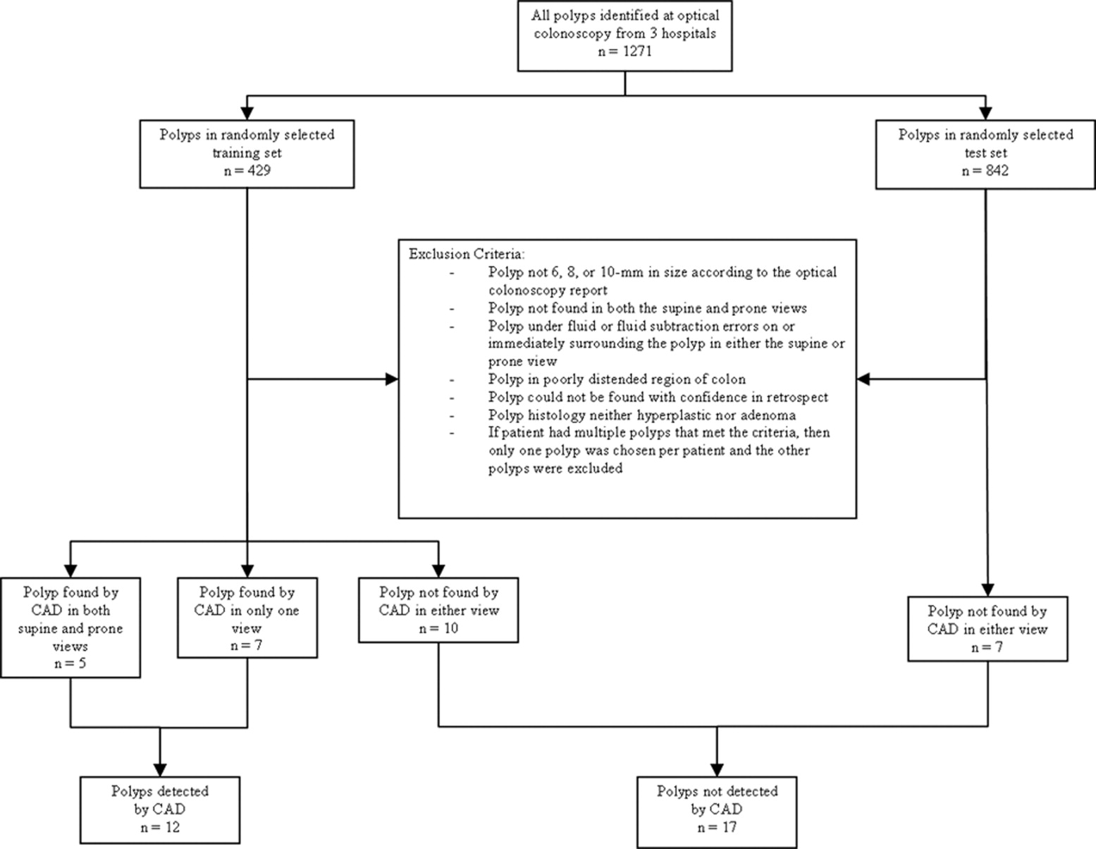
The factors that influence the conspicuity of polyps on computed tomographic (CT) colonography (CTC) are poorly understood. The aim of this study is to compare radiologists’ visual assessment of polyp conspicuity to quantitative image features and show the relationship between visual conspicuity and the detection of colonic polyps by computer-aided detection (CAD) on CTC.
Methods
One polyp (size range 6—10 mm) was selected from the CTC examination of each of 29 patients from a larger cohort. All patients underwent oral contrast-enhanced CTC with same-day optical colonoscopy with segmental unblinding. The polyps were analyzed by a previously validated CAD system and placed into one of two groups (detected [ n = 12] or not detected [ n = 17] by CAD). The study population was intentionally enriched with polyps that were not detected by the CAD system. Four board-certified radiologists, blinded to the CAD results, reviewed two- and three-dimensional CTC images of the polyps and scored the conspicuity of the polyps using a 4-point scale (0 = least conspicuous, 3 = most conspicuous). Polyp height and width were measured by a trained observer. A t -test (two-tailed, unpaired equal variance) was done to determine statistical significance. Intra- and interobserver variabilities of the conspicuity scores were assessed using the weighted κ test. Regression analysis was used to investigate the relationship of conspicuity to polyp height and width.
Results
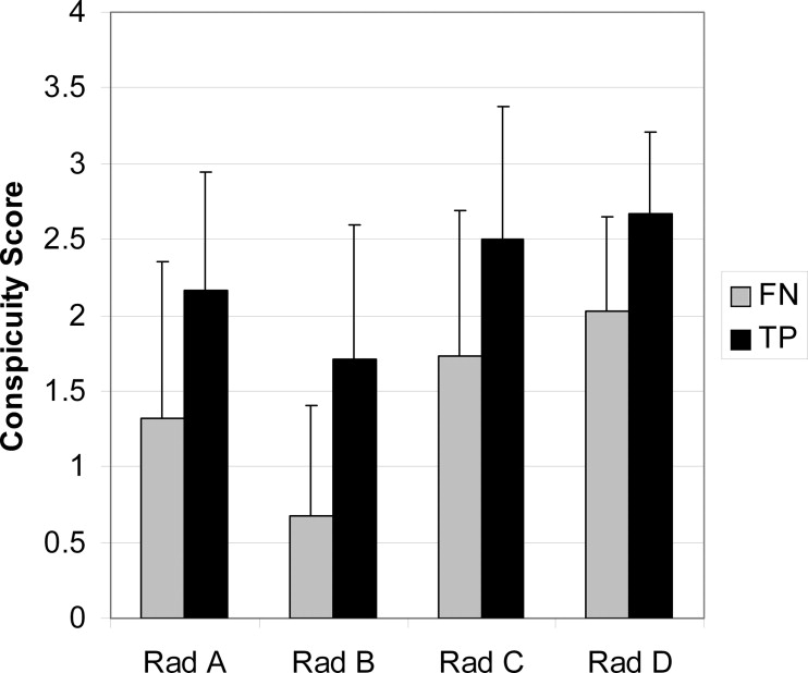
A statistically significant difference was found between the average conspicuity scores for polyps that were detected by CAD compared to those that were not (2.3 ± 0.6 vs. 1.4 ± 0.8) ( P = .004). There was moderate intraobserver agreement of the conspicuity scores (weighted κ 0.57 ± 0.09). Interobserver agreement was fair (average weighted κ for six pair-wise comparisons, 0.38 ± 0.15). Conspicuity was correlated with manual measurement of polyp height ( r 2 = 0.38–0.56, P < .001).
Conclusions
This CAD system tends to detect 6—10 mm polyps that are more visually conspicuous. Polyp height is a major determinant of visual conspicuity. The generalizability of these findings to other CAD systems is currently unknown. Nevertheless, CAD developers may need to specifically target flatter and less conspicuous polyps for CAD to better assist the radiologist to find polyps in this clinically important size category.
Accurate detection and diagnosis of colonic polyps using computed tomographic (CT) colonography (CTC) is dependent upon many factors. One of the most important factors is the ability of a human observer to visually locate and properly classify polyp candidates. Unfortunately, this ability is inconsistent as demonstrated by studies that reported high intra- and interobserver variabilities among radiologists ( ).
To ensure that patients are properly diagnosed, it is desirable to both reduce observer variability and increase average diagnostic performance. One way to achieve these goals is through the use of computer-aided detection (CAD) systems. For example, CAD systems are available to help detect breast cancers on mammography, pulmonary nodules on chest computed tomography and polyps on CTC.
Get Radiology Tree app to read full this article<
Get Radiology Tree app to read full this article<
Materials and methods
Get Radiology Tree app to read full this article<
Patient Population
Get Radiology Tree app to read full this article<
Get Radiology Tree app to read full this article<
Get Radiology Tree app to read full this article<
Get Radiology Tree app to read full this article<
Get Radiology Tree app to read full this article<
Patient Preparation
Get Radiology Tree app to read full this article<
CT Scanning
Get Radiology Tree app to read full this article<
Polyp Identification
Get Radiology Tree app to read full this article<
Get Radiology Tree app to read full this article<
Get Radiology Tree app to read full this article<
Computer-aided Polyp Detection
Get Radiology Tree app to read full this article<
Data Preparation
Get Radiology Tree app to read full this article<
Get Radiology Tree app to read full this article<
Get Radiology Tree app to read full this article<
Data Collection—Observer
Get Radiology Tree app to read full this article<
Get Radiology Tree app to read full this article<
Get Radiology Tree app to read full this article<
Quantitative Information
Get Radiology Tree app to read full this article<
Qualitative Information
Get Radiology Tree app to read full this article<
Data Collection—Measurements
Get Radiology Tree app to read full this article<
Get Radiology Tree app to read full this article<
Get Radiology Tree app to read full this article<
Statistical Analysis
Get Radiology Tree app to read full this article<
Get Radiology Tree app to read full this article<
Get Radiology Tree app to read full this article<
Results
Polyp Size
Get Radiology Tree app to read full this article<
Reader Agreement
Get Radiology Tree app to read full this article<
Table 1
Intraobserver Agreement of Conspicuity Score
Reader Supine Prone Supine and Prone A 0.58 0.64 0.62 B 0.51 0.63 0.48 C 0.30 0.47 0.50 D 0.68 0.67 0.67 Mean ± SD 0.52 ± 0.16 0.60 ± 0.09 0.57 ± 0.09
SD, standard deviation.
Table values are weighted κ for each of the four radiologist’s conspicuity scores comparing the first and second reads. Weighted κ is shown separately for the supine, prone, and combined (supine and prone) reads.
Table 2
Interobserver Agreement of Conspicuity Score
Reader B C D A 0.53 0.38 0.26 B — 0.30 0.23 C — — 0.60
Table values are weighted κ for each pairwise comparison of radiologist conspicuity scores for the first of the two reads, when using the supine and prone images together (the “combined read”) to grade conspicuity.
Get Radiology Tree app to read full this article<
Conspicuity and CAD
Get Radiology Tree app to read full this article<
Get Radiology Tree app to read full this article<
Conspicuity and Image Features
Get Radiology Tree app to read full this article<
Get Radiology Tree app to read full this article<
Conspicuity and True Polyp Size
Get Radiology Tree app to read full this article<
Qualitative Features
Get Radiology Tree app to read full this article<
Table 3
Readers’ Description of Less Conspicuous Polyps
Physical Characteristics of Polyps Polyps Not Found by CAD Polyps Found by CAD Flat 21%, 12/56 9%, 2/23 Edges not prominent (ie, less well defined) 50%, 28/56 22%, 5/23 Difficult to distinguish from a normal fold 21%, 12/56 35%, 8/23 Looks like noise or a normal bump on the colonic surface 68%, 38/56 57%, 13/23 Looks like stool 16%, 9/56 9%, 2/23
Data are numbers of readings having each description for less conspicuous polyps (conspicuity scores <3). Assessments are for the first reading session on review of the supine and prone images together.
Get Radiology Tree app to read full this article<
Discussion
Get Radiology Tree app to read full this article<
Get Radiology Tree app to read full this article<
Get Radiology Tree app to read full this article<
Get Radiology Tree app to read full this article<
Get Radiology Tree app to read full this article<
Get Radiology Tree app to read full this article<
Get Radiology Tree app to read full this article<
Get Radiology Tree app to read full this article<
Get Radiology Tree app to read full this article<
Get Radiology Tree app to read full this article<
Get Radiology Tree app to read full this article<
Get Radiology Tree app to read full this article<
Get Radiology Tree app to read full this article<
Get Radiology Tree app to read full this article<
Acknowledgments
Get Radiology Tree app to read full this article<
Get Radiology Tree app to read full this article<
References
1. Johnson C.D., Toledano A.Y., Herman B.A., et. al.: Computerized tomographic colonography: performance evaluation in a retrospective multicenter setting. Gastroenterology 2003; 125: pp. 688-695.
2. Petrick N., Haider M., Summers R.M., et. al.: CT Colonography and computer-aided detection as a second reader: observer performance study. Radiology 2008; 246: pp. 148-156.
3. Kundel H.L., Revesz G.: Lesion conspicuity, structured noise, and film reader error. Am J Roentgenol 1976; 126: pp. 1233-1238.
4. Revesz G., Kundel H.L., Graber M.A.: The influence of structured noise on the detection of radiologic abnormalities. Invest Radiol 1974; 9: pp. 479-486.
5. Revesz G., Kundel H.L.: Psycholophysical studies of detection errors in chest radiology. Radiology 1977; 123: pp. 559-562.
6. Krupinski E.A., Berger W.G., Dallas W.J., Roehrig H.: Searching for nodules: what features attract attention and influence detection?. Acad Radiol 2003; 10: pp. 861-868.
7. Mulhall B.P., Veerappan G.R., Jackson J.L.: Meta-analysis: computed tomographic colonography. Ann Intern Med 2005; 142: pp. 635-650.
8. Shi R., Schraedley-Desmond P., Napel S., et. al.: CT colonography: influence of 3D viewing and polyp candidate features on interpretation with computer-aided detection. Radiology 2006; 239: pp. 768-776.
9. Summers R.M., Yao J., Pickhardt P.J., et. al.: Computed tomographic virtual colonoscopy computer-aided polyp detection in a screening population. Gastroenterology 2005; 129: pp. 1832-1844.
10. Soetikno R.M., Kaltenbach T., Rouse R.V., et. al.: Prevalence of nonpolypoid (flat and depressed) colorectal neoplasms in asymptomatic and symptomatic adults. JAMA 2008; 299: pp. 1027-1035.
11. Pickhardt P.J., Choi J.R., Hwang I., et. al.: Computed tomographic virtual colonoscopy to screen for colorectal neoplasia in asymptomatic adults. N Engl J Med 2003; 349: pp. 2191-2200.
12. Pickhardt P.J., Choi J.H.: Electronic cleansing and stool tagging in CT colonography: advantages and pitfalls with primary three-dimensional evaluation. AJR Am J Roentgenol 2003; 181: pp. 799-805.
13. Altman D.G.: Practical statistics for medical research.1999.Chapman & Hall/CRCBoca Raton, FL
14. Park S.H., Ha H.K., Kim A.Y., et. al.: Flat polyps of the colon: detection with 16-MDCT colonography—preliminary results. AJR Am J Roentgenol 2006; 186: pp. 1611-1617.
15. Park S.H., Ha H.K., Kim M.J., et. al.: False-negative results at multi-detector row CT colonography: multivariate analysis of causes for missed lesions. Radiology 2005; 235: pp. 495-502.
16. Pickhardt P.J., Nugent P.A., Choi J.R., Schindler W.R.: Flat colorectal lesions in asymptomatic adults: implications for screening with CT virtual colonoscopy. AJR Am J Roentgenol 2004; 183: pp. 1343-1347.
17. Li J, Franaszek M, Petrick N, Yao J, Huang A, Summers RM. Wavelet method for CT colonography computer-aided polyp detection. In: 2006 3rd IEEE International Symposium on Biomedical Imaging: From Nano to Macro - Proceedings, 2006; 2006:1316–1319.
18. Li J., Van Uitert R., Yao J., et. al.: Wavelet method for CT colonography computer-aided polyp detection. Med Phys 2008; 35: pp. 3527-3538.
19. Pickhardt P.J., Choi J.R., Nugent P.A., Schindler W.R.: The effect of diagnostic confidence on the probability of optical colonoscopic confirmation of potential polyps detected on CT colonography: prospective assessment in 1,339 asymptomatic adults. AJR Am J Roentgenol 2004; 183: pp. 1661-1665.
20. Krupinski E.A.: Visual search of mammographic images: influence of lesion subtlety. Acad Radiol 2005; 12: pp. 965-969.
21. Kundel H.L., Revesz G., Toto L.: Contrast gradient and the detection of lung nodules. Invest Radiol 1979; 14: pp. 18-22.
22. Doshi T., Rusinak D., Halvorsen R.A., Rockey D.C., Suzuki K., Dachman A.H.: CT colonography: false-negative interpretations. Radiology 2007; 244: pp. 165-173.
23. Gluecker T.M., Fletcher J.G., Welch T.J., et. al.: Characterization of lesions missed on interpretation of CT colonography using a 2D search method. AJR Am J Roentgenol 2004; 182: pp. 881-889.
24. Arnesen R.B., Adamsen S., Svendsen L.B., Raaschou H.O., von Benzon E., Hansen O.H.: Missed lesions and false-positive findings on computed-tomographic colonography: a controlled prospective analysis. Endoscopy 2005; 37: pp. 937-944.
25. Fidler J.L., Fletcher J.G., Johnson C.D., et. al.: Understanding interpretive errors in radiologists learning computed tomography colonography. Acad Radiol 2004; 11: pp. 750-756.