
Rationale and Objectives
To predict the T stage of nonrectal colon cancer using contrast-enhanced computed tomography colonography.
Materials and Methods
Sixty-one patients with 67 nonrectal colon cancers consecutively underwent contrast-enhanced computed tomography colonography after an incomplete colonoscopy. Two readers evaluated wall deformity and perilesional fat abnormality on three-dimensional double contrast enema-like views and multiplanar reconstructions. Pathology was used as the standard of reference. McNemar, Fisher, and Cohen κ statistics were used.
Results
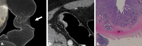
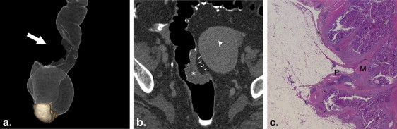
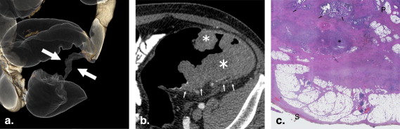
At pathologic examination, we found the following stages: T1 ( n = 5), T2 ( n = 10), T3 ( n = 41), T4a ( n = 6), and T4b ( n = 5). Intraobserver and interobserver reproducibilities were almost perfect for wall deformity (κ = 1.00 and κ = 0.88, respectively), substantial for perilesional fat abnormality (κ = 0.79 and κ = 0.74, respectively). Using the results of the more experienced reader, accuracy of wall deformity ≥50% (apple-core) alone for T ≥ 3 was 62 of 67 (0.93, 95% confidence interval [CI] 0.83–0.97) and that of perilesional fat abnormality alone was 37 of 67 (0.55, 95% CI 0.43–0.67) ( P < .001). Predictive value for ≥ T3 of the association wall deformity ≥50% with perilesional fat abnormality was 22 of 22 (1.00, 95% CI 0.85–1.00), higher, but not significantly, than that of wall deformity ≥50% with normal perilesional fat 29 of 33 (0.88, 95% CI 0.72–0.97) ( P = .148, Fisher exact test).
Conclusions
The presence of apple-core wall deformity, regardless of perilesional fat abnormality, is highly predictive of stage T3 or higher.
Computed tomography (CT) colonography (CTC) represents a good alternative to optical colonoscopy in diagnosing polyps and colorectal cancers because it has been shown to have similar accuracy, a higher patient compliance, and a lower rate of complications . Moreover, patients with a colorectal cancer detected on optical colonoscopy still benefit from CTC if optical colonoscopy is incomplete ; since contrast-enhanced CT is usually performed for staging, added benefit can be obtained by converting the routine staging CT into a contrast-enhanced CTC instead. Contrast-enhanced CT for preoperative T staging of colorectal cancer was first reported in 1986 but an acceptable accuracy was reached only with the advent of spiral CT using pneumocolon in 1998 . With the development of CTC, also known as virtual colonoscopy, different reports have described the usefulness of CTC in patients with known colorectal cancer and incomplete optical colonoscopy. Only a few studies have evaluated the accuracy of CTC in preoperative T staging of colorectal cancer, reporting values between 0.78 and 0.84 .
Preoperative T staging of rectal cancers has been well established, and magnetic resonance imaging (MRI) is the standard examination, while the same issue, not currently valid for colon cancers, may soon become more relevant. In particular, preoperative contrast-enhanced CTC staging could help with decisions concerning an appropriate type of surgery or chemoradiation.
Get Radiology Tree app to read full this article<
Materials and methods
Population
Get Radiology Tree app to read full this article<
Bowel Preparation
Get Radiology Tree app to read full this article<
Get Radiology Tree app to read full this article<
Get Radiology Tree app to read full this article<
CTC Protocol
Get Radiology Tree app to read full this article<
Get Radiology Tree app to read full this article<
Get Radiology Tree app to read full this article<
Get Radiology Tree app to read full this article<
Get Radiology Tree app to read full this article<
Image Analysis
Get Radiology Tree app to read full this article<
Get Radiology Tree app to read full this article<
Reference Standard
Get Radiology Tree app to read full this article<
Statistical Analysis
Get Radiology Tree app to read full this article<
Results
Population and Colon Cancer
Get Radiology Tree app to read full this article<
Get Radiology Tree app to read full this article<
Technical Quality of CE-CTC
Get Radiology Tree app to read full this article<
CE-CTC Findings
Get Radiology Tree app to read full this article<
Get Radiology Tree app to read full this article<
Get Radiology Tree app to read full this article<
Get Radiology Tree app to read full this article<
Table 1
Sensitivity, Specificity, Predictive Values, and Accuracy of Apple-core WD Alone and Perilesional Fat Abnormality Alone for T Stage ≥ 3 at CE-CTC
Apple-core WD Perilesional Fat Abnormality Ratio Point Estimate 95% CI Ratio Point Estimate 95% CI Sensitivity 51/52 0.98 0.90–1.00 22/52 0.42 0.29–0.57 Specificity 11/15 0.73 0.45–0.92 15/15 1.00 0.78–1.00 Positive predictive value 51/55 0.93 0.82–0.98 22/22 1.00 0.85–1.00 Negative predictive value 11/12 0.92 0.62–1.00 15/45 0.33 0.20–0.49 Accuracy ∗ 62/67 0.92 0.83–0.97 37/67 0.55 0.43–0.67
Get Radiology Tree app to read full this article<
Get Radiology Tree app to read full this article<
Get Radiology Tree app to read full this article<
Get Radiology Tree app to read full this article<
Get Radiology Tree app to read full this article<
Discussion
Get Radiology Tree app to read full this article<
Get Radiology Tree app to read full this article<
Get Radiology Tree app to read full this article<
Get Radiology Tree app to read full this article<
Get Radiology Tree app to read full this article<
Get Radiology Tree app to read full this article<
Get Radiology Tree app to read full this article<
Get Radiology Tree app to read full this article<
Get Radiology Tree app to read full this article<
Acknowledgments
Get Radiology Tree app to read full this article<
Get Radiology Tree app to read full this article<
References
1. Pickhardt P.J., Choi J.R., Hwang I., et. al.: Computed tomographic virtual colonoscopy to screen for colorectal neoplasia in asymptomatic adults. N Engl J Med 2003; 349: pp. 2191-2200.
2. Halligan S., Altman D.G., Taylor S.A., et. al.: CT colonography in the detection of colorectal polyps and cancer: Systematic review, meta-analysis, and proposed minimum data set for study level reporting. Radiology 2005; 237: pp. 893-904.
3. Kim D.H., Pickhardt P.J., Taylor A.J., et. al.: CT colonography versus colonoscopy for the detection of advanced neoplasia. N Engl J Med 2007; 57: pp. 1403-1412.
4. Johnson C.D., Chen M.H., Toledano A.Y., et. al.: Accuracy of CT colonography for detection of large adenomas and cancers. N Engl J Med 2008; 359: pp. 1207-1217.
5. Regge D., Laudi C., Galatola G., et. al.: Diagnostic accuracy of computed tomographic colonography for the detection of advanced neoplasia in individuals at increased risk of colorectal cancer. JAMA 2009; 301: pp. 2453-2461.
6. Graser A., Stieber P., Nagel D., et. al.: Comparison of CT colonography, colonoscopy, sigmoidoscopy and faecal occult blood tests for the detection of advanced adenoma in an average risk population. Gut 2009; 58: pp. 241-248.
7. Chaparro M., Gisbert J.P., del Campo L., et. al.: Accuracy of computed tomographic colonography for the detection of polyps and colorectal tumors: A systematic review and meta-analysis. Digestion 2009; 80: pp. 1-17.
8. Pickhardt P.J., Hassan C., Halligan S., et. al.: Colorectal cancer: CT colonography and colonoscopy for detection–systematic review and meta-analysis. Radiology 2011; 259: pp. 393-405.
9. Fenlon H.M., David B., McAneny , et. al.: Occlusive colon carcinoma: Virtual colonoscopy in the preoperative evaluation of the proximal colon. Radiology 1999; 210: pp. 423-428.
10. Macari M., Berman P., Dicker M., et. al.: Usefulness of CT colonography in patients with incomplete colonoscopy. AJR Am J Roentgenol 1999; 173: pp. 561-564.
11. Morrin M., Farrell R.J., Raptopoulos V., et. al.: Tomographic colonography in patients with colorectal cancers and obstructing colorectal lesions. Dis Colon Rectum 2000; 43: pp. 303-311.
12. Neri E., Giusti P., Battolla L., et. al.: Colorectal Cancer: Role of CT colonography in preoperative evaluation after incomplete colonoscopy. Radiology 2002; 223: pp. 615-619.
13. Kim J.H., Kim W.H., Kim T.I., et. al.: Incomplete colonoscopy in patients with occlusive colorectal cancer: Usefulness of CT colonography according to tumor location. Yonsei Med J 2007; 48: pp. 934-941.
14. Freeny P.C., Marks W.M., Ryan J.A., et. al.: Colorectal carcinoma evaluation with CT: Preoperative staging and detection of postoperative recurrence. Radiology 1986; 158: pp. 347-353.
15. Thompson W.M., Halvorsen R.A., Foster W.L., et. al.: Preoperative and postoperative CT staging of recto-sigmoid carcinoma. AJR Am J Roentgenol 1986; 146: pp. 703-710.
16. Harvey C.J., Amin Z., Hare C.B., et. al.: Helical CT pneumocolon to assess colonic tumor: Radiologic–pathologic correlation. AJR Am J Roentgenol 1998; 170: pp. 1439-1443.
17. Filippone A., Ambrosini R., Fuschi M., et. al.: Preoperative T and N staging of colorectal cancer: accuracy of contrast-enhanced multi-detector row CT colonography. Radiology 2004; 231: pp. 83-90.
18. Nagata K., Endo S., Kudo S.E., et. al.: CT air-contrast enema as a preoperative examination for colorectal cancer. Dig Surg 2004; 21: pp. 352-358.
19. Jin K.N., Lee J.M., Kim S.H., et. al.: The diagnostic value of multiplanar reconstruction on MDCT colonography for the preoperative staging of colorectal cancer. Eur Radiol 2006; 16: pp. 2284-2291.
20. Utano K., Endo K., Togashi K., et. al.: Preoperative T staging of colorectal cancer by CT colonography. Dis Colon Rectum 2008; 51: pp. 875-881.
21. Laghi A., Iannaccone R., Trenna S., et. al.: Multislice spiral CT colonography in the evaluation of colorectal neoplasms. Radiol Med 2002; 104: pp. 394-403.
22. Edge S.B.: American Joint Committee on Cancer: AJCC Cancer Staging Manual.7th ed.2009.SpringerNew York
23. Lacy A.M., Garcia-Valdecasas J.C., Delgado S., et. al.: Laparoscopy-assisted colectomy versus open colectomy for treatment of non-metastatic colon cancer, a randomized trial. Lancet 2002; 359: pp. 2224-2229.
24. The Clinical Outcomes of Surgical Therapy (COST) Study Group: A comparison of laparoscopically assisted and open colectomy for colon cancer. N Engl J Med 2004; 350: pp. 2050-2059.
25. Hasegawa H., Kabeshima Y., Watanabe M., et. al.: Randomized controlled trial of laparoscopic versus open colectomy for advanced colorectal cancer. Surg Endosc 2003; 17: pp. 636-640.
26. Hemandas A.K., Abdelrahman T., Flashman K.G., et. al.: Laparoscopic colorectal surgery produces better outcomes for high risk cancer patients compared to open surgery. Ann Surg 2010; 252: pp. 84-89.
27. Ng C.S., Doyle T.C., Dixon A.K., et. al.: Histopathological correlates of abnormal pericolic fat on CT in the assessment of colorectal carcinoma. Br J Radiol 2002; 75: pp. 31-37.
28. Baca B., Selçuk D., Kilic I.E., et. al.: The contributions of virtual colonoscopy to laparoscopic colorectal surgery. Hepatogastroenterology 2007; 54: pp. 1976-1982.
29. Matsuki M., Okuda J., Kanazava S., et. al.: Virtual CT colectomy by three-dimensional imaging using multidetector-row CT for laparoscopic colorectal surgery. Abdom Imaging 2005; 30: pp. 698-707.
30. Israel O., Yefremov N., Bar-Shalom R., et. al.: PET/CT detection of unexpected gastrointestinal foci of 18F-FDG uptake: Incidence, localization patterns, and clinical significance. J Nucl Med 2005; 46: pp. 758-762.
31. Kayani I., Groves A.M., Syed R., et. al.: Combined F-18 FDG positron emission tomography/computed tomography in the diagnosis of colonic polyps: The potential and limitations of the technique. Clin Nucl Med 2005; 30: pp. 116-117.
32. Nagata K., Ota Y., Okawa T., et. al.: PET/CT colonography for the preoperative evaluation of the colon proximal to the obstructive colorectal cancer. Dis Colon Rectum 2008; 51: pp. 882-890.
33. Taylor S.A., Bomanji J.B., Manpanzure L., et. al.: Nonlaxative PET/CT colonography: Feasibility, acceptability, and pilot performance in patients at higher risk of colonic neoplasia. J Nucl Med 2010; 51: pp. 854-861.