
Rationale and Objectives
The objective of this study was to determine the diagnostic accuracy of contrast-enhanced magnetic resonance angiography (MRA) when used in the preoperative evaluation of hepatic vascular anatomy in living liver donors.
Materials and Methods
A computer-assisted literature searching of EMBASE, PubMed (MEDLINE), and the Cochrane library databases was conducted to identify potentially relevant articles which primarily examined the utility of contrast-enhanced MRA in the preoperative evaluation of hepatic vascular anatomy in living liver donors. We used the Q statistic of chi-squared value test and inconsistency index (I-squared, I 2 ) to estimate the heterogeneity of the data extracted from all selected studies. Meta-Disc software (version 1.4) ( ftp://ftp.hrc.es/pub/programas/metadisc/Metadisc_update.htm ) was used to perform our analysis.
Results
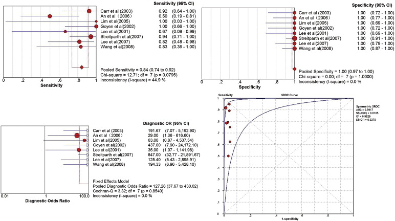
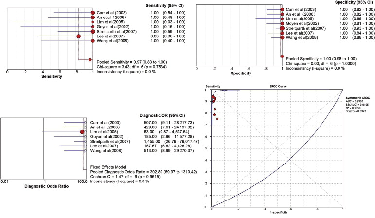
Eight studies were included in the present meta-analysis. A total of 289 living liver donor candidates and 198 patients who underwent liver harvesting were included in the present study. The pooled sensitivities of hepatic artery (HA), portal vein (PV), and hepatic vein (HV) in this meta-analysis were 0.84, 0.97, and 0.94, respectively. The pooled specificities of HA, PV, and HV were 1.00, 1.00, and 1.00, respectively. The pooled diagnostic odds ratios of HA, PV, and HV were 127.28, 302.80, and 256.59, respectively. The area under the summary receiver-operating characteristic curves of HA, PV, and HV were 0.9917, 0.9960, and 0.9813, respectively.
Conclusions
The high sensitivity and specificity demonstrated in this meta-analysis suggest that contrast-enhanced MRA was a promising test for the preoperative evaluation of hepatic vascular anatomy in living liver donors.
Living donors liver transplantation (LDLT) is becoming more and more common recently because of the shortage of donor livers . One of the main clinical issues of this procedure is the risk to the donors who are healthy until the date of transplantation. To reduce the morbidity and mortality and to ensure a successful LDLT, careful preoperative evaluations of the hepatic vascular and biliary anatomy, as well as the parenchyma, are essential for us to get useful information and help us in the surgical planning for vascular and biliary anastomoses .
Digital subtraction angiography (DSA) is considered as the diagnostic criterion standard for the preoperative evaluation of hepatic vascular anatomy in living liver donors. However, DSA is not an ideal screening test. One of the most important reasons is because DSA is not only associated with high cost but also have an invasive nature. Because of this, DSA is associated with some potential complications . Computed tomographic (CT) angiography has also been used recently in the preoperative evaluation of the hepatic vasculature of living liver donors . In particular, multidetector CT can now provide detailed images of the arterial system in a short breath-hold. However, because CT requires using ionizing radiation and potentially nephrotoxic iodinated contrast material, it could also potentially lead to nephrotoxicity, thus contraindicated in those patients with decreased renal function.
Get Radiology Tree app to read full this article<
Get Radiology Tree app to read full this article<
Materials and methods
Literature Searching Strategy
Get Radiology Tree app to read full this article<
Study Selection
Get Radiology Tree app to read full this article<
Inclusion and Exclusion Criteria
Get Radiology Tree app to read full this article<
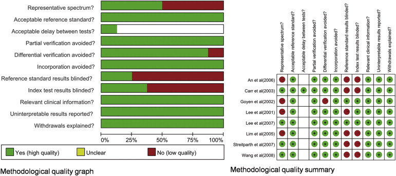
Data Extraction and Quality Assessment
Get Radiology Tree app to read full this article<
Meta-analysis
Get Radiology Tree app to read full this article<
Get Radiology Tree app to read full this article<
Results
Literature Search
Get Radiology Tree app to read full this article<
Excluded Studies
Get Radiology Tree app to read full this article<
Included Studies
Demographics
Get Radiology Tree app to read full this article<
Table 1
Characteristics of Included Studies
Study (year) Potential Donor Patients Hepatectomy Age Range (Mean) Percentage of Men Brand Tesla Contrast Agents Reference Standard Hepatic Vascular TP FP FN TN Sens Spec Carr et al. (2003) 25 24 20–54 (35) 48.0 Siemens, 1.5 Gd-DTPA, Berlex Laboratories 24 Surgery, 2 DSA HA 12 0 1 11 92.3 100 PV 6 0 0 19 100 100 HV 15 0 0 10 100 100 An et al. (2006) 44 24 20–58 (28) 66.7 Siemens, 1.5 Gd-BOPTA, Bracco Imaging 24 Surgery HA 5 0 5 14 50 100 PV 5 0 0 19 100 100 HV 8 0 1 15 88.9 100 Lim et al. (2005) 43 11 16–52 (31.6) 69.8 Philips, 1.5 Gd-BOPTA, Bracco Imaging 11 Surgery HA 1 0 0 10 100 100 PV 1 0 0 10 100 100 HV 9 0 0 2 100 100 Goyen et al. (2002) 38 16 20–50 (38) 60.5 Siemens, 1.5 Gd-BOPTA, Bracco Imaging 20 DSA, 16 surgery HA 9 0 0 11 100 100 PV 2 0 0 18 100 100 HV 1 0 0 19 100 100 Lee et al. (2001) 25 9 18–51 (35) NA Siemens, 1.5 Gd-DTPA; Berlex Laboratories 13 DSA, 9 surgery HA 2 0 1 10 66.7 100 PV 0 0 0 9 NA 100 HV 1 0 0 8 100 100 Streitparth et al. (2007) 55 55 18–68 (42) 34.5 Philips, 1.5 Gd-DTPA, Schering 55 Surgery HA 16 0 1 38 94.1 100 PV 7 0 0 48 100 100 HV 20 0 3 32 86.9 100 Lee et al. (2007) 27 27 16–44 (31) 85.2 Siemens, 1.5 Gd-BOPTA, Bracco Imaging 27 Surgery HA 9 0 2 16 81.8 100 PV 5 0 1 21 83.3 100 Wang et al. (2008) 32 32 21–63 (35) 56.3 Siemens, 3.0 Gd-BOPTA, Bracco Imaging 32 Surgery HA 5 0 1 26 83.3 100 PV 4 0 0 28 100 100 HV 8 0 0 24 100 100Total289198 HA5901113684.3100 PV300117296.8100 HV620411093.9100
DSA, digital subtraction angiography; FN, false negative; FP, false positive; Gd-BOPTA, gadobenate dimeglumine; Gd-DTPA, gadopentetate dimeglumine; HA, hepatic artery; HV, hepatic vein; NA, not available; PV, portal vein; TN, true negative; TP, true positive; Sens, sensitivity; Spec, specificity.
Get Radiology Tree app to read full this article<
Assessment of study quality
Get Radiology Tree app to read full this article<
Get Radiology Tree app to read full this article<
Diagnostic accuracy and the evaluation of clinical utility
Get Radiology Tree app to read full this article<

Get Radiology Tree app to read full this article<
Potential biases
Get Radiology Tree app to read full this article<
Discussion
Get Radiology Tree app to read full this article<
Get Radiology Tree app to read full this article<
Get Radiology Tree app to read full this article<
Get Radiology Tree app to read full this article<
Get Radiology Tree app to read full this article<
References
1. Strong R.W., Lynch S.V., Ong T.H., et. al.: Successful liver transplantation from a living donor to her son. N Engl J Med 1990; 322: pp. 1505-1507.
2. Mueller A.R., Pascher A., Platz K.P., et. al.: Immunosuppressive management following intestinal transplantation in adult patients. Transplant Proc 2003; 35: pp. 2075-2077.
3. Lee S.Y., Ko G.Y., Gwon D.I., et. al.: Living donor liver transplantation: complications in donors and interventional management. Radiology 2004; 230: pp. 443-449.
4. Yuan Y., Gotoh M.: Biliary complications in living liver donors. Surg Today 2010; 40: pp. 411-417.
5. Orons P., Zajko A.: Angiography and interventional procedures in liver transplantation. Radiol Clin North Am 1995; 33: pp. 541-558.
6. Duran C., Uraz S., Kantarci M., et. al.: Hepatic arterial mapping by multidetector computer tomographic angiography in living donor liver transplantation. J Comput Assist Tomogr 2009; 33: pp. 618-625.
7. Zhuang Z.G., Qian L.J., Gong H.X., et. al.: Multidetector computed tomography angiography in the evaluation of potential living donors for liver transplantation: single-center experience in China. Transplant Proc 2008; 40: pp. 2466-2477.
8. Chen W.H., Xin W., Wang J., et. al.: Multi-slice spiral CT angiography in evaluating donors of living-related liver transplantation. Hepatobiliary Pancreat Dis Int 2007; 6: pp. 364-369.
9. Laissy J.P., Trillaud H., Douek P.: MR angiography: noninvasive vascular imaging of the abdomen. Abdom Imaging 2002; 27: pp. 488-506.
10. Griffin M., Grist T.M., François C.J.: Dynamic four-dimensional MR angiography of the chest and abdomen. Magn Reson Imaging Clin N Am 2009; 17: pp. 77-90.
11. Carr J.C., Nemcek A.A., Abecassis M., et. al.: Preoperative evaluation of the entire hepatic vasculature in living liver donors with use of contrast-enhanced MR angiography and true fast imaging with steady-state precession. J Vasc Interv Radiol 2003; 14: pp. 441-449.
12. An S.K., Lee J.M., Suh K.S., et. al.: Gadobenate dimeglumine-enhanced liver MRI as the sole preoperative imaging technique: a prospective study of living liver donors. AJR Am J Roentgenol 2006; 187: pp. 1223-1233.
13. Lim J.S., Kim M.J., Kim J.H., et. al.: Preoperative MRI of potential living-donor-related liver transplantation using a single dose of gadobenate dimeglumine. AJR Am J Roentgenol 2005; 185: pp. 424-431.
14. Goyen M., Barkhausen J., Debatin J.F., et. al.: Right-lobe living related liver transplantation: evaluation of a comprehensive magnetic resonance imaging protocol for assessing potential donors. Liver Transpl 2002; 8: pp. 241-250.
15. Lee V.S., Morgan G.R., Teperman L.W., et. al.: MR imaging as the sole preoperative imaging modality for right hepatectomy: a prospective study of living adult-to-adult liver donor candidates. AJR Am J Roentgenol 2001; 176: pp. 1475-1482.
16. Streitparth F., Pech M., Figolska S., et. al.: Living related liver transplantation: preoperative magnetic resonance imaging for assessment of hepatic vasculature of donor candidates. Acta Radiol 2007; 48: pp. 20-26.
17. Lee M.W., Lee J.M., Lee J.Y., et. al.: Preoperative evaluation of hepatic arterial and portal venous anatomy using the time resolved echo-shared MR angiographic technique in living liver donors. Eur Radiol 2007; 17: pp. 1074-1080.
18. Wang H., Mu X.T., Wu C.N., et. al.: Preoperative evaluation of hepatic vasculature with gadobenate dimeglumine three dimensional dynamic contrast-enhanced MR angiography in living liver donors. Chin J Med Imaging Technol 2008; 24: pp. 1749-1752.
19. Whiting P., Rutjes A.W., Reitsma J.B., et. al.: The development of QUADAS: a tool for the quality assessment of studies of diagnostic accuracy included in systematic reviews. BMC Med Res Methodol 2003; 3: pp. 25.
20. Higgins J.P., Thompson S.G., Deeks J.J., et. al.: Measuring inconsistency in meta-analyses. BMJ 2003; 327: pp. 557-560.
21. Zamora J., Abraira V., Muriel A., et. al.: Meta-DiSc: a software for meta-analysis of test accuracy data. BMC Med Res Methodol 2006; 12: pp. 31.
22. Lee M.W., Lee J.M., Lee J.Y., et. al.: Preoperative evaluation of the hepatic vascular anatomy in living liver donors: comparison of CT angiography and MR angiography. J Magn Reson Imaging 2006; 24: pp. 1081-1087.
23. Mangold S, Bretschneider C, Fenchel M, et al. MRI for evaluation of potential living liver donors: a new approach including contrast-enhanced magnetic resonance cholangiography. Abdom Imaging 37: 244–251
24. Cheng Y.F., Chen C.L., Huang T.L., et. al.: Single imaging modality evaluation of living donors in liver transplantation: magnetic resonance imaging. Transplantation 2001; 72: pp. 1527-1733.
25. Fulcher A.S., Szucs R.A., Bassignani M.J., et. al.: Right lobe living donor liver transplantation: preoperative evaluation of the donor with MR imaging. AJR Am J Roentgenol 2001; 176: pp. 1483-1491.
26. Schroeder T., Malagó M., Debatin J.F., et. al.: “All-in-one” imaging protocols for the evaluation of potential living liver donors: comparison of magnetic resonance imaging and multidetector computed tomography. Liver Transpl 2005; 11: pp. 776-787.
27. Artioli D., Tagliabue M., Aseni P., et. al.: Detection of biliary and vascular anatomy in living liver donors: value of gadobenate dimeglumine enhanced MR and MDCT angiography. Eur J Radiol 2010; 76: pp. e1-e5.
28. Huang B.Y., Castillo M.: Neurovascular imaging at 1.5 tesla versus 3.0 tesla. Magn Reson Imaging Clin N Am 2009; 17: pp. 29-46.
29. Bernstein M.A., Huston J., Lin C., et. al.: High-resolution intracranial and cervical MRA at 3.0T: technical considerations and initial experience. Magn Reson Med 2001; 46: pp. 955-962.
30. Voth M., Michaely H.J., Schwenke C., et. al.: Contrast-enhanced magnetic resonance angiography (MRA): evaluation of three different contrast agents at two different doses (0.05 and 0.1 mmol/kg) in pigs at 1.5 Tesla. Eur Radiol 2011; 21: pp. 337-344.
31. Reimer P., Schneider G., Schima W.: Hepatobiliary contrast agents for contrast-enhanced MRI of the liver: properties, clinical development and applications. Eur Radiol 2004; 14: pp. 559-578.