
Rationale and Objectives
Contrast-enhanced spectral mammography (CESM) uses full field digital mammography with the added benefit of intravenous contrast administration to significantly reduce false-positive and false-negative results and improve specificity while maintaining high sensitivity. For CESM to fulfill its purpose, one should be aware of possible artifacts and other factors which may interfere with image quality, and attention should be taken to minimize these factors.
Materials and Methods
This pictorial demonstration will depict types of artifacts detected and other factors that interfere with image acquisition in our practice since CESM implementation.
Results
Many of the artifacts and other factors we have encountered while using CESM have simple solutions to resolve them.
Conclusion
The illustrated artifacts and other factors interfering with image quality will serve as a useful reference to anyone using CESM.
Introduction
Contrast-enhanced spectral mammography (CESM) is a relatively new tool in breast imaging. It uses full field digital mammography (FFDM) with the added benefit of intravenous contrast administration to assess for the physiological properties of breast cancer . This technique received FDA clearance in 2011. At our institution, CESM has been in clinical use since November 2012 and serves as a valuable tool in high-risk screening, further evaluation of extremely dense breast tissue, diagnostic assessment of suspicious lesions, breast cancer staging, surgical planning, and assessment of chemotherapy response. The current article presents the artifacts and other factors that interfere with image acquisition we have encountered thus far and highlights the necessary steps to improve image quality. Although some of these artifacts are similar to those seen with FFDM, many are unique to CESM. As CESM use becomes more widespread, it is important to be able to recognize how image quality is affected, understand the significance, and know the necessary steps to take to mitigate the level of image degradation created by these artifacts and other factors .
Materials and Methods
This pictorial demonstration will display the types of artifacts and other factors detected in our practice since CESM implementation and are divided into three categories: factors related to FFDM, contrast administration, and CESM (Germaine et al., presented at the 2015 63rd annual meeting of the Association of University Radiologists).
CESM Modality-Specific Factors Affecting Image Quality
FFDM-Related Factors
Get Radiology Tree app to read full this article<
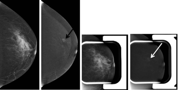
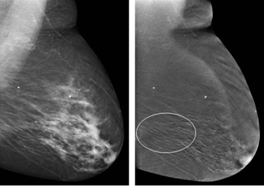
Motion
Get Radiology Tree app to read full this article<
Get Radiology Tree app to read full this article<
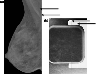
Subject-Related Factors
Get Radiology Tree app to read full this article<
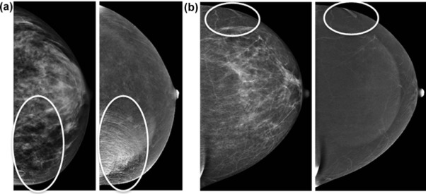
Get Radiology Tree app to read full this article<
Air Artifact
Get Radiology Tree app to read full this article<
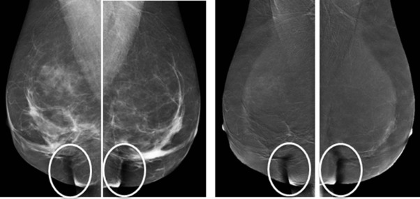
Get Radiology Tree app to read full this article<
Contrast-Related Factors
Get Radiology Tree app to read full this article<
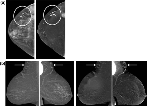
Contrast Splatter
Get Radiology Tree app to read full this article<
Get Radiology Tree app to read full this article<
Abnormal Timing of Contrast Bolus
Get Radiology Tree app to read full this article<
Get Radiology Tree app to read full this article<
Transient Retention of Contrast in Vein
Get Radiology Tree app to read full this article<
Get Radiology Tree app to read full this article<
CESM-Related Factors
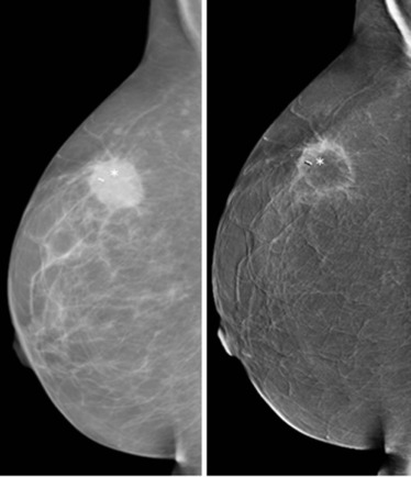
Negative Contrast Enhancement
Get Radiology Tree app to read full this article<
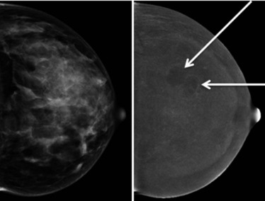
Get Radiology Tree app to read full this article<
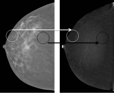
Halo Artifact
Get Radiology Tree app to read full this article<
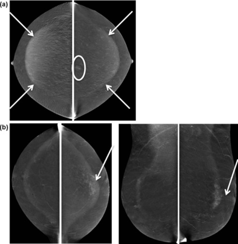
Get Radiology Tree app to read full this article<
Ripple Artifact
Get Radiology Tree app to read full this article<
Get Radiology Tree app to read full this article<
Aborted Study
Get Radiology Tree app to read full this article<
Get Radiology Tree app to read full this article<
Discussion
Get Radiology Tree app to read full this article<
Get Radiology Tree app to read full this article<
Get Radiology Tree app to read full this article<
References
1. Daniaux M., Zordo T., Zantner W., et. al.: Dual-energy contrast-enhanced spectral mammography (CESM). Arch Gynecol Obstet 2015; 292: pp. 739-747.
2. Dromain C., Balleyguier C., Adler G., et. al.: Contrast-enhanced digital mammography. Eur J Radiol 2009; 69: pp. 34-42.
3. Dromain C., Thibault F., Muller S., et. al.: Dual-energy contrast-enhanced digital mammography: initial clinical results. Eur J Radiol 2011; 21: pp. 565-574.
4. Fallenberg E., Dromain C., Diekmann F., et. al.: Contrast-enhanced spectral mammography versus MRI: initial results in the detection of breast cancer and assessment of tumour size. Eur J Radiol 2014; 24: pp. 256-264.
5. Jochelson M.S., Dershaw D.D., Sung J.S., et. al.: Bilateral contrast-enhanced dual-energy digital mammography: feasibility and comparison with conventional digital mammography and MR imaging in women with known breast carcinoma. Radiology 2013; 266: pp. 743-751.
6. Lobbes M.B., Smidt M.L., Houwers J., et. al.: Contrast-enhanced mammography: techniques, current results, and potential indications. Clin Radiol 2013; 68: pp. 935-944.
7. Ayyala R.S., Chorlton M., Behrman R.H., et. al.: Digital mammographic artifacts on full-field systems: what are they and how do I fix them?. Radiographics 2008; 28: pp. 1999-2008.
8. Geiser W.R., Haygood T.M., Santiago L., et. al.: Challenges in mammography: part 1, artifacts in digital mammography. Am J Roentgenol 2011; 197: pp. W1023-W1030.
9. Blum K.S., Rubbert C., Mathys B., et. al.: Use of contrast-enhanced spectral mammography for intramammary cancer staging. Acad Radiol 2014; 21: pp. 1363-1369.