
Rationale and Objectives
To compare contrast-enhanced coronary magnetic resonance angiography (MRA) at 3.0 T with the same technique performed at 1.5 T using the contrast agent gadofosveset.
Materials and Methods
In this prospective randomized study, 19 healthy male volunteers (mean age 28 years, mean weight 79.8 kg), after signing informed consents, underwent contrast-enhanced inversion recovery three-dimensional fast low angle shot (FLASH) MRA at 1.5 and at 3.0 T. Prospective electrocardiogram-triggering was combined with adaptive respiratory gating. For contrast-enhanced images, the intravascular contrast agent gadofosveset was used. Acquisition time, signal-to-noise ratio (SNR) of coronary blood, contrast-to-noise ratio (CNR) between coronaries and adjacent myocardium or epicardial fat and image quality were analyzed for statistical differences by using a two-tailed paired-sample t -test. The ratio calculations were based on measurements performed on the raw data and the image quality was blinded and independently evaluated by two experienced radiologists using a five-point scale.
Results
The mean values for the acquisition time were 14.58 ± 0.1 minutes at 1.5 T and 16.40 ± 0.2 minutes at 3.0 T. Overall SNR of all evaluated coronary segments proved higher at 3.0 T compared to 1.5 T (74.0 ± 42.1 at 3.0 T vs. 50.2 ± 20.2 at 1.5 T, P = .04). Overall CNR between coronaries and myocardium was significantly increased at 3.0 T in comparison to 1.5 T (40.1 ± 21.9 at 3.0 T vs. 24.4 ± 17.2 at 1.5 T, P = .01). Between the two methods, no significant difference in overall CNR between coronaries and epicardial fat was observed ( P = .08, NS). The 3.0 T MRA demonstrated superior overall image quality with respect to 1.5 T (2.28 ± 0.71 at 3.0 T vs. 1.92 ± 0.38 at 1.5T, P = .004).
Conclusion
The use of higher field strength, 3.0 T instead of 1.5 T, resulted in similar CNR between coronaries and epicardial fat, higher SNR values and CNR between blood and myocardium, as well as an improved overall image quality, when gadofosveset in combination with electrocardiogram and respiratory triggering for coronary MRA was used.
Coronary artery disease (CAD) remains one of the leading causes of morbidity and mortality in developed countries. Recent surveys suggest that three-dimensional whole-heart coronary magnetic resonance angiography (MRA) at 1.5 T is a promising technique for non-invasive assessment of the coronary arteries . When a contrast-enhanced whole-heart approach is used, electrocardiogram (ECG) and respiratory triggering require a contrast agent that enables increased blood signal intensity for a longer period than the first pass of an extravascular contrast agent. In theory, field strength of 3.0 T offers an almost twofold higher signal intensity and signal-to-noise ratio (SNR), than a 1.5 T system, if the same pulse sequence technique is used . In contrast to a steady-state free-precession (SSFP) readout technique, a spoiled gradient-echo sequence, such as fast low-angle shot (FLASH), proved relatively insensitive to the increased field inhomogeneities of 3.0 T .
Various contrast agents have been used at 3.0T in previous investigations for coronary MRA, such as gadopentetate dimeglumine or gadobenate dimeglumine . The intravascular contrast media provide long time windows for coronary MRA acquisition because of prolonged plasma half-life and slow extravasation in the interstitial space .
Get Radiology Tree app to read full this article<
Get Radiology Tree app to read full this article<
Get Radiology Tree app to read full this article<
Materials and methods
Get Radiology Tree app to read full this article<
Study Population
Get Radiology Tree app to read full this article<
Table 1
Characteristics of the Volunteers Group
No Volunteer Age (Years) Weight (kg) Body Mass Index 1 31 70 22.3 2 37 82 22.7 3 30 68 21.2 4 36 73 22.5 5 33 75 23.7 6 25 52 17.2 7 24 74 21.6 8 28 83 25.6 9 24 72 19.5 10 35 90 26.0 11 21 74 23.4 12 26 73 21.8 13 34 86 22.4 14 22 115 31.9 15 28 93 27.8 16 25 108 32.6 17 30 91 29.7 18 22 80 22.6 19 23 62 20.0
Get Radiology Tree app to read full this article<
MRI
Get Radiology Tree app to read full this article<
Get Radiology Tree app to read full this article<
Get Radiology Tree app to read full this article<
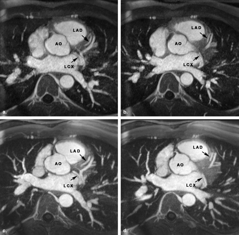
Contrast-enhanced Coronary MRA at 1.5 and 3.0 T
Get Radiology Tree app to read full this article<
Get Radiology Tree app to read full this article<
Contrast Agent
Get Radiology Tree app to read full this article<
Quantitative Analysis
Get Radiology Tree app to read full this article<
Get Radiology Tree app to read full this article<
SNR =SI/SDnoise; SNR =
SI
/
S
D
noise;
CNRcor/myo=(SIcor-SImyo)/SDnoise CN
R
cor
/
myo
=
(
S
I
cor
-S
I
myo
)
/
S
D
noise
CNRcor/fat=(SIcor-SIfat)/SDnoise CN
R
cor
/
fat
=
(
S
I
cor
-S
I
fat
)
/
S
D
noise
Get Radiology Tree app to read full this article<
Get Radiology Tree app to read full this article<
Qualitative Analysis
Get Radiology Tree app to read full this article<
Get Radiology Tree app to read full this article<
Statistical Analysis
Get Radiology Tree app to read full this article<
Results
Get Radiology Tree app to read full this article<
SNR and CNR Measurements
Get Radiology Tree app to read full this article<
Table 2
Measurements Result of Both Methods Concerning the Mean SNR Values
Mean SNR_P_ Value Evaluated
Regions 1.5 T 3.0 T 1.5 T vs. 3.0 T LMS 54.2 ± 20.6 78.5 ± 40.6 .04 Prox. LAD 50.1 ± 18.4 81.2 ± 49.1 .02 Prox. LCX 48.2 ± 17.4 71.4 ± 45.6 .06 Prox. RCA 48.3 ± 24.6 65.0 ± 32.9 .10 Overall segments 50.2 ± 20.2 74.0 ± 42.1 .04 Fat 15.2 ± 7.4 21.1 ± 11.0 .06 Myocardium 25.8 ± 10.4 34.2 ± 34.2 .32
LMS, left main stem coronary artery; LAD, left anterior descending coronary artery; RCA, right coronary artery; LCX, left circumflex artery; prox, proximal coronary segment.
Get Radiology Tree app to read full this article<
Get Radiology Tree app to read full this article<
Table 3a
Measurements Results of Both Methods Concerning the Values of CNR cor/fat
CNR cor/fat P Value Coronary
Segments 1.5 T 3.0 T 1.5 T vs. 3.0 T LMS 39.0 ± 14.5 57.4 ± 38.0 .07 Prox. LAD 34.9 ± 13.3 60.0 ± 46.5 .04 Prox. LCX 32.9 ± 12.0 50.2 ± 43.2 .12 Prox. RCA 33.1 ± 18.4 43.8 ± 31.3 .23 Overall 35.0 ± 14.7 52.9 ± 39.9 .08
Table 3b
Measurements Results of Both Methods Concerning the Values of CNR cor/myo
CNR cor/myo P Value Coronary
Segments 1.5 T 3.0 T 1.5 T vs. 3.0 T LMS 28.4 ± 17.7 44.3 ± 21.1 .02 Prox. LAD 24.2 ± 14.0 47.0 ± 23.6 .002 Prox. LCX 22.3 ± 13.7 37.1 ± 21.9 .02 Prox. RCA 22.5 ± 22.7 31.8 ± 19.0 .16 Overall 24.4 ± 17.2 40.1 ± 21.9 .01
LMS, left main stem coronary artery; LAD, left anterior descending coronary artery; RCA, right coronary artery; LCX, left circumflex artery; CNR cor/myo , CNR between coronary blood and myocardium; prox, proximal coronary segment.
Get Radiology Tree app to read full this article<
Image Quality
Get Radiology Tree app to read full this article<
Get Radiology Tree app to read full this article<
Get Radiology Tree app to read full this article<
Table 4a
Image Quality Results of Both Readers for Both Methods using Paired t-test
Mean Image Quality_P_ Value Coronary
Segments 1.5 T 3.0 T 1.5 T vs. 3.0 T LMS 2.2 ± 0.7 2.5 ± 0.5 .14 Proximal 2.2 ± 0.5 2.6 ± 0.4 .01 Middle 1.8 ± 0.3 2.3 ± 0.4 .002 Distal 1.5 ± 0.3 1.8 ± 0.4 .02 Overall 1.9 ± 0.7 2.2 ± 0.7 .004
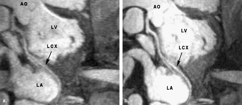
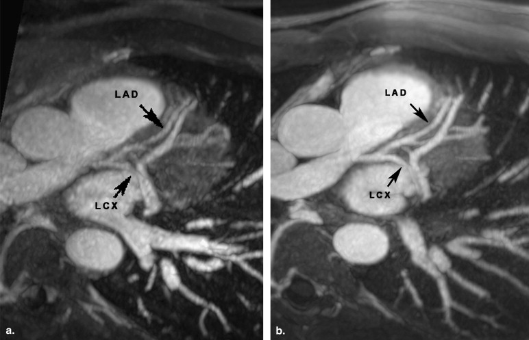
Table 4b
Image Quality Results of Both Methods for Each Reader Individually using Wilcoxon Test
Wilcoxon Test
1.5 T vs 3.0 T Coronary
Segments Reader 1 Reader 2 LMS 0.19 0.26 Proximal 0.003 0.04 Middle 0.003 0.009 Distal 0.03 0.04 Overall 0.004 0.01
LMS, left main stem coronary artery; proximal, middle, distal, and overall, proximal, middle, distal, and all coronary segments of left anterior descending coronary artery, left circumflex artery, and right coronary artery.
Get Radiology Tree app to read full this article<
Discussion
Get Radiology Tree app to read full this article<
Get Radiology Tree app to read full this article<
Get Radiology Tree app to read full this article<
Get Radiology Tree app to read full this article<
Get Radiology Tree app to read full this article<
Get Radiology Tree app to read full this article<
Get Radiology Tree app to read full this article<
Get Radiology Tree app to read full this article<
Get Radiology Tree app to read full this article<
Get Radiology Tree app to read full this article<
Limitations
Get Radiology Tree app to read full this article<
Conclusion
Get Radiology Tree app to read full this article<
References
1. Bi X., Li D.: Coronary arteries at 3.0 T: contrast-enhanced magnetization-prepared three-dimensional breathhold MR angiography. J Magn Reson Imaging 2005; 21: pp. 133-139.
2. Li D., Zheng J., Weinmann H.J.: Contrast-enhanced MR imaging of coronary arteries: comparison of intra- and extravascular contrast agents in swine. Radiology 2001; 218: pp. 670-678.
3. Kelle S., Thouet T., Tangcharoen T., et. al.: Whole-heart coronary magnetic resonance angiography with MS-325 (Gadofosveset). Med Sci Monit 2007; 13: pp. CR469-CR474.
4. Bi X., Carr J.C., Li D.: Whole-heart coronary magnetic resonance angiography at 3 Tesla in 5 minutes with slow infusion of Gd-BOPTA, a high-relaxivity clinical contrast agent. Magn Reson Med 2007; 58: pp. 1-7.
5. Finn J.P., Nael K., Deshpande V., et. al.: Cardiac MR imaging: state of the technology. Radiology 2006; 241: pp. 338-354.
6. Kim W.Y., Danias P.G., Stuber M., et. al.: Coronary magnetic resonance angiography for the detection of coronary stenoses. N Engl J Med 2001; 345: pp. 1863-1869.
7. Bi X., Deshpande V., Simonetti O., et. al.: Three-dimensional breathhold SSFP coronary MRA: a comparison between 1.5T and 3.0T. J Magn Reson Imaging 2005; 22: pp. 206-212.
8. Sommer T., Hackenbroch M., Hofer U., et. al.: Coronary MR angiography at 3.0 T versus that at 1.5 T: initial results in patients suspected of having coronary artery disease. Radiology 2005; 234: pp. 718-725.
9. Kaul M.G., Stork A., Bansmann P.M., et. al.: Evaluation of balanced steady-state free precession (TrueFISP) and K-space segmented gradient echo sequences for 3D coronary MR angiography with navigator gating at 3 Tesla. Rofo 2004; 176: pp. 1560-1565.
10. Bi X, MS, Li D. Coronary arteries at 3.0 T: contrast-enhanced magnetization-prepared three-dimensional breathhold MR angiography. J Magn Reson Imaging 2005; 21:133–139.
11. Liu X., Bi X., Huang J., et. al.: Contrast-enhanced whole-heart coronary magnetic resonance angiography at 3.0 T: comparison with steady-state free precession technique at 1.5 T. Invest Radiol 2008; 43: pp. 663-668.
12. Zheng J., Bae K.T., Woodard P.K., et. al.: Efficacy of slow infusion of gadolinium contrast agent in three-dimensional MR coronary artery imaging. J Magn Reson Imaging 1999; 10: pp. 800-805.
13. Goldfarb J.W., Edelman R.R.: Coronary arteries: breath-hold, gadolinium-enhanced, three-dimensional MR angiography. Radiology 1998; 206: pp. 830-834.
14. Prompona M., Cyran C., Nikolaou K., et. al.: Contrast-enhanced whole-heart MR coronary angiography at 3.0 T using the intravascular contrast agent gadofosveset. Invest Radiol 2009; 44: pp. 369-374.
15. Nassenstein K., Waltering K.U., Kelle S., et. al.: Magnetic resonance coronary angiography with Vasovist: in-vivo T1 estimation to improve image quality of navigator and breath-hold techniques. Eur Radiol 2008; 18: pp. 103-109.
16. Nassenstein K., Waltering K.U., Eggebrecht H., et. al.: [MR coronary angiography with MS-325, a blood pool contrast agent: comparison of an inversion recovery steady-state free precession with an inversion recovery fast low angle shot sequence in volunteers]. Rofo 2006; 178: pp. 508-514.
17. Lauffer R.B., Parmelee D.J., Dunham S.U., et. al.: MS-325: albumin-targeted contrast agent for MR angiography. Radiology 1998; 207: pp. 529-538.
18. Weber O.M., Martin A.J., Higgins C.B.: Whole-heart steady-state free precession coronary artery magnetic resonance angiography. Magn Reson Med 2003; 50: pp. 1223-1228.
19. Li D., Carr J.C., Shea S.M., et. al.: Coronary arteries: magnetization-prepared contrast-enhanced three-dimensional volume-targeted breath-hold MR angiography. Radiology 2001; 219: pp. 270-277.
20. Frenzel T., Lengsfeld P., Schirmer H., et. al.: Stability of gadolinium-based magnetic resonance imaging contrast agents in human serum at 37 degrees C. Invest Radiol 2008; 43: pp. 817-828.
21. Scanlon P.J., Faxon D.P., Audet A.M., et. al.: ACC/AHA guidelines for coronary angiography. A report of the American College of Cardiology/American Heart Association Task Force on practice guidelines (Committee on Coronary Angiography). Developed in collaboration with the Society for Cardiac Angiography and Interventions. J Am Coll Cardiol 1999; 33: pp. 1756-1824.
22. Spuentrup E., Bornert P., Botnar R.M., et. al.: Navigator-gated free-breathing three-dimensional balanced fast field echo (TrueFISP) coronary magnetic resonance angiography. Invest Radiol 2002; 37: pp. 637-642.
23. Deshpande V.S., Shea S.M., Laub G., et. al.: 3D magnetization-prepared true-FISP: a new technique for imaging coronary arteries. Magn Reson Med 2001; 46: pp. 494-502.
24. Maintz D., Aepfelbacher F.C., Kissinger K.V., et. al.: Coronary MR angiography: comparison of quantitative and qualitative data from four techniques. Am J Roentgenol 2004; 182: pp. 515-521.
25. Zagrosek A., Noeske R., Abdel-Aty H., et. al.: MR coronary angiography using 3D-SSFP with and without contrast application. J Cardiovasc Magn Reson 2005; 7: pp. 809-814.
26. Schar M., Kozerke S., Fischer S.E., et. al.: Cardiac SSFP imaging at 3 Tesla. Magn Reson Med 2004; 51: pp. 799-806.
27. Deshpande V.S., Shea S.M., Li D.: Artifact reduction in true-FISP imaging of the coronary arteries by adjusting imaging frequency. Magn Reson Med 2003; 49: pp. 803-809.
28. Hoult D.I.: Sensitivity and power deposition in a high-field imaging experiment. J Magn Reson Imaging 2000; 12: pp. 46-67.
29. Fischer S.E., Wickline S.A., Lorenz C.H.: Novel real-time R-wave detection algorithm based on the vectorcardiogram for accurate gated magnetic resonance acquisitions. Magn Reson Med 1999; 42: pp. 361-370.
30. Herborn C.U., Barkhausen J., Paetsch I., et. al.: Coronary arteries: contrast-enhanced MR imaging with SH L 643A–experience in 12 volunteers. Radiology 2003; 229: pp. 217-223.
31. Stuber M., Botnar R.M., Fischer S.E., et. al.: Preliminary report on in vivo coronary MRA at 3 Tesla in humans. Magn Reson Med 2002; 48: pp. 425-429.
32. Parmelee D.J., Walovitch R.C., Ouellet H.S., et. al.: Preclinical evaluation of the pharmacokinetics, biodistribution, and elimination of MS-325, a blood pool agent for magnetic resonance imaging. Invest Radiol 1997; 32: pp. 741-747.
33. Taupitz M., Schnorr J., Wagner S., et. al.: Coronary magnetic resonance angiography: experimental evaluation of the new rapid clearance blood pool contrast medium P792. Magn Reson Med 2001; 46: pp. 932-938.
34. Stehning C., Börnert P., Nehrke K., et. al.: Free-breathing whole-heart coronary MRA with 3D radial SSFP and self-navigated image reconstruction. Magn Reson Med 2005; 54: pp. 476-480.
35. Hofman M.B., Wickline S.A., Lorenz C.H.: Quantification of in-plane motion of the coronary arteries during the cardiac cycle: implications for acquisition window duration for MR flow quantification. J Magn Reson Imaging 1998; 8: pp. 568-576.
36. Wang Y., Vidan E., Bergman G.W.: Cardiac motion of coronary arteries: variability in the rest period and implications for coronary MR angiography. Radiology 1999; 213: pp. 751-758.