
Rationale and Objectives
Atherosclerosis is a systemic process associated with arterial calcification in multiple vascular beds. This study investigated correlations between aortic root calcification (ARC) quantified using Agatston and volumetric scoring methods with coronary atherosclerotic markers (coronary artery calcification [CAC], calcified plaques, and luminal stenosis).
Materials and Methods
This cross-sectional study was carried out between January and December 2013. One hundred ninety-six consecutive patients with intermediate pretest probability of ischemic heart disease who underwent 64-slice multidetector computed tomography angiography were recruited, with 175 patients being eligible to enroll in the study.
Results
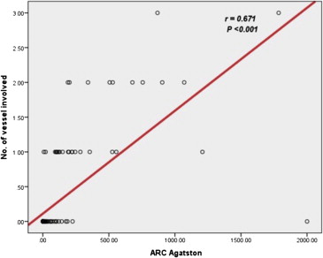
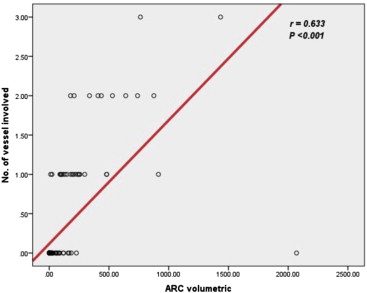
A significant correlation was observed between ARC and total CAC using the Agatston and volumetric scoring methods (r = 0.225; P = .003 and r = 0.243; P = .001, respectively). With regard to individual coronary vessel calcification and ARC, a significant correlation was observed between ARC and left main stem artery calcification calculated using the volumetric and Agatston scoring methods (P < .05). A significant correlation was observed between high ARC and presence of coronary calcified plaque measured using the Agatston and volumetric scoring methods. A strong correlation was also observed between ARC and number of coronary stenotic vessels measured using the Agatston and volumetric scoring methods (r = 0.67; P < .001 and r = 0.63; P < .001, respectively).
Conclusions
ARC can be used as an additional marker to assess coronary atherosclerosis and may have a complementary role with CAC for detection of coronary artery disease.
Recently, multidetector computed tomography (MDCT) has become the preferred imaging tool for coronary artery disease (CAD) and aortic root morphology assessment before aortic valve replacement because of its high temporal and spatial resolution and its reportedly high reproducibility . Various methods and scores have been used to measure arterial calcification by MDCT including Agatston, volume, and mass scores, with significant variability between theses scoring methods in terms of reproducibility and interscan agreement .
Coronary artery calcification (CAC) assessed by MDCT has incremental prognostic values beyond those of traditional cardiac risk factors and scores for cardiovascular disease (CVD) morbidity and mortality reported in several large follow-up studies, and it may help in reclassification of patients at increased risk .
Get Radiology Tree app to read full this article<
Get Radiology Tree app to read full this article<
Get Radiology Tree app to read full this article<
Materials and methods
Get Radiology Tree app to read full this article<
Get Radiology Tree app to read full this article<
Get Radiology Tree app to read full this article<
Get Radiology Tree app to read full this article<
Get Radiology Tree app to read full this article<
CT Scan Protocol
Get Radiology Tree app to read full this article<
Get Radiology Tree app to read full this article<
Aortic Root Calcification Analysis
Get Radiology Tree app to read full this article<
Statistical Analysis
Get Radiology Tree app to read full this article<
Results
Get Radiology Tree app to read full this article<
Table 1
Patient Characteristics
Parameter Number (%) Age (mean ± SD), years 56.3 ± 6 Male 86 (49.1) Female 89 (50.9) Hypertension 95 (54.2) Diabetes 19 (10.8) Hyperlipidemia 32 (18) Smoking 53 (30.2) Family history 20 (11.4) BMI ≥ 30 kg/m 2 46 (26) CAC 0–9 121 (69.1) 10–99 17 (9.7) 100–399 25 (14.3) >400 12 (6.9) ARC 0–9 143 (81.7) 10–99 17 (9.7) 100–399 9 (5.1) >400 6 (3.4)
ARC, aortic root calcification; BMI, body mass index; CAC, coronary artery calcification; SD, standard deviation.
Get Radiology Tree app to read full this article<
Get Radiology Tree app to read full this article<
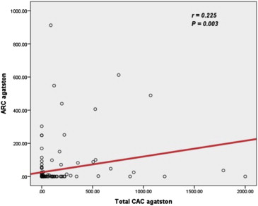
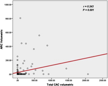
Table 2
Correlations Between ARC and Individual Coronary Artery Calcification Measured Using the Agatston Scoring Method
Agatston Scoring Grade Coronary Artery ARC_P_ Value Total ( n = 175), % ARC ( n = 175), % <10 121 (69.1) 143 (81.7) .006 10–99 17 (9.7) 17 (9.7) 1 100–399 25 (14.3) 9 (5.1) .004 ≥400 12 (6.9) 6 (3.4) .146LCXARC <10 155 (88.6) 143 (81.7) .071 10–99 13 (7.4) 17 (9.7) .445 100–399 5 (2.9) 9 (5.1) .275 ≥400 2 (1.1) 6 (3.4) .153LADARC <10 124 (70.9) 143 (81.7) .017 10–99 21 (12.0) 17 (9.7) .492 100–399 22 (12.6) 9 (5.1) .014 ≥400 8 (4.6) 6 (3.4) .585LMARC <10 170 (97.1) 143 (81.7) <.001 10–99 4 (2.3) 17 (9.7) .003 100–399 1 (0.6) 9 (5.1) .01 ≥400 0 (0.0) 6 (3.4) .013RCAARC <10 155 (88.6) 143 (81.7) .071 10–99 14 (8.0) 17 (9.7) .572 100–399 5 (2.9) 9 (5.1) .275 ≥400 1 (0.6) 6 (3.4) .056
ARC, aortic root calcification; LAD, left anterior descending artery; LCX, left circumflex artery; LM, left main stem artery; RCA, right coronary artery.
Table 3
Correlations Between ARC and Individual Coronary Artery Calcification Measured Using the Volumetric Scoring Method
Volumetric Scoring Grade Coronary Artery ARC_P_ Value Total ( n = 175), n (%) ARC ( n = 175), n (%) <10 119 (68.0) 140 (80.0) .01 10–99 21 (12.0) 18 (10.3) .61 100–399 23 (13.1) 12 (6.9) .05 ≥400 12 (6.9) 5 (2.9) .082LCXARC <10 153 (87.4) 140 (80.0) .06 10–99 13 (7.4) 18 (10.3) .347 100–399 8 (4.6) 12 (6.9) .357 ≥400 1 (0.6) 5 (2.9) .1LADARC <10 122 (69.7) 140 (80.0) .027 10–99 27 (15.4) 18 (10.3) .151 100–399 19 (10.9) 12 (6.9) .188 ≥400 7 (4.0) 5 (2.9) .557LMARC <10 168 (96.0) 140 (80.0) <.001 10–99 6 (3.4) 18 (10.3) .574 100–399 1 (0.6) 12 (6.9) .002 ≥400 0 (0.0) 5 (2.9) .024RCAARC <10 152 (86.9) 140 (80.0) .085 10–99 18 (10.3) 18 (10.3) 1 100–399 4 (2.3) 12 (6.9) .085 ≥400 1 (0.6) 5 (2.9) .1
ARC, aortic root calcification; LAD, left anterior descending artery; LCX, left circumflex artery; LM, left main stem artery; RCA, right coronary artery.
Table 4
Correlations Between ARC and Coronary Calcified Plaques Measured Using the Agatston Method
Calcium Grade Calcified Plaque_P_ Value No ( n = 153), n (%) Yes ( n = 22), n (%) <10 130 (85.0) 13 (59.1) .003 10–99 12 (7.8) 5 (22.7) .028 100–399 8 (5.2) 1 (4.5) .892 ≥400 3 (2.0) 3 (13.6) .03
ARC, aortic root calcification.
Table 5
Correlations Between ARC and Coronary Calcified Plaques Measured Using the Volumetric Scoring Method
Calcium Grade Calcified Plaque_P_ Value No ( n = 153), n (%) Yes ( n = 22), n (%) <10 127 (83.0) 13 (59.1) .009 10–99 14 (9.2) 4 (18.2) .192 100–399 9 (5.9) 3 (13.6) .178 ≥400 3 (2.0) 2 (9.1) .05
ARC, aortic root calcification.
Get Radiology Tree app to read full this article<
Discussion
Get Radiology Tree app to read full this article<
Get Radiology Tree app to read full this article<
Get Radiology Tree app to read full this article<
Get Radiology Tree app to read full this article<
Get Radiology Tree app to read full this article<
Get Radiology Tree app to read full this article<
Get Radiology Tree app to read full this article<
Get Radiology Tree app to read full this article<
Get Radiology Tree app to read full this article<
Conclusion
Get Radiology Tree app to read full this article<
References
1. Feuchtner G.: Imaging of cardiac valves by computed tomography. Scientifica (Cairo) 2013; 2013: pp. 270579.
2. Elkeles R.S.: Coronary artery calcium and cardiovascular risk in diabetes. Atherosclerosis 2010; 210: pp. 331-336.
3. McCollough C.H., Ulzheimer S., Halliburton S.S., et. al.: Coronary artery calcium: a multi-institutional, multimanufacturer international standard for quantification at cardiac CT. Radiology 2007; 243: pp. 527-538.
4. Nasir K., Rubin J., Blaha M.J., et. al.: Interplay of coronary artery calcification and traditional risk factors for the prediction of all-cause mortality in asymptomatic individuals. Circ Cardiovasc Imaging 2012; 5: pp. 467-473.
5. Silverman M.G., Blaha M.J., Krumholz H.M., et. al.: Impact of coronary artery calcium on coronary heart disease events in individuals at the extremes of traditional risk factor burden: the Multi-Ethnic Study of Atherosclerosis. Eur Heart J 2013;
6. Hartmann M., von Birgelen C.: Is there a role for thoracic aortic calcium to fine-tune cardiovascular risk prediction?. Int J Cardiovasc Imaging 2013; 29: pp. 217-219.
7. Wasilewski J., Głowacki J., Poloński L.: Not at random location of atherosclerotic lesions in thoracic aorta and their prognostic significance in relation to the risk of cardiovascular events. Pol J Radiol 2013; 78: pp. 38-42.
8. Kanjanauthai S., Nasir K., Katz R., et. al.: Relationships of mitral annular calcification to cardiovascular risk factors: the Multi-Ethnic Study of Atherosclerosis (MESA). Atherosclerosis 2011; 213: pp. 558-562.
9. Allison M.A., Hsi S,Wassel C.L., Morgan C., et. al.: Calcified atherosclerosis in different vascular beds and the risk of mortality. Arterioscler Thromb Vasc Biol 2012; 32: pp. 140-146.
10. Nasir K., Katz R., Al-Mallah M., et. al.: Relationship of aortic valve calcification with coronary artery calcium severity: the Multi-Ethnic Study of Atherosclerosis (MESA). J Cardiovasc Comput Tomogr 2010; 4: pp. 41-46.
11. Eisen A., Tenenbaum A., Koren-Morag N., et. al.: Calcification of the thoracic aorta as detected by spiral computed tomography among stable angina pectoris patients: association with cardiovascular events and death. Circulation 2008; 118: pp. 1328-1334.
12. Erbel R., MÖhlenkamp S., Moebus S., Heinz Nixdorf Recall Study Investigative Group , et. al.: Coronary risk stratification, discrimination, and reclassification improvement based on quantification of subclinical coronary atherosclerosis: the Heinz Nixdorf Recall study. J Am Coll Cardiol 2010; 56: pp. 1397-1406.
13. Min J.K., Labounty T.M., Gomez M.J., et. al.: Incremental prognostic value of coronary computed tomographic angiography over coronary artery calcium score for risk prediction of major adverse cardiac events in asymptomatic diabetic individuals. Atherosclerosis 2014; 232: pp. 298-304.
14. Kälsch H., Lehmann N., Möhlenkamp S., et. al.: Prevalence of thoracic aortic calcification and its relationship to cardiovascular risk factors and coronary calcification in an unselected population-based cohort: the Heinz Nixdorf Recall Study. Int J Cardiovasc Imaging 2013; 29: pp. 207-216.
15. Takasu J., Budoff M.J., O’Brien K.D., et. al.: Relationship between coronary artery and descending thoracic aortic calcification as detected by computed tomography: the Multi-Ethnic Study of Atherosclerosis. Atherosclerosis 2009; 204: pp. 440-446.
16. Budoff M.J., Nasir K., Katz R., et. al.: Thoracic aortic calcification and coronary heart disease events: the multi-ethnic study of atherosclerosis (MESA). Atherosclerosis 2011; 215: pp. 196-202.
17. Santos R.D., Rumberger J.A., Budoff M.J., et. al.: Thoracic aorta calcification detected by electron beam tomography predicts all-cause mortality. Atherosclerosis 2010; 209: pp. 131-135.
18. Jacobs P.C., Prokop M., van der Graaf Y., et. al.: Comparing coronary artery calcium and thoracic aorta calcium for prediction of all-cause mortality and cardiovascular events on low-dose non-gated computed tomography in a high-risk population of heavy smokers. Atherosclerosis 2010; 209: pp. 455-462.
19. Erbel R., Möhlenkamp S., Kerkhoff G., et. al.: Non-invasive screening for coronary artery disease: calcium scoring. Heart 2007; 93: pp. 1620-1629.
20. Shahzad R., van Walsum T., Schaap M., et. al.: Vessel specific coronary artery calcium scoring: an automatic system. Acad Radiol 2013; 20: pp. 1-9.
21. Qian Z., Anderson H., Marvasty I., et. al.: Lesion- and vessel-specific coronary artery calcium scores are superior to whole-heart Agatston and volume scores in the diagnosis of obstructive coronary artery disease. J Cardiovasc Comput Tomogr 2010; 4: pp. 391-399.
22. Kim J., Bravo P.E., Gholamrezanezhad A., et. al.: Coronary artery and thoracic aorta calcification is inversely related to coronary flow reserve as measured by ⁸²Rb PET/CT in intermediate risk patients. J Nucl Cardiol 2013; 20: pp. 375-384.
23. Ann S.H., Jung J.I., Jung H.O., et. al.: Aortic valve calcium score is associated with coronary calcified plaque burden. Int Heart J 2013; 54: pp. 355-361.
24. Mahabadi A.A., Bamberg F., Toepker M., et. al.: Association of aortic valve calcification to the presence, extent, and composition of coronary artery plaque burden: from the Rule Out Myocardial Infarction using Computer Assisted Tomography (ROMICAT) trial. Am Heart J 2009; 158: pp. 562-568.
25. Utsunomiya H., Yamamoto H., Kunita E., et. al.: Combined presence of aortic valve calcification and mitral annular calcification as a marker of the extent and vulnerable characteristics of coronary artery plaque assessed by 64-multidetector computed tomography. Atherosclerosis 2010; 213: pp. 166-172.
26. Jang S., Yong H.S., Doo K.W., et. al.: Relation of aortic calcification, wall thickness, and distensibility with severity of coronary artery disease: evaluation with coronary CT angiography. Acta Radiol 2012; 53: pp. 839-844.
27. Zweig B.M., Sheth M., Simpson S., et. al.: Association of abdominal aortic calcium with coronary artery calcium and obstructive coronary artery disease: a pilot study. Int J Cardiovasc Imaging 2012; 28: pp. 399-404.
28. Lee H.Y., Song I.S., Yoo S.M., et. al.: Can the extent of epicardial adipose tissue thickness or the presence of descending thoracic aortic calcification predict significant coronary artery stenosis in patients with a zero coronary calcium score on multi-detector CT?. Atherosclerosis 2010; 212: pp. 495-500.
29. Callister T.Q., Cooil B., Raya S.P., et. al.: Coronary artery disease: improved reproducibility of calcium scoring with an electron-beam CT volumetric method. Radiology 1998; 208: pp. 807-814.
30. Hong C., Bae K.T., Pilgram T.K.: Coronary artery calcium: accuracy and reproducibility of measurements with multi-detector row CT–assessment of effects of different thresholds and quantification methods. Radiology 2003; 227: pp. 795-801.
31. Budoff M.J., Achenbach S., Blumenthal R.S., et. al.: Assessment of coronary artery disease by cardiac computed tomography: a scientific statement from the American Heart Association Committee on Cardiovascular Imaging and Intervention, Council on Cardiovascular Radiology and Intervention, and Committee on Cardiac Imaging, Council on Clinical Cardiology. Circulation 2006; 114: pp. 1761-1791.