
Rationale and Objectives
This study evaluated the altered regional cerebral blood flow (rCBF) in resting state in patients with acute posttraumatic stress disorder (PTSD) 3 months after trauma.
Materials and Methods
The rCBF was measured in 30 patients with acute PTSD and 36 healthy controls.
Results
Survivors with acute PTSD showed decreased rCBF, the Clinician-Administered PTSD Scale score correlated negatively with the rCBF, and rCBF at resting state decreased in acute PTSD.
Conclusions
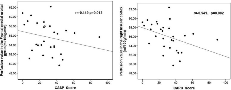
PTSD symptom severity was associated with diminished cerebral blood flow in the right insular cortex and right orbital medial frontal gyrus. The rCBF may predict PTSD symptom severity.
Introduction
Posttraumatic stress disorder (PTSD) is a condition that develops from a sudden, life-threatening, or catastrophic event causing delayed emergence and long-term persistence of mental disorders. Patients experience mental disorders in various ways, including intrusive memories, disturbing recollections, nightmares, flashbacks, distress, and physiological reactions on exposure to reminders of the traumatic event. These disorders seriously affect the patients’ quality of life.
Many functional imaging studies have been performed in patients with chronic PTSD. For example, Semple et al. found increased regional cerebral blood flow (rCBF) in the right amygdala, left parahippocampal gyrus, and occipital cortex when patients performed an auditory continuous performance task. Further, patients with PTSD showed decreased frontal cortex and anterior cingulate activity compared to healthy controls. In a similar study, the researchers found a significantly increased rCBF in the orbitofrontal cortex and a reduced left/right hippocampal perfusion ratio in the PTSD group . The subjects in the foregoing studies included veterans with combat-related PTSD and a history of substance abuse, associated with major effects on brain structure and function . Indeed, many patients with PTSD are often heavily medicated or have current or past alcohol and substance abuse issues .
Get Radiology Tree app to read full this article<
Get Radiology Tree app to read full this article<
Get Radiology Tree app to read full this article<
Get Radiology Tree app to read full this article<
Get Radiology Tree app to read full this article<
Materials and Methods
Subjects
Get Radiology Tree app to read full this article<
Get Radiology Tree app to read full this article<
Imaging Data Acquisition
Structural MRI Data
Get Radiology Tree app to read full this article<
Perfusion Data
Get Radiology Tree app to read full this article<
Image Processing
Structural MRI Data
Get Radiology Tree app to read full this article<
Perfusion Data
Get Radiology Tree app to read full this article<
Smoothing
Get Radiology Tree app to read full this article<
Masking
Get Radiology Tree app to read full this article<
ROI Generation
Get Radiology Tree app to read full this article<
Statistical Analyses
Get Radiology Tree app to read full this article<
Get Radiology Tree app to read full this article<
Results
Patient Demographics
Get Radiology Tree app to read full this article<
Table 1
Demographics of Physically Healthy Trauma Survivors and Normal Controls
Characteristics PTSD ( n = 30) NC ( n = 36)P Sex, n (%) 13:17 16:20 0.90 Female 13(43) 16(44) Male 17(57) 20(56) Mean age (y) 33.3 ± 9.8 34.4 ± 7.8 0.57 Mean education (y) 7.6 ± 2.2 10.5 ± 1.3 0.01 \* Days after trauma 91.5 ± 1.1 CAPS Mean 49.9 ± 27.4 Female 34.021.8 Male 26.3 ± 15.9 Ethnicity Han (Chinese) Han (Chinese)
CAPS, Clinician-Administered PTSD Scale; NC, normal controls; PTSD, posttraumatic stress disorder.
Data are presented as mean ± SD. Age and gender had no significant difference between subjects with PTSD and healthy controls by independent t tests analysis of covariance (ANCOVA) ( P > 0.05).
Get Radiology Tree app to read full this article<
Get Radiology Tree app to read full this article<
Get Radiology Tree app to read full this article<
Table 2
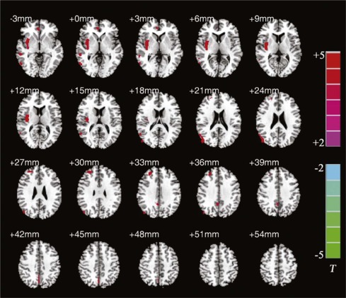
Decreased rCBF in Various Brain Regions of Patients with PTSD
Regions Mean ± SD in mL/100 g/min Talairach_T__Z_ PTSD ( n = 30) HC ( n = 36) x yy zz R TL 55.877 ± 3.043 107.793 ± 19.020 58 32 2 4.72 4.34 MFG 53.062 ± 8.963 66.450 ± 15.719 28 44 34 4.54 4.19 OMFG 55.666 ± 3.267 85.186 ± 19.068 4 46 −2 4.20 3.92 INS 56.085 ± 2.991 138.226 ± 22.917 36 −10 10 5.24 4.73 MOG 58.097 ± 3.351 96.672 ± 15.239 48 −74 24 5.40 4.85 Pcu 60.466 ± 3.899 123.065 ± 18.058 4 −72 44 4.26 3.97 L TL 33.605 ± 2.810 59.331 ± 10.512 −66 −18 −14 4.22 3.93
INS, insula; L, left; MFG, middle frontal gyrus; MOG, middle occipital gyrus; OMFG, orbital medial frontal gyrus; Pcu, precuneus; R, right; TL, temporal lobe.
Group comparisons were performed by analysis of two-sample t tests in SPM5 with the PTSD group as a fixed factor and age and gender as covariates. The results were compared to a threshold using a false discovery rate (FDR)–corrected P value < 0.05.
Get Radiology Tree app to read full this article<
Correlation of Symptom Severity with Perfusion
Get Radiology Tree app to read full this article<
Table 3
Correlation Between rCBF Values and CAPS Score
Regions CAPS r_P_ R TL 0.027 0.886 MFG 0.132 0.488 OMFG −0.231 \* 0.219 \* INS −0.396 \* 0.030 \* MOG 0.040 0.832 Pcu 0.112 0.556 L TL 0.312 0.094
INS, insula; L, left; MFG, middle frontal gyrus; MOG, middle occipital gyrus; OMFG, orbital medial frontal gyrus; Pcu, precuneus; R, right; TL, temporal lobe.
Region of in (ROI)-based correlational analysis was performed using Pearson partial correlation analysis. Age and gender were treated as controlling covariates. Significance levels were set at P < 0.05.
Get Radiology Tree app to read full this article<
Get Radiology Tree app to read full this article<
Discussion
Get Radiology Tree app to read full this article<
Get Radiology Tree app to read full this article<
Get Radiology Tree app to read full this article<
Get Radiology Tree app to read full this article<
Get Radiology Tree app to read full this article<
Get Radiology Tree app to read full this article<
Get Radiology Tree app to read full this article<
Conclusions
Get Radiology Tree app to read full this article<
Acknowledgments
Get Radiology Tree app to read full this article<
Get Radiology Tree app to read full this article<
References
1. Semple W.E., Goyer P.F., McCormick R., et. al.: Higher brain blood flow at amygdala and lower frontal cortex blood flow in PTSD patients with comorbid cocaine and alcohol abuse compared with normals. Psychiatry 2000; 63: pp. 65-74.
2. Semple W.E., Goyer P., McCormick R., et. al.: Preliminary report: brain blood flow using PET in patients with posttraumatic stress disorder and substance-abuse histories. Biol Psychiatry 1993; 34: pp. 115-118.
3. Franklin T.R., Acton P.D., Maldjian J.A., et. al.: Decreased gray matter concentration in the insular, orbitofrontal, cingulate, and temporal cortices of cocaine patients. Biol Psychiatry 2002; 51: pp. 134-142.
4. Chung Y.A., Kim S.H., Chung S.K., et. al.: Alterations in cerebral perfusion in posttraumatic stress disorder patients without re-exposure to accident-related stimuli. Clin Neurophysiol 2006; 117: pp. 637-642.
5. Bonne O., Gilboa A., Louzoun Y., et. al.: Resting regional cerebral perfusion in recent posttraumatic stress disorder. Biol Psychiatry 2003; 54: pp. 1077-1086.
6. Lucey J.V., Costa D.C., Adshead G., et. al.: Brain blood flow in anxiety disorders. OCD, panic disorder with agoraphobia, and post-traumatic stress disorder on 99mTcHMPAO single photon emission tomography (SPET). Br J Psychiatry 1997; 171: pp. 346-350.
7. Lindauer R.J., Booij J., Habraken J.B., et. al.: Cerebral blood flow changes during script-driven imagery in police officers with posttraumatic stress disorder. Biol Psychiatry 2004; 56: pp. 853-861.
8. Pagani M., Hogberg G., Salmaso D., et. al.: Regional cerebral blood flow during auditory recall in 47 subjects exposed to assaultive and non-assaultive trauma and developing or not posttraumatic stress disorder. Eur Arch Psychiatry Clin Neurosci 2005; 255: pp. 359-365.
9. Zubieta J.K., Chinitz J.A., Lombardi U., et. al.: Medial frontal cortex involvement in PTSD symptoms: a SPECT study. J Psychiatr Res 1999; 33: pp. 259-264.
10. Bremner J.D., Staib L.H., Kaloupek D., et. al.: Neural correlates of exposure to traumatic pictures and sound in Vietnam combat veterans with and without posttraumatic stress disorder: a positron emission tomography study. Biol Psychiatry 1999; 45: pp. 806-816.
11. Bremner J.D., Vermetten E., Vythilingam M., et. al.: Neural correlates of the classic color and emotional Stroop in women with abuse-related posttraumatic stress disorder. Biol Psychiatry 2004; 55: pp. 612-620.
12. Shin L.M., Kosslyn S.M., McNally R.J., et. al.: Visual imagery and perception in posttraumatic stress disorder. A positron emission tomographic investigation. Arch Gen Psychiatry 1997; 54: pp. 233-241.
13. Schuff N., Zhang Y., Zhan W., et. al.: Patterns of altered cortical perfusion and diminished subcortical integrity in posttraumatic stress disorder: an MRI study. Neuroimage 2011; 54: pp. S62-S68.
14. Chen Y., Fu K., Feng C., et. al.: Different regional gray matter loss in recent onset PTSD and non PTSD after a single prolonged trauma exposure. PLoS ONE 2012; 7: e48298
15. Detre J.A., Wang J., Wang Z., et. al.: Arterial spin-labeled perfusion MRI in basic and clinical neuroscience. Curr Opin Neurol 2009; 22: pp. 348-355.
16. Petersen E.T., Mouridsen K., Golay X., et. al.: The QUASAR reproducibility study, Part II: results from a multi-center Arterial Spin Labeling test-retest study. Neuroimage 2010; 49: pp. 104-113.
17. Pfefferbaum A., Chanraud S., Pitel A.L., et. al.: Volumetric cerebral perfusion imaging in healthy adults: regional distribution, laterality, and repeatability of pulsed continuous arterial spin labeling (PCASL). Psychiatry Res 2010; 182: pp. 266-273.
18. Guidelines for psychiatric practice in public sector psychiatric inpatient facilities. Committee on State and Community Psychiatric Systems of the Council on Psychiatric Services. American Psychiatric Association. Am J Psychiatry 1994; 151: pp. 797-798.
19. Loewy R.L., Bearden C.E., Johnson J.K., et. al.: The prodromal questionnaire (PQ): preliminary validation of a self-report screening measure for prodromal and psychotic syndromes. Schizophr Res 2005; 79: pp. 117-125.
20. Detre J.A., Leigh J.S., Williams D.S., et. al.: Perfusion imaging. Magn Reson Med 1992; 23: pp. 37-45.
21. Good C.D., Johnsrude I.S., Ashburner J., et. al.: A voxel-based morphometric study of ageing in 465 normal adult human brains. Neuroimage 2001; 14: pp. 21-36.
22. Rousset O.G., Ma Y., Evans A.C.: Correction for partial volume effects in PET: principle and validation. J Nucl Med 1998; 39: pp. 904-911.
23. Quarantelli M., Berkouk K., Prinster A., et. al.: Integrated software for the analysis of brain PET/SPECT studies with partial-volume-effect correction. J Nucl Med 2004; 45: pp. 192-201.
24. Rauch S.L., van der Kolk B.A., Fisler R.E., et. al.: A symptom provocation study of posttraumatic stress disorder using positron emission tomography and script-driven imagery. Arch Gen Psychiatry 1996; 53: pp. 380-387.
25. Geschwind N., Galaburda A.M.: Cerebral lateralization. Biological mechanisms, associations, and pathology: I. A hypothesis and a program for research. Arch Neurol 1985; 42: pp. 428-459.
26. Nitschke J.B., Heller W., Palmieri P.A., et. al.: Contrasting patterns of brain activity in anxious apprehension and anxious arousal. Psychophysiology 1999; 36: pp. 628-637.
27. Davis M., Whalen P.J.: The amygdala: vigilance and emotion. Mol Psychiatry 2001; 6: pp. 13-34.
28. Chen S., Xia W., Li L., et. al.: Gray matter density reduction in the insula in fire survivors with posttraumatic stress disorder: a voxel-based morphometric study. Psychiatry Res 2006; 146: pp. 65-72.
29. Molina M.E., Isoardi R., Prado M.N., et. al.: Basal cerebral glucose distribution in long-term post-traumatic stress disorder. World J Biol Psychiatry 2010; 11: pp. 493-501.
30. Vermetten E., Schmahl C., Southwick S.M., et. al.: Positron tomographic emission study of olfactory induced emotional recall in veterans with and without combat-related posttraumatic stress disorder. Psychopharmacol Bull 2007; 40: pp. 8-30.
31. Augustine J.R.: Circuitry and functional aspects of the insular lobe in primates including humans. Brain Res Brain Res Rev 1996; 22: pp. 229-244.
32. Dalgleish T.: The emotional brain. Nat Rev Neurosci 2004; 5: pp. 583-589.
33. Mullan S., Penfield W.: Illusions of comparative interpretation and emotion; production by epileptic discharge and by electrical stimulation in the temporal cortex. AMA Arch Neurol Psychiatry 1959; 81: pp. 269-284.
34. Damasio A.R., Grabowski T.J., Bechara A., et. al.: Subcortical and cortical brain activity during the feeling of self-generated emotions. Nat Neurosci 2000; 3: pp. 1049-1056.
35. Hull A.M.: Neuroimaging findings in post-traumatic stress disorder. Systematic review. Br J Psychiatry 2002; 181: pp. 102-110.
36. Bremner J.D.: Does stress damage the brain?. Biol Psychiatry 1999; 45: pp. 797-805.
37. Zhang X., Zhang J., Wang L., et. al.: Altered resting-state functional connectivity of the amygdala in Chinese earthquake survivors. Prog Neuropsychopharmacol Biol Psychiatry 2016; 65: pp. 208-214.
38. Margulies D.S., Vincent J.L., Kelly C., et. al.: Precuneus shares intrinsic functional architecture in humans and monkeys. Proc Natl Acad Sci USA 2009; 106: pp. 20069-20074.
39. Gusnard D.A., Raichle M.E., Raichle M.E.: Searching for a baseline: functional imaging and the resting human brain. Nat Rev Neurosci 2001; 2: pp. 685-694.
40. Vogt B.A., Finch D.M., Olson C.R.: Functional heterogeneity in cingulate cortex: the anterior executive and posterior evaluative regions. Cereb Cortex 1992; 2: pp. 435-443.
41. Yin Y., Li L., Jin C., et. al.: Abnormal baseline brain activity in posttraumatic stress disorder: a resting-state functional magnetic resonance imaging study. Neurosci Lett 2011; 498: pp. 185-189.
42. Barkay G., Freedman N., Lester H., et. al.: Brain activation and heart rate during script-driven traumatic imagery in PTSD: preliminary findings. Psychiatry Res 2012; 204: pp. 155-160.