
Rationale and Objectives
Diffraction-enhanced imaging (DEI) is a new x-ray imaging modality that differs from conventional radiography in its use of three physical mechanisms to generate contrast. DEI is able to generate contrast from x-ray absorption, refraction, and ultra-small-angle scatter rejection (extinction) to produce high-contrast images with a much lower radiation dose compared to conventional radiography.
Materials and Methods
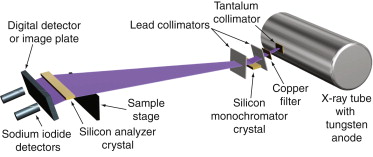
A prototype DEI system was constructed using a 1-kW tungsten x-ray tube and a single silicon monochromator and analyzer crystal. The monochromator crystal was aligned to reflect the combined Kα1 (59.32 keV) and Kα2 (57.98 keV) characteristic emission lines of tungsten using a tube voltage of 160 kV. System performance and demonstration of contrast were evaluated using a nylon monofilament refraction phantom, full-thickness breast specimens, a human thumb, and a live mouse.
Results
Images acquired using this system successfully demonstrated all three DEI contrast mechanisms. Flux measurements acquired using this 1-kW prototype system demonstrated that this design can be scaled to use a more powerful 60-kW x-ray tube to generate similar images with an image time of approximately 30 seconds. This single–crystal pair design can be further modified to allow for an array of crystals to reduce clinical image times to <3 seconds.
Conclusions
This paper describes the design, construction, and performance of a new DEI system using a commercially available tungsten anode x-ray tube and includes the first high-quality low-dose diffraction-enhanced images of full-thickness human tissue specimens.
Since Roentgen’s discovery in 1895, x-ray imaging has relied on the varied attenuation of photons to generate images. Diffraction-enhanced imaging (DEI) is a new x-ray imaging modality that uses two additional contrast mechanisms, refraction and ultra-small-angle scatter rejection (extinction) . Previous imaging of breast cancer specimens , articular cartilage , and small animals with synchrotron-based DEI systems has shown improvement in fine detail in images compared to conventional radiography, with applications ranging from industrial inspection to medical imaging .
In conventional radiography, contrast between different tissues is provided by their differential x-ray attenuation. For soft tissues, with adjacent materials having similar electron densities, achieving adequate contrast with conventional x-ray imaging requires imaging at low x-ray energies (approximately 20 keV) to take maximal advantage of small differences in the effective atomic number through the photoelectric effect. Unfortunately, the attenuation of x-rays increases as energy is lowered. To obtain adequate transmission through the body and produce an image with an acceptable signal-to-noise ratio, more incident radiation must be used when imaging at low energies, resulting in a higher absorbed dose to patients.
Get Radiology Tree app to read full this article<
Get Radiology Tree app to read full this article<
Get Radiology Tree app to read full this article<
Get Radiology Tree app to read full this article<
Get Radiology Tree app to read full this article<
Get Radiology Tree app to read full this article<
Get Radiology Tree app to read full this article<
Get Radiology Tree app to read full this article<
Get Radiology Tree app to read full this article<
Materials and methods
Get Radiology Tree app to read full this article<
Get Radiology Tree app to read full this article<
Get Radiology Tree app to read full this article<
Get Radiology Tree app to read full this article<
Get Radiology Tree app to read full this article<
Get Radiology Tree app to read full this article<
Get Radiology Tree app to read full this article<
Get Radiology Tree app to read full this article<
Get Radiology Tree app to read full this article<
Results
Get Radiology Tree app to read full this article<
![Figure 2, Nylon refraction phantom and measured postanalyzer rocking curves. The diameters of the nylon monofilaments from largest to smallest are 560, 360, 200, and 100 μm. Phantom images were acquired at −0.6 microradians (a) and +0.6 microradians (c) on the rocking curve using a tube voltage of 160 kV and 6.2 mA with a surface dose of 0.04 mGy. A measured postanalyzer rocking curve (b) using the Bragg [111] and Bragg [333] reflections is presented to demonstrate system performance.](https://storage.googleapis.com/dl.dentistrykey.com/clinical/DesignandImplementationofaCompactLowDoseDiffractionEnhancedMedicalImagingSystem/1_1s20S1076633209001366.jpg)
Get Radiology Tree app to read full this article<
Get Radiology Tree app to read full this article<
Get Radiology Tree app to read full this article<
Get Radiology Tree app to read full this article<
Get Radiology Tree app to read full this article<
Get Radiology Tree app to read full this article<
Discussion
Get Radiology Tree app to read full this article<
Get Radiology Tree app to read full this article<
Get Radiology Tree app to read full this article<
Get Radiology Tree app to read full this article<
Conclusion
Get Radiology Tree app to read full this article<
Acknowledgments
Get Radiology Tree app to read full this article<
Get Radiology Tree app to read full this article<
References
1. Chapman D., Thomlinson W., Johnston R.E., et. al.: Diffraction enhanced x-ray imaging. Phys Med Biol 1997; 42: pp. 2015-2025.
2. Bravin A., Keyrilainen J., Fernandez M., et. al.: High-resolution CT by diffraction-enhanced x-ray imaging: mapping of breast tissue samples and comparison with their histo-pathology. Phys Med Biol 2007; 52: pp. 2197-2211.
3. Fiedler S., Bravin A., Keyrilainen J., et. al.: Imaging lobular breast carcinoma: comparison of synchrotron radiation DEI-CT technique with clinical CT, mammography and histology. Phys Med Biol 2004; 49: pp. 175-188.
4. Kiss M.Z., Sayers D.E., Zhong Z., Parham C., Pisano E.D.: Improved image contrast of calcifications in breast tissue specimens using diffraction enhanced imaging. Phys Med Biol 2004; 49: pp. 3427-3439.
5. Pisano E.D., Johnston R.E., Chapman D., et. al.: Human breast cancer specimens: diffraction-enhanced imaging with histologic correlation—improved conspicuity of lesion detail compared with digital radiography. Radiology 2000; 214: pp. 895-901.
6. Mollenhauer J., Aurich M.E., Zhong Z., et. al.: Diffraction-enhanced x-ray imaging of articular cartilage. Osteoarthritis Cartilage 2002; 10: pp. 163-171.
7. Muehleman C., Li J., Zhong Z.: Preliminary study on diffraction enhanced radiographic imaging for a canine model of articular cartilage. Osteoarthritis Cartilage 2006; 14: pp. 882-888.
8. Kitchen M.J., Lewis R.A., Yagi N., et. al.: Phase contrast x-ray imaging of mice and rabbit lungs: a comparative study. Br J Radiol 2005; 78: pp. 1018-1027.
9. Mannan K.A., Schultke E., Menk R.H., et. al.: Synchrotron supported DEI/KES of a brain tumor in an animal model: the search for a microimaging modality. Nucl Instrum Methods Phys Res A 2004; 548: pp. 106-110.
10. Zhong Z., Thomlinson W., Chapman D., Sayers D.: Implementation of diffraction-enhanced imaging experiments at the NSLS and APS. Nucl Instrum Methods Phys Res A 2000; 450: pp. 556-567.
11. Hasnah M.O., Zhong Z., Oltulu O., et. al.: Diffraction enhanced imaging contrast mechanisms in breast cancer specimens. Med Phys 2002; 29: pp. 2216-2221.
12. Lewis R.A., Hall C.J., Hufton A.P., et. al.: X-ray refraction effects: application to the imaging of biological tissues. Br J Radiol 2003; 76: pp. 301-308.
13. Muehleman C., Sumner D.R., Zhong Z.: Refraction effects of diffraction enhanced radiographic imaging—a new look at bone. J Am Podiatr Med Assoc 2004; 94: pp. 453-455.
14. Pagot E., Fiedler S., Cloetens P., et. al.: Quantitative comparison between two phase contrast techniques: diffraction enhanced imaging and phase propagation imaging. Phys Med Biol 2005; 50: pp. 709-724.
15. Ingal V.N., Beliaevskaya E.A., Brianskaya A.P., Merkurieva R.D.: Phase mammography—a new technique for breast investigation. Phys Med Biol 1998; 43: pp. 2555-2567.
16. Pfeiffer F., Weitkamp T., Bunk O., David C.: Phase retrieval and differential phase-contrast imaging with low-brilliance x-ray sources. Nature Phys 2006; 2: pp. 258-261.
17. Pfeiffer F., Bech M., Bunk O., et. al.: Hard-x-ray dark-field imaging using a grating interferometer. Nature Mater 2008; 7: pp. 134-137.
18. David C., Bruder J., Rohbeck T., et. al.: Fabrication of diffraction gratings for hard x-ray phase contrast imaging. Microelectron Eng 2007; 84: pp. 1172-1177.
19. Myers G.R., Mayo S.C., Gureyev T.E., Paganin D.M., Wilkins S.W.: Polychromatic cone-beam phase-contrast tomography. Phys Rev A 2007; 76: pp. 1-4.
20. Keyrilainen J., Fernandez M., Fiedler S., et. al.: Visualization of calcifications and thin collagen strands in human breast tumour specimens by the diffraction-enhanced imaging technique: a comparison with conventional mammography and histology. Eur J Radiol 2005; 53: pp. 226-237.
21. Boone J.M.: Glandular breast dose for monoenergetic and high-energy x-ray beams: Monte Carlo assessment. Radiology 1999; 213: pp. 23-37.
22. Yaffe M.J.: Direct digital mammography using a scanned-slot CCD imaging-system. Med Progr Technol 1993; 19: pp. 13-21.
23. Nishikawa R.M., Mawdsley G.E., Fenster A., Yaffe M.J.: Scanned-projection digital mammography. Med Phys 1987; 14: pp. 717-727.
24. Amis E.S., Butler P.F., Applegate K.E., et. al.: American College of Radiology white paper on radiation dose in medicine. J Am Coll Radiol 2007; 4: pp. 272-284.
25. Brenner D.J., Hall E.J.: Computed tomography—an increasing source of radiation exposure. N Engl J Med 2007; 357: pp. 2277-2284.
26. Dilmanian F.A., Zhong Z., Ren B., et. al.: Computed tomography of x-ray index of refraction using the diffraction enhanced imaging method. Phys Med Biol 2000; 45: pp. 933-946.
27. Hashimoto E., Maksimenko A., Sugiyama H., et. al.: First application of x-ray refraction-based computed tomography to a biomedical object. Zoolog Sci 2006; 23: pp. 809-813.