
Rational and Objectives
We evaluated the ability of 64-slice computed tomography (CT), conventional cine-angiography, and intravascular ultrasound (IVUS) to detect stent fractures under ideal conditions. Coronary stent fracture has been implicated as one of the mechanisms of stent thrombosis and, perhaps, in-stent restenosis. However, the preferred imaging modality in detecting fractures in coronary stents has not been well established.
Materials and Methods
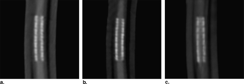
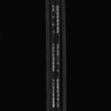
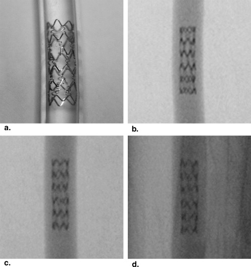
Four different types of commonly used coronary stents (Cypher, Taxus, Vision, Hepacoat) each with three strut fractures (Cypher, 5; Taxus, 5; Vision, 4; Hepacoat, 5) were nominally deployed in polyurethane tubes and imaged with 64-slice CT, conventional cine-angiography, and IVUS. For each stent type, an unfractured control stent was also imaged.
Results
Overall accuracy (84.1% vs. 73.9%), sensitivity (80.7 vs. 77.2%), and specificity (100% vs. 58.3%) for stent fracture detection was higher with 64 multislice CT compared to conventional cine-angiography. Stent fractures were not accurately detected by IVUS. Fracture detection by multislice CT was best when the stents were imaged at 45° to the z-axis.
Conclusions
Under ideal in vitro conditions, CT has a high accuracy when used to evaluate coronary stent fractures. The overall accuracy, sensitivity, and specificity of detecting stent fractures are lower by conventional cine-angiography. Stent fractures were not detected using IVUS.
The medical use of stents has had a significant impact on the treatment of a wide array of medical conditions. Stents can be used in both vascular and nonvascular structures (eg, gastrointestinal/biliary tract, bronchial tree); stent fracture is a known complication in both types of settings . Excessive mechanical stress from extreme flexion of the vessel or compression by tissue has been proposed as a possible cause of some of these fractures . Coronary stents, which were reserved for bailout situations when they were first developed, are now used in more than 80% of the approximately 1,000,000 percutaneous coronary interventions performed annually in the United States . The efficacy of bare metal stents is limited by the occurrence of in-stent restenosis, which ranges from 15% to 35% in unselected cases, depending on lesion morphology and concomitant medical conditions .
The development of drug-eluting stents has proven to be very effective in suppressing neo-intimal proliferation and reducing subsequent in-stent restenosis . However, stent fracture is increasingly being reported as a complication of drug-eluting stent placement, and is believed to play a significant role in some cases of stent thrombosis as well as in-stent restenosis . Although most coronary stent fractures reported in the literature represent complete avulsion fractures detected by intravascular ultrasound (IVUS), there are a paucity of data on the preferred imaging modality for detection of individual strut fractures, which likely represent precursor events before complete stent avulsion. We therefore sought to compare the ability of several different imaging modalities to detect stent fractures.
Materials and methods
Get Radiology Tree app to read full this article<
Table 1
List of Stent Types Used
Stent type Size (mm) Strut thickness (mm) Hounsfield Density Vision 3.0 × 23 0.08 1091 ± 72 Hepacoat 3.5 × 8, 3.5 × 13 0.14 700 ± 22 Taxus 2.75 × 12 0.13 937 ± 50 Cypher 3.0 × 23 0.18 1025 ± 35
Get Radiology Tree app to read full this article<
Get Radiology Tree app to read full this article<
CT Angiography
Get Radiology Tree app to read full this article<
Conventional Cine-angiography
Get Radiology Tree app to read full this article<
Intravascular Ultrasound
Get Radiology Tree app to read full this article<
Image Analysis
Get Radiology Tree app to read full this article<
Statistical Analysis
Get Radiology Tree app to read full this article<
Results
Get Radiology Tree app to read full this article<
Table 2
Detection Rate of Any Stent Fracture by Stent Type and Imaging Modality
64-slice Computed Tomography Cypher Taxus Vision Hepacoat Combined Overall accuracy 83.3% 100% 66.7% 83.3% 84.1% Sensitivity 80.0% 100% 58.3% 80.0% 80.7% Specificity 100% 100% 100% 100% 100% Cine-angiography Overall accuracy 66.7% 100% 80% 50% 73.9% Sensitivity 80.0% 100% 75.0% 53.3% 77.2% Specificity 0% 100% 100% 33.3% 58.3% Intravascular ultrasound Nonvisible Nonvisible Nonvisible Nonvisible
Table 3
Detection Rate of Exactly Three Strut Fractures by Stent Type and Imaging Modality
64-Slice Computed Tomography Cypher Taxus Vision Hepacoat Combined Overall accuracy 61.1% 44.4% 27.8% 27.8% 42% Sensitivity 53.3% 33.3% 16.7% 13.3% 29.8% Specificity 100% 100% 100% 100% 100% Cine-angiography Overall accuracy 11.1% 44.4% 26.7% 16.7% 24.6% Sensitivity 13.3% 33.3% 8.3% 13.3% 17.5% Specificity 0% 100% 100% 33.3% 58.3% intravascular ultrasound Nonvisible Nonvisible Nonvisible Nonvisible
Table 4
Composite Accuracy of Detection of Stent Fracture by Varying Scan Angle Using Computed Tomography
Scan Angle (z) Sensitivity Specificity Accuracy 0° 78.9% 100% 82.6% 45° 84.2% 100% 87.0% 90° 78.9% 100% 82.6
Table 5
Accuracy of Detection of Strut Fracture by Varying Scan Angle and Stent Type Using Multislice Computed Tomography
Scan Angle (z) Cypher Taxus Vision Hepacoat Combined Accuracy 0° 73.0% 73.0% 50.0% 47.0% 66.7% 45° 73.0% 87.0% 42.0% 73.0% 73.9% 90° 60.0% 80.0% 42.0% 40.0% 63.8%
Get Radiology Tree app to read full this article<
Discussion
Get Radiology Tree app to read full this article<
Get Radiology Tree app to read full this article<
Get Radiology Tree app to read full this article<
Get Radiology Tree app to read full this article<
Get Radiology Tree app to read full this article<
Get Radiology Tree app to read full this article<
Get Radiology Tree app to read full this article<
Get Radiology Tree app to read full this article<
Get Radiology Tree app to read full this article<
References
1. Donahue D.G., Saltzman J.R., Krims P.: Stent fracture in malignant biliary obstruction. Gastrointest Endosc 1993; 39: 864–685
2. Noppen M., Stratakos G., D’haese J., et. al.: Removal of covered self-expandable metallic airway stents in benign disorders: indications, technique, and outcomes. Chest 2005; 127: pp. 482-487.
3. Laird J.R.: Limitations of percutaneous transluminal angioplasty and stenting for the treatment of disease of the superficial femoral and popliteal arteries. J Endovasc Ther 2006; 13: pp. II30-II40.
4. Iida O., Nanto S., Uematsu M., et. al.: Effect of exercise on frequency of stent fracture in the superficial femoral artery. Am J Cardiol 2006; 98: pp. 272-274.
5. Mitka M.: Progress in percutaneous heart procedures leads to update in clinical guidelines. JAMA 2006; 295: pp. 263-264.
6. Kimura T., Yokoi H., Nakagawa Y., et. al.: Three-year follow-up after implantation of metallic coronary-artery stents. N Engl J Med 1996; 334: pp. 561-566.
7. Moses J.W., Leon M.B., Popma J.J., et. al.: Sirolimus-eluting stents versus standard stents in patients with stenosis in a native coronary artery. N Engl J Med 2003; 349: pp. 1315-1323.
8. Lee M.S., Jurewitz D., Aragon J., et. al.: Stent fracture associated with drug-eluting stents: clinical characteristics and implications. Cathet Cardiovasc Interv 2007; 69: pp. 387-394.
9. Sun Z., Davidson R., Lin C.H.: Multi-detector row CT angiography in the assessment of coronary in-stent restenosis: a systematic review. Eur J Radiol 2007; Dec 25 (Epub ahead of print)
10. Seifarth H., Ozgun M., Raupach R., et. al.: 64- Versus 16-slice CT angiography for coronary artery stent assessment: in vitro experience. Invest Radiol 2006; 41: pp. 22-27.
11. Maintz D., Juergens K.U., Wichter T., et. al.: Imaging of coronary artery stents using multislice computed tomography: in vitro evaluation. Eur Radiol 2003; 13: pp. 830-835.
12. Rist C., Nikolaou K., Flohr T., et. al.: High-resolution ex vivo imaging of coronary artery stents using 64-slice computed tomography—initial experience. Eur Radiol 2006; 16: pp. 1564-1569.