
Rationale and Objectives
The purpose of this study was to assess the implementation of a digital anatomy lecture series based largely on annotated, radiographic images and the utility of the Radiological Society of North America–developed Medical Imaging Resource Center (MIRC) for providing an online educational resource.
Materials and Methods
A series of digital teaching images were collected and organized to correspond to lecture and dissection topics. MIRC was used to provide the images in a Web-based educational format for incorporation into anatomy lectures and as a review resource. A survey assessed the impressions of the medical students regarding this educational format.
Results
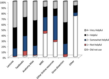
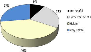
MIRC teaching files were successfully used in our teaching approach. The lectures were interactive with questions to and from the medical student audience regarding the labeled images used in the presentation. Eighty-five of 120 students completed the survey. The majority of students (87%) indicated that the MIRC teaching files were “somewhat useful” to “very useful” when incorporated into the lecture. The students who used the MIRC files were most likely to access the material from home (82%) on an occasional basis (76%). With regard to areas for improvement, 63% of the students reported that they would have benefited from more teaching files, and only 9% of the students indicated that the online files were not user friendly.
Conclusions
The combination of electronic radiology resources available in lecture format and on the Internet can provide multiple opportunities for medical students to learn and revisit first-year anatomy. MIRC provides a user-friendly format for presenting radiology education files for medical students.
Over the past century, there has been seemingly exponential growth in the breadth of material that has been incorporated into modern medical school curricula. The addition of new material and courses has led to limited instruction time available for existing basic science courses . Although the importance of basic science in the medical curriculum remains undisputed, there is a recognized need for training that is efficient yet thorough. A number of medical schools across the country have begun to examine their instructional methods and to revise and reform curricula. Modern electronic resources provide unique capabilities for presenting and disseminating educational information for course work and for study and have served as a platform for much of this reform.
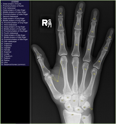
Among the basic science courses, gross anatomy represents a unique opportunity for the incorporation of technology and electronic dissemination of information because of the visual nature of the course material. Since the time that radiography of cadavers was first proposed as a teaching tool for anatomy more than 15 years ago , the role of radiology in supplementing anatomy teaching has continued to expand as digitization has allowed greater access to images. Electronic presentations of radiographs, computed tomographic images, ultrasonographic images, and magnetic resonance images have fast become a standard platform for teaching medical students first-year gross anatomy . In addition to the technologies that have enabled the electronic display of radiographic imaging, other new technologies have been developed that allow manipulation of the images with annotations and labeling and provide the capability to distribute the images on Web-based platforms that can be viewed by medical students at various locations, such as on-campus medical student computer labs, stations that are incorporated into anatomy dissection stations, and home personal computers. Although electronic formats for anatomy education are being incorporated into gross anatomy at medical schools across the country, few studies have reported the utility of these resources and medical students’ impressions of these resources.
Get Radiology Tree app to read full this article<
Get Radiology Tree app to read full this article<
Materials and methods
Get Radiology Tree app to read full this article<
Table 1
Categories Used to Characterize the Electronic Radiographic Teaching Files
Indexed Content Indexed Field Name Anatomy Appendicular Axial Organ system Abdomen Cardiovascular Chest Gastrointestinal Genitourinary Musculoskeletal Neurology Pulmonary Vascular Modalities Angiography Radiography Computed tomography Magnetic resonance Ultrasound
Get Radiology Tree app to read full this article<
Get Radiology Tree app to read full this article<
Get Radiology Tree app to read full this article<
Get Radiology Tree app to read full this article<
Get Radiology Tree app to read full this article<
Get Radiology Tree app to read full this article<
Get Radiology Tree app to read full this article<
Get Radiology Tree app to read full this article<
Get Radiology Tree app to read full this article<
Get Radiology Tree app to read full this article<
Results
Get Radiology Tree app to read full this article<
Get Radiology Tree app to read full this article<
Get Radiology Tree app to read full this article<
Get Radiology Tree app to read full this article<
Get Radiology Tree app to read full this article<
Get Radiology Tree app to read full this article<
Get Radiology Tree app to read full this article<
Get Radiology Tree app to read full this article<
Get Radiology Tree app to read full this article<
Discussion
Get Radiology Tree app to read full this article<
Get Radiology Tree app to read full this article<
Get Radiology Tree app to read full this article<
Get Radiology Tree app to read full this article<
Get Radiology Tree app to read full this article<
Get Radiology Tree app to read full this article<
Get Radiology Tree app to read full this article<
Get Radiology Tree app to read full this article<
Get Radiology Tree app to read full this article<
Get Radiology Tree app to read full this article<
Appendix
Get Radiology Tree app to read full this article<
Get Radiology Tree app to read full this article<
 Open full size image
Get Radiology Tree app to read full this article<
Get Radiology Tree app to read full this article<
 Open full size image
Get Radiology Tree app to read full this article<
References
1. Drake R.L., McBride J.M., Lachman N., et. al.: Medical education in the anatomical sciences: the winds of change continue to blow. Anat Sci Educ 2009; 2: pp. 253-259.
2. McNiesh L.M., Madewell J.E., Allman R.M.: Cadaver radiography in the teaching of gross anatomy. Radiology 1983; 148: pp. 73-74.
3. Miles K.A.: Diagnostic imaging in undergraduate medical education: an expanding role. Clin Radiol 2005; 60: pp. 742-745.
4. Turmezei T.D., Tam M.D., Loughna S.: A survey of medical students on the impact of a new digital imaging library in the dissection room. Clin Anat 2009; 22: pp. 761-769.
5. Siegel E., Reiner B.: The Radiological Society of North America’s Medical Image Resource Center: an update. J Digit Imaging 2001; 14: pp. 77-79.
6. Roth C.J., Weadock W.J., Dipietro M.A.: A novel application of the MIRC repository in medical education. J Digit Imaging 2005; 18: pp. 85-90.
7. Siegel E., Channin D., Perry J., et. al.: Medical Image Resource Center 2002: an update on the RSNA’s Medical Image Resource Center. J Digit Imaging 2002; 15: pp. 2-4.
8. Tellis W.M., Andriole K.P.: Implementing a MIRC interface for a database driven teaching file. AMIA Annu Symp Proc 2003; 2003:1029
9. McLachlan J.C., Bligh J., Bradley P., Searle J.: Teaching anatomy without cadavers. Med Educ 2004; 38: pp. 418-424.
10. Erkonen W.E., Albanese M.A., Smith W.L., et. al.: Effectiveness of teaching radiologic image interpretation in gross anatomy. A long-term follow-up. Invest Radiol 1992; 27: pp. 264-266.
11. Choi A.R., Tamblyn R., Stringer M.D.: Electronic resources for surgical anatomy. Aust N Z J Surg 2008; 78: pp. 1082-1091.
12. Magid D., Hudson D.W., Feigin D.S.: Chest radiographic anatomy retention: the impact of preclinical groundwork on clinical recall in two schools. Acad Radiol 2009; 16: pp. 1443-1447.
13. Baruch Y., Holtom B.C.: Survey response rate levels and trends in organizational research. Hum Relat 2008; 61: pp. 1139-1160.