
Rationale and Objectives
The primary role of radiology in the preclinical setting is the use of imaging to improve students’ understanding of anatomy. Many currently available Web-based anatomy programs include either suboptimal or overwhelming levels of detail for medical students. Our objective was to develop a user-friendly software program that anatomy instructors can completely tailor to match the desired level of detail for their curriculum, meets the unique needs of the first- and the second-year medical students, and is compatible with most Internet browsers and tablets.
Materials and Methods
RadStax is a Web-based application developed using free, open-source, ubiquitous software. RadStax was first introduced as an interactive resource for independent study and later incorporated into lectures. First- and second-year medical students were surveyed for quantitative feedback regarding their experience.
Results
RadStax was successfully introduced into our medical school curriculum. It allows the creation of learning modules with labeled multiplanar (MPR) image sets, basic anatomic information, and a self-assessment feature. The program received overwhelmingly positive feedback from students. Of 115 students surveyed, 87.0% found it highly effective as a study tool and 85.2% reported high user satisfaction with the program.
Conclusions
RadStax is a novel application for instructors wishing to create an atlas of labeled MPR radiologic studies tailored to meet the specific needs their curriculum. Simple and focused, it provides an interactive experience for students similar to the practice of radiologists. This program is a robust anatomy teaching tool that effectively aids in educating the preclinical medical student.
For many medical students, their first exposure to the field of radiology occurs in gross anatomy and related courses. The quality of this experience has long-term impact on medical students’ competence in and opinions about radiology . The instructor’s goal in this setting is not to teach clinical radiology but rather to use imaging to demonstrate anatomy and provide students with the basics of interpretation . Although a didactic approach is often necessary to disseminate key information within a limited time frame, clinical correlates and interactive experiences are preferred by students and are thought to improve their understanding of anatomy . Digital technology can help create more interactive experiences in medical education , as evidenced by the increasing use of the iPad (Apple Inc., Cupertino, CA); however, there is often an underutilization of this technology in radiology courses . Although there are many applications and online radiology resources already available, such as E-anatomy (IMAIOS, Montpellier, France) or the Medical Resource Imaging Center (MIRC; RSNA, Oak Brook, IL), none is optimized for preclinical medical education . Lack of time and resources is a significant limitation for faculty who seek to provide the most rewarding educational experience possible .
After reviewing the currently available resources, we identified key characteristics of an ideal teaching tool that can be tailored to meet the specific needs of the preclinical anatomy or radiology instructor. The ideal resource would include 1) a fast and intuitive way to create labels for all anatomy of interest; 2) the incorporation of basic information about each labeled region of interest; 3) the ability to view all three planes of imaging simultaneously; 4) a guide bar for localization to facilitate three-dimensional (3D) understanding; 5) a search function; 6) the ability to be easily incorporated into lectures; 7) availability for independent study; and 8) a means for self-assessment. Therefore, the purpose of our endeavor was to develop a free, user-friendly, Web-based program for instructors to create an institution-specific tool compatible with both Web browsers and the iPad, which is becoming ubiquitous in the classroom.
Get Radiology Tree app to read full this article<
 Open full size image
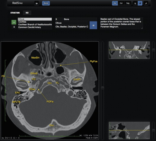
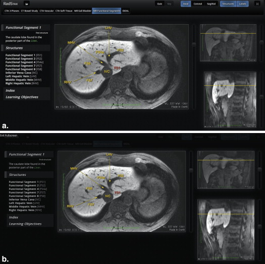
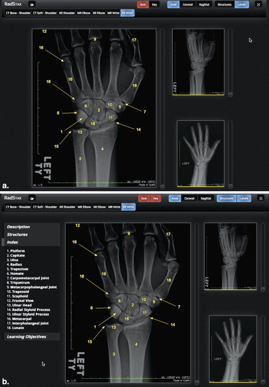
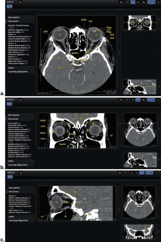
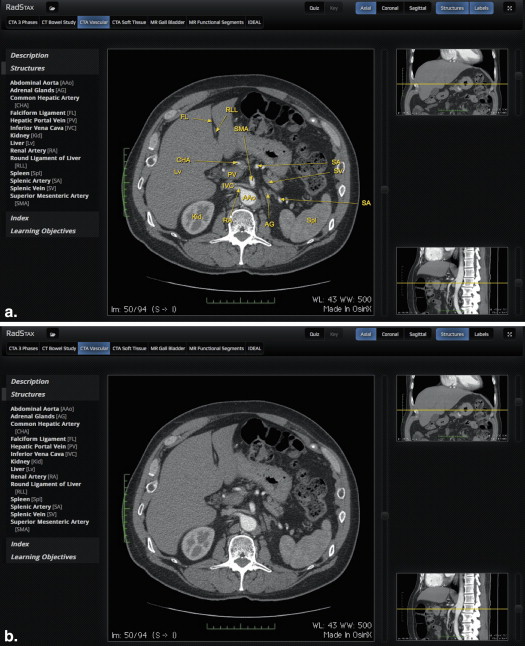
Figure 1
Screenshot of RadStax, an interactive Web-based program for viewing imaging studies that allows 1) a fast and intuitive way to create labels for all anatomy of interest; 2) the incorporation of basic information about each labeled region of interest; 3) the ability to view all three planes of imaging simultaneously; 4) a guide bar for localization to facilitate 3D understanding; 5) a search function; 6) the ability to be easily incorporated into lectures; 7) availability for independent study; and 8) a means for self-assessment.
Get Radiology Tree app to read full this article<
Methods
Technical Development
Get Radiology Tree app to read full this article<
Get Radiology Tree app to read full this article<
Table 1
Sample Spreadsheet
Set Name Formal Name Common Names Structure Description Vascular Basilar artery BA, vessel, artery Origin: confluence of vertebral artery at the junction of the pons and medulla oblongata branches: anterior inferior cerebellar artery and pontine arteries
Terminal branches: superior cerebellar artery and posterior cerebral artery Viscera Common bile duct CBD, Vater Origin: the common bile duct is formed from the confluence of the cystic duct and common hepatic duct
Drains to: joins the main pancreatic duct to form the ampulla of Vater Bone Coracoid process CP Superior to the glenoid cavity, this anterolateral projection of the scapula gives rise to the attachment of three muscles: coracobrachialis, short head of the biceps, and pectoralis minor. Nerve Facial nerve FN, CN VII, nerve, inner ear Arises from the pons. Controls muscles of facial expression, conveys taste from anterior 2/3 tongue. Supplies preganglionic parasympathetic fibers to submandibular and lacrimal glands Muscle Flexor hallucis longus FHL Proximal attachment: the inferior two-thirds of the posterior surface of the fibula and the inferior part of the interosseous membrane
Distal attachment: the base of the distal phalanx of the great toe Level Functional segment 1 FS1, liver, portal vein, hepatic, division, divisions, FS, FD1, FD The caudate lobe found in the posterior part of the liver Gland Pituitary gland PGl, hypophysis Rests in the hypophysial fossa. Composed of the anterior pituitary and posterior pituitary Space Rectouterine pouch RuP, Douglas Separates the body of the uterus and the supravaginal cervix from the sigmoid colon Connective tissue Thecal sac ThSac A membrane of dura mater that surrounds the spinal cord and cauda equina and is filled with cerebrospinal fluid
Sample of the information entered into a spreadsheet and then converted into XML format. The columns “Set Name” and “Common Names” were created with a future version in mind, which will provide a text search function.
Get Radiology Tree app to read full this article<
Get Radiology Tree app to read full this article<
Get Radiology Tree app to read full this article<
Get Radiology Tree app to read full this article<
Get Radiology Tree app to read full this article<
Program Implementation and Assessment
Get Radiology Tree app to read full this article<
Get Radiology Tree app to read full this article<
Table 2
Survey of First- and Second-Year Medical Students Who Used RadStax
First-Year Students; N = 55 Second-Year Students; N = 60 Combined percentage; N = 115 How helpful was it to see all three planes of imaging simultaneously (with a guide in the other two windows)? 8.42/10 8.20/10 83.1 How helpful was it to be able to scroll through all three planes of imaging simultaneously? 8.37/10 8.11/10 82.4 How helpful was it to have the label follow structures while scrolling through the main window? 9.25/10 9.71/10 94.8 How useful was the quiz function? 7.08/10 8.07/10 75.8 How useful were the descriptions associated with labeled structures? 6.73/10 7.47/10 71.0 How helpful was it to have multiple modalities of the same study? 8.45/10 8.27/10 83.6 Overall, did you find RadStax to be an EFFECTIVE study resource? 8.47/10 8.47/10 84.7 Overall, did you find RadStax to be an EFFICIENT study resource? 7.73/10 8.16/10 79.5 Overall, how satisfied were you with RadStax? 7.93/10 8.42/10 81.8
Medical students were asked to participate in a voluntary and anonymous electronic survey. Subjects responded on a 10-point scale ranging from (1) “Not helpful / Did not use” to (10) “Extremely helpful.” The data mean is displayed.
Get Radiology Tree app to read full this article<
Get Radiology Tree app to read full this article<
Results
Technical Product
Get Radiology Tree app to read full this article<
Get Radiology Tree app to read full this article<
Get Radiology Tree app to read full this article<
Get Radiology Tree app to read full this article<
Implementation
Get Radiology Tree app to read full this article<
Survey Results
Get Radiology Tree app to read full this article<
Get Radiology Tree app to read full this article<
Discussion
Get Radiology Tree app to read full this article<
Get Radiology Tree app to read full this article<
Get Radiology Tree app to read full this article<
Get Radiology Tree app to read full this article<
Get Radiology Tree app to read full this article<
Get Radiology Tree app to read full this article<
Get Radiology Tree app to read full this article<
Get Radiology Tree app to read full this article<
Acknowledgments
Get Radiology Tree app to read full this article<
Get Radiology Tree app to read full this article<
Get Radiology Tree app to read full this article<
References
1. Branstetter B.F., Humphrey A.L., Schumann J.B.: The long-term impact of preclinical education on medical students’ opinions about radiology. Acad Radiol 2008; 15: pp. 1331-1339.
2. Feigin D.S., Magid D., Smirniotopoulos J.G., et. al.: Learning and retaining normal radiographic chest anatomy: does preclinical exposure improve student performance?. Acad Radiol 2007; 14: pp. 1137-1142.
3. Bloomfield J.: Radiology–focus of the medical curriculum?. AAJR Am J Roentgenol 1982; 138: pp. 980-981.
4. Erinjeri J.P., Bhalla S.: Redefining radiology education for first-year medical students: shifting from a passive to an active case-based approach. Acad Radiol 2006; 13: pp. 789-796.
5. Zou L., King A., Soman S., et. al.: Medical students’ preferences in radiology education a comparison between the Socratic and didactic methods utilizing powerpoint features in radiology education. Acad Radiol 2011; 18: pp. 253-256. Epub 2010 Nov 13
6. Phillips A.W., Smith S.G., Straus C.M.: The role of radiology in preclinical anatomy: a critical review of the past, present, and future. Acad Radiol 2013; 20: pp. 297-304.e1.
7. Phillips A.W., Smith S.G., Ross C.F., et. al.: Improved understanding of human anatomy through self-guided radiological anatomy modules. Acad Radiol 2012; 19: pp. 902-907. Epub 2012 Apr 24
8. Roubidoux M.A., Chapman C.M., Piontek M.E.: Development and evaluation of an interactive Web-based breast imaging game for medical students. Acad Radiol 2002; 9: pp. 1169-1178.
9. Durfee S.M., Jain S., Shaffer K.: Incorporating electronic media into medical student education: a survey of AMSER members on computer and web use in radiology courses. Alliance of Medical Student Educators in Radiology. Acad Radiol 2003; 10: pp. 205-210.
10. Marker D., Juluru K., Long C., et. al.: Strategic improvements for gross anatomy Web-based teaching. Anat Res Int 2012; 2012: pp. 146262. Epub 2011 Dec 14
11. Dashevsky B, Gorovoy M, Weadock W, Juluru K. Radiology teaching files: an assessment of their role and desired features based on a national survey. [ahead of print]
12. Lewis P.J., Chen J.Y., Lin D.J., et. al.: Radiology ExamWeb: development and implementation of a national web-based examination system for medical students in radiology. Acad Radiol 2013; 20: pp. 290-296.