
Rationale and Objectives
In brain MRI of multiple sclerosis (MS) patients, enhancement of the lesions is usually evaluated in early contrast-enhanced T1-weighted images (CE-T1WI). The objective of this study is to determine the sensitivity of contrast-enhanced fluid-attenuated-inversion-recovery (CE-FLAIR) and delayed contrast-enhanced MRI in evaluation of MS brain lesions.
Materials and Methods
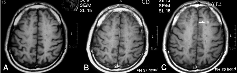
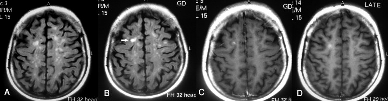
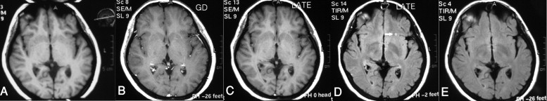
Brain MRI examination including early and delayed CE-T1WI and early and delayed CE-FLAIR images was performed for 46 patients with clinically definite MS disease. Number, size, location, degree, and pattern of enhancement of the enhanced lesions in each sequence were recorded separately.
Results
A total number of 87 enhanced lesions was detected in 30 patients. Early CE-T1WI could detect only 63 lesions (72.4% of total) in 24 patients, while delayed CE-T1WI and early and delayed CE-FLAIR images showed 85 (97.7%), 84 (96.6%), and 81 (93.1%) lesions in 28, 28, and 26 patients, respectively. A greater degree of enhancement and larger lesion size were observed in the additional sequences compared with the early CE-T1WI.
Conclusions
The sensitivity of early CE-T1WI for the detection of enhanced MS lesions is significantly lower than that for other additional sequences. Delayed CE-FLAIR images could not add significant information to other sequences. Therefore, early CE-FLAIR and delayed CE-T1WI brain MRI can be considered as part of the evaluation of MS patients, especially if, despite clinically suspected active disease, no enhanced lesion is found in the routine CE-T1WI.
Multiple sclerosis (MS) is one of the most common diseases of the central nervous system and the most common disabling neurologic disease of young adults ( ). The diagnostic criteria of MS are clinical: two episodes of aggravating symptoms and evidence of temporal and spatial dissemination. However, cerebrospinal fluid (CSF) tests and MRI have expanding roles in the early diagnosis of MS ( ).
Contrast-enhanced brain MRI is a part of baseline evaluation of suspected and clinically definite MS patients who have shown brain lesions in precontrast MR studies. Contrast-enhanced MRI is regarded as the best indicator of disease activity and response to therapy, being used during the treatment course and follow-up of MS patients ( ). As active MS lesions are associated with focal disruption of the blood-brain barrier (BBB) due to perivascular inflammation, injection of contrast leads to significant shortening of T1-relaxation time of these lesions with subsequent increased signal intensity. Thus, evaluation of enhancement of acute lesions is usually made in early contrast-enhanced T1-weighted images (CE-T1WI) ( ). Some researchers believe that obtaining delayed T1-weighted images (20 minutes to 1 hour after contrast administration) in MS patients will add valuable information to early CE-T1WI ( ), although diagnostic value of delayed imaging has been refuted by some others ( ).
Get Radiology Tree app to read full this article<
Get Radiology Tree app to read full this article<
Methods
Subjects
Get Radiology Tree app to read full this article<
Get Radiology Tree app to read full this article<
MRI Acquisitions
Get Radiology Tree app to read full this article<
Get Radiology Tree app to read full this article<
Get Radiology Tree app to read full this article<
Get Radiology Tree app to read full this article<
Evaluations
Get Radiology Tree app to read full this article<
Get Radiology Tree app to read full this article<
Get Radiology Tree app to read full this article<
Get Radiology Tree app to read full this article<
Data Analysis
Get Radiology Tree app to read full this article<
Results
Get Radiology Tree app to read full this article<
Get Radiology Tree app to read full this article<
Get Radiology Tree app to read full this article<
Number of Lesions
Get Radiology Tree app to read full this article<
Get Radiology Tree app to read full this article<
Degree of Enhancement
Get Radiology Tree app to read full this article<
Get Radiology Tree app to read full this article<
Size of the Lesions
Get Radiology Tree app to read full this article<
Get Radiology Tree app to read full this article<
Table 1
Number of patients, total number of enhancing lesions and lesion characteristics in different pulse sequences
Sequence Early CE-T1 Delayed CE-T1 Early CE-FLAIR Delayed CE-FLAIR Number of patients with enhancing lesions 24 28 28 26 Total number of enhancing lesions 63 (72.4%) 85 (97.7%) 84 (96.6%) 81 (93.1%) Mean degree of enhancement 1.96 ± 0.99 2.6 ± 0.88 3.54 ± 1.3 3.6 ± 1.5 Mean size of the lesions (mm) 8.8 ± 5.2 9.5 ± 4.7 9.8 ± 5.5 10.1 ± 5.5
Get Radiology Tree app to read full this article<
Lesion Location
Get Radiology Tree app to read full this article<
Get Radiology Tree app to read full this article<
Table 2
Number and percentage of enhancing lesions in all acquired sequences differentiated by their location
Location P.V S.C C.S B.S CE CC Total Early CE-T1 24 (38%) 17 (27%) 17 (27%) 2 (3%) 3 (5%) 0 63 Delayed CE-T1 38 (45%) 22 (26%) 19 (22%) 3 (3.5%) 3 (3.5%) 0 85 Early CE-FLAIR 37 (44%) 22 (26%) 19 (22%) 3 (3.5%) 3 (3.5%) 0 84 Delayed CE-FLAIR 35 (43%) 21 (26%) 19 (23%) 3 (4%) 3 (4%) 0 81 Not enhanced in early CE-T1 15 (62.5%) 6 (25%) 2 (0.08%) 1 (0.04%) 0 0 24
Note- P.V: Paraventricular, S.C: Subcortical, C.S: Centrum semiovale, B.S: Brain stem, CE: Cerebellum, CC: Corpus callosum.
Get Radiology Tree app to read full this article<
Pattern of Enhancement of Lesions
Get Radiology Tree app to read full this article<
Discussion
Get Radiology Tree app to read full this article<
Get Radiology Tree app to read full this article<
Get Radiology Tree app to read full this article<
Get Radiology Tree app to read full this article<
Get Radiology Tree app to read full this article<
Get Radiology Tree app to read full this article<
Get Radiology Tree app to read full this article<
Get Radiology Tree app to read full this article<
Get Radiology Tree app to read full this article<
Get Radiology Tree app to read full this article<
Get Radiology Tree app to read full this article<
Get Radiology Tree app to read full this article<
Get Radiology Tree app to read full this article<
Get Radiology Tree app to read full this article<
Get Radiology Tree app to read full this article<
Get Radiology Tree app to read full this article<
Get Radiology Tree app to read full this article<
Get Radiology Tree app to read full this article<
Get Radiology Tree app to read full this article<
Get Radiology Tree app to read full this article<
Get Radiology Tree app to read full this article<
Conclusion
Get Radiology Tree app to read full this article<
Get Radiology Tree app to read full this article<
References
1. Gebarski S.S., Gabrielsen T.O., Gilman S., Knake J.E., Latack J.T., Aisen A.M.: The initial diagnosis of multiple sclerosis: Clinical impact of magnetic resonance imaging. Ann Neurol 1985; 17: pp. 469-474.
2. McDonald W.I., Compston A., Edan G., et. al.: Recommended diagnostic criteria for multiple sclerosis: Guidelines from the International Panel on the Diagnosis of MS. Ann Neurol 2001; 50: pp. 121-127.
3. Miller D.H., Filippi M., Fazekas F., et. al.: Role of MRI within diagnostic criteria for multiple sclerosis. Ann Neurol 2004; 56: pp. 273-278.
4. Silver N.C., Good C.D., Barker G.J., et. al.: Sensitivity of contrast enhanced MRI in multiple sclerosis: Effects of gadolinium dose, magnetization transfer contrast and delayed imaging. Brain 1997; 120: pp. 1149-1161.
5. Silver N.C., Tofts P.S., Symms M.R., Barker G.J., Thompson A.J., Miller D.H.: Quantitative contrast-enhanced MRI to evaluate blood-brain barrier integrity in multiple sclerosis: A preliminary study. Multiple Sclerosis 2001; 7: pp. 75-82.
6. Filippi M., Yousry T., Rocca M.A., Fesl G., Voltz R., Comi G.: Sensitivity of delayed gadolinium-enhanced MRI in multiple sclerosis. Acta Neurol Scand 1997; 95: pp. 331-334.
7. Wolansky L.J., Bardini J.A., Cook S.D., Zimmer A.E., Sheffet A., Lee H.J.: Triple-dose versus single-dose gadoteridol in multiple sclerosis patients. J Neuroimaging 1994; 4: pp. 141-145.
8. He J., Grossman R.I., Ge Y., Mannon L.J.: Enhancing patterns in multiple sclerosis: Evolution and persistence. AJNR Am J Neuroradiol 2001; 22: pp. 664-669.
9. Goo H.W., Choi C.G.: Post-contrast FLAIR imaging of the brain in children: Normal and abnormal intracranial enhancement. Pediatr Radiol 2003; 33: pp. 843-849.
10. Mathews V.P., Caldemeyer K.S., Lowe M.J., Greenspan S.L., Weber D.M., Ulmer J.L.: Brain: Gadolinium-enhanced fast fluid-attenuated inversion-recovery MR imaging. Radiology 1999; 211: pp. 257-263.
11. Essig M., Knopp M.V., Schoenberg S.O., et. al.: Cerebral gliomas and metastases: Assessment with contrast-enhanced fast fluid-attenuated inversion-recovery MR imaging. Radiology 1999; 210: pp. 551-557.
12. Griffiths P.D., Coley S.C., Romanowski C.A., Hodgson T., Wilkinson I.D.: Contrast-enhanced fluid–attenuated inversion recovery imaging for leptomeningeal disease in children. AJNR Am J Neuroradiol 2003; 24: pp. 719-723.
13. Mihara F., Gupta K.L., Righi A.M.: Non-T1 weighted spin-echo MR imaging with contrast material: Experimental and preliminary clinical assessment. Radiat Med 1994; 12: pp. 209-212.
14. Jackson E.F., Hayman L.A.: Meningeal enhancement on fast FLAIR images. Radiology 2000; 215: pp. 922-924.
15. Kanamalla U.S., Baker K.B., Boyko O.B.: Gadolinium diffusion into subdural space: Visualization with FLAIR MR imaging. AJR Am J Roentgenol 2001; 176: pp. 1604-1605.
16. Hirota T., Ishihara K., Akazawa K., Kubota T., Yamada K., Nishimura T.: Delayed post-contrast FLAIR image for depicting meningeal carcinomatosis. Br J Radiol 2004; 77: pp. 528-531.
17. Ercan N., Gultekin S., Celik H., Tali T.E., Oner Y.A., Erbas G.: Diagnostic value of contrast enhanced fluid inversion recovery MR imaging of intracranial metastases. AJNR Am J Neuroradiol 2004; 25: pp. 761-765.
18. Rumpel H., Chan L.L.: Serial FLAIR imaging after Gd-DTPA contrast, pitfalls in stroke trial MRI. Stroke 2003; 34: pp. 797-798.
19. Singh S.K., Leeds N.E., Ginsberg L.E.: MR imaging of leptomeningeal metastases: Comparison of three sequences. AJNR Am J Neuroradiol 2002; 23: pp. 817-821.
20. Zheng-rong Z., Tian-zhen S., Xing-rong C., Wei-jun P.: Diagnostic value of contrast-enhanced fluid attenuated inversion recovery MRI for intracranial tumors in comparison with post contrast T1W spin-echo MRI. Chin Med J 2006; pp. 467-473. 119-6
21. Essig M., Schoenberg S.O., Debus J., Van Kaich G.: Disappearance of tumor contrast on contrast-enhanced FLAIR imaging of cerebral gliomas. Magn Reson Imaging 2000; 18: pp. 513-518.
22. Melhem E.R., Bert R.J., Walker R.E.: Usefulness of optimized gadolinium-enhanced fast fluid-attenuated inversion recovery MR imaging in revealing lesions of the brain. AJR Am J Roentgenol 1998; 171: pp. 803-807.
23. Pyhtinen J., Karttunen A., Tikkakoski T.: Increasing benefit of MRI in multiple sclerosis. Acta Radiol 2006; 47: pp. 960-971.
24. Maravilla K.R.: Enhancing our understanding of multiple sclerosis: Tracking contrast-enhanced plaques with MR imaging. AJNR Am J Neuroradiol 2001; 22: pp. 601-603.
25. McFarland H.F., Stone L.A., Calabresi P.A., Maloni H., Bash C.N., Frank J.A.: MRI studies of multiple sclerosis: Implications for the natural history of the disease and for monitoring effectiveness in experimental therapies. Multiple Sclerosis 1996; 2: pp. 198-205.
26. Ge Y.: Multiple sclerosis: The role of MR imaging. AJNR Am J Neuroradiol 2006; 27: pp. 1165-1176.
27. Brex P.A., O’Riordan J.I., Miszkiel K.A., et. al.: Multisequence MRI in clinically isolated syndromes and the early development of MS. Neurology 1999; 53: pp. 1184-1190.
28. Tan I.L., van Schijndel R.A., Pouwels P.J., Adèr H.J., Barkhof F.: Serial isotropic three-dimensional fast FLAIR imaging: Using image registration and subtraction to reveal active multiple sclerosis lesions. AJR Am J Roentgenol 2002; 179: pp. 777-782.
29. Bakshi R., Ariyaratana S., Benedict R.H., Jacobs L.: Fluid-attenuated inversion recovery magnetic resonance imaging detects cortical and juxtacortical multiple sclerosis lesions. Arch Neurol 2001; 58: pp. 742-748.
30. Uysal E., Erturk M., Yildirim H., Seleker F., Basak M.: Sensitivity of immediate and delayed gadolinium-enhanced MRI after injection of 0.5 M and 1.0 M gadolinium chelates for detecting multiple sclerosis lesions. AJR Am J Roentgenol 2007; 188: pp. 697-702.
31. Woo J.H., Henry L.P., Krejza J., Melhem E.R.: Detection of simulated multiple sclerosis lesions on T2-weighted and FLAIR images of the brain: Observer performance. Radiology 2006; 241: pp. 206-212.
32. Filippi M., Capra R., Campi A., et. al.: Triple dose of gadolinium-DTPA and delayed MRI in patients with benign multiple sclerosis. J Neurol Neurosurg Psychiatry 1996; 60: pp. 526-530.