
Rationale and Objectives
Diffraction-enhanced imaging (DEI) is a type of phase contrast x-ray imaging that has improved image contrast at a lower dose than conventional radiography for many imaging applications, but no studies have been done to determine if DEI might be useful for diagnosing lung injury. The goals of this study were to determine if DEI could differentiate between healthy and injured lungs for a rat model of gastric aspiration and to compare diffraction-enhanced images with chest radiographs.
Materials and Methods
Radiographs and diffraction-enhanced chest images of adult Sprague Dawley rats were obtained before and 4 hours after the aspiration of 0.4 mL/kg of 0.1 mol/L hydrochloric acid. Lung damage was confirmed with histopathology.
Results
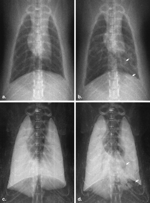
The radiographs and diffraction-enhanced peak images revealed regions of atelectasis in the injured rat lung. The diffraction-enhanced peak images revealed the full extent of the lung with improved clarity relative to the chest radiographs, especially in the portion of the lower lobe that extended behind the diaphragm on the anteroposterior projection.
Conclusions
For a rat model of gastric acid aspiration, DEI is capable of distinguishing between a healthy and an injured lung and more clearly than radiography reveals the full extent of the lung and the lung damage.
Diffraction-enhanced imaging (DEI), sometimes called analyzer-based imaging, is a type of phase contrast x-ray imaging that produces contrast not only from the x-ray attenuation in the subject but also from the refraction and small-angle scattering. It has been shown that DEI has significantly improved contrast resolution relative to radiography for a variety of imaging applications, including mammography , cartilage imaging , and bone and bone implant imaging . To date, the vast majority of DEI studies have been conducted at synchrotron research facilities, but recently, x-ray tube–based DEI systems have been developed by Parham et al and Nesch et al ; these systems provide a realistic pathway to clinical DEI, and thus a new emphasis on determining clinical imaging targets for DEI is warranted. Although several studies have shown DEI’s ability to resolve the lung with high contrast , its ability to distinguish between healthy and injured lung has not yet been fully explored. The goal of this study was to determine if DEI, using realistic imaging parameters for a future clinical DEI system, is capable of resolving differences between healthy and injured lung in a rat model of gastric acid aspiration.
Rat Model of Lung Injury
Aspiration of gastric acid into the lungs and the resulting acute pneumonitis was first described by Mendelson in 1946. Small animal models were used from the outset to develop an understanding of the nature of the lung injury. The temporal response of the rat lung to the aspiration of acids of different volumes and pH values was studied by Kennedy et al . The rat lung was found to have a biphasic response to the acid aspiration, with the maximum lung injury, as measured by permeability index, occurring 4 hours after acid aspiration and accompanied by an acute inflammatory response. During this acute inflammatory phase, one would expect to see radiographic evidence of alveolar atelectasis . The combination of this well-understood animal model and the known radiographic findings for this injury makes this an appropriate model of lung injury to study the applicability of DEI in this setting.
DEI
Get Radiology Tree app to read full this article<
Get Radiology Tree app to read full this article<
Get Radiology Tree app to read full this article<
Materials and methods
Animal Experimental Protocol
Get Radiology Tree app to read full this article<
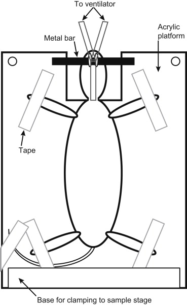
Get Radiology Tree app to read full this article<
Image Acquisition
Get Radiology Tree app to read full this article<
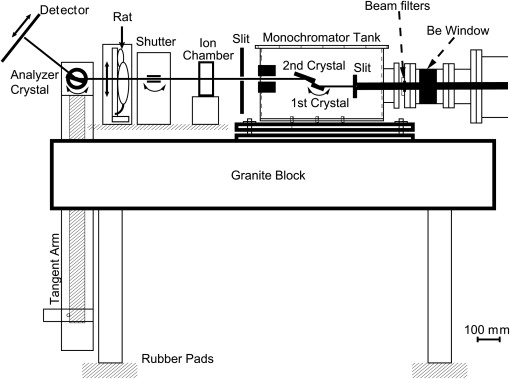
Get Radiology Tree app to read full this article<
Histology
Get Radiology Tree app to read full this article<
Image Processing
Get Radiology Tree app to read full this article<
Results
Histology
Get Radiology Tree app to read full this article<
Image Comparison
Get Radiology Tree app to read full this article<
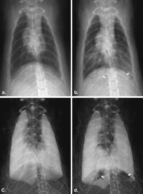
Get Radiology Tree app to read full this article<
Get Radiology Tree app to read full this article<
Get Radiology Tree app to read full this article<
Discussion
Get Radiology Tree app to read full this article<
Get Radiology Tree app to read full this article<
Get Radiology Tree app to read full this article<
Get Radiology Tree app to read full this article<
Get Radiology Tree app to read full this article<
Acknowledgments
Get Radiology Tree app to read full this article<
Get Radiology Tree app to read full this article<
References
1. Faulconer L.S., Parham C.A., Connor D.M., et. al.: Effect of breast compression on lesion characteristic visibility with diffraction-enhanced imaging. Acad Radiol 2010; 17: pp. 433-440.
2. Kao T., Connor D., Dilmanian F.A., et. al.: Characterization of diffraction-enhanced imaging contrast in breast cancer. Phys Med Biol 2009; 54: pp. 3247-3256.
3. Faulconer L., Parham C., Connor D.M., et. al.: Radiologist evaluation of an X-ray tube-based diffraction-enhanced imaging prototype using full-thickness breast specimens. Acad Radiol 2009; 16: pp. 1329-1337.
4. Rocha H.S., Pereira G.R., Faria P., et. al.: Diffraction-enhanced imaging microradiography applied in breast samples. Eur J Radiol 2008; 68: pp. S37-S40.
5. Liu C., Yan X., Zhang X., et. al.: Evaluation of x-ray diffraction enhanced imaging in the diagnosis of breast cancer. Phys Med Biol 2007; 52: pp. 419-427.
6. Kiss M.Z., Sayers D.E., Zhong Z., et. al.: Improved image contrast of calcifications in breast tissue specimens using diffraction enhanced imaging. Phys Med Biol 2004; 49: pp. 3427-3439.
7. Fiedler S., Bravin A., Keyrilainen J., et. al.: Imaging lobular breast carcinoma: comparison of synchrotron radiation DEI-CT technique with clinical CT, mammography and histology. Phys Med Biol 2004; 49: pp. 175-188.
8. Pisano E.D., Johnston R.E., Chapman D., et. al.: Human breast cancer specimens: diffraction-enhanced imaging with histologic correlation—improved conspicuity of lesion detail compared with digital radiography. Radiology 2000; 214: pp. 895-901.
9. Keyrilainen J., Bravin A., Fernandez M., et. al.: Phase-contrast X-ray imaging of breast. Acta Radiol 2010; 51: pp. 866-884.
10. Keyrilainen J., Fernandez M., Karjalainen-Lindsberg M.L., et. al.: Toward high-contrast breast CT at low radiation dose. Radiology 2008; 249: pp. 321-327.
11. Li J., Williams J.M., Zhong Z., et. al.: Reliability of diffraction enhanced imaging for assessment of cartilage lesions, ex vivo. Osteoarthritis Cartilage 2005; 13: pp. 187-197.
12. Li J., Zhong Z., Lidtke R., et. al.: Radiography of soft tissue of the foot and ankle with diffraction enhanced imaging. J Anat 2003; 202: pp. 463-470.
13. Majumdar S., Issever A.S., Burghardt A., et. al.: Diffraction enhanced imaging of articular cartilage and comparison with micro-computed tomography of the underlying bone structure. Eur Radiol 2004; 14: pp. 1440-1448.
14. Mollenhauer J., Aurich M.E., Zhong Z., et. al.: Diffraction-enhanced X-ray imaging of articular cartilage. Osteoarthritis Cartilage 2002; 10: pp. 163-171.
15. Muehleman C., Chapman L.D., Kuettner K.E., et. al.: Radiography of rabbit articular cartilage with diffraction-enhanced imaging. Anat Rec A Discov Mol Cell Evol Biol 2003; 272: pp. 392-397.
16. Muehleman C., Fogarty D., Reinhart B., et. al.: In-laboratory diffraction-enhanced X-ray imaging for articular cartilage. Clin Anat 2010; 23: pp. 530-538.
17. Muehleman C., Li J., Connor D., et. al.: Diffraction-enhanced imaging of musculoskeletal tissues using a conventional x-ray tube. Acad Radiol 2009; 16: pp. 918-923.
18. Connor D.M., Sayers D., Sumner D.R., et. al.: Diffraction enhanced imaging of controlled defects within bone, including bone-metal gaps. Phys Med Biol 2006; 51: pp. 3283-3300.
19. Wagner A., Sachse A., Keller M., et. al.: Qualitative evaluation of titanium implant integration into bone by diffraction enhanced imaging. Phys Med Biol 2006; 51: pp. 1313-1324.
20. Cooper DM, Bewer B, Wiebe S, et al. Diffraction enhanced x-ray imaging of the distal radius: a novel approach for visualization of trabecular bone architecture. Can Assoc Radiol J. In press.
21. Muehleman C., Sumner D.R., Zhong Z.: Refraction effects of diffraction-enhanced radiographic imaging: a new look at bone. J Am Podiatr Med Assoc 2004; 94: pp. 453-455.
22. Connor D.M., Hallen H.D., Lalush D.S., et. al.: Comparison of diffraction-enhanced computed tomography and monochromatic synchrotron radiation computed tomography of human trabecular bone. Phys Med Biol 2009; 54: pp. 6123-6133.
23. Parham C., Zhong Z., Connor D.M., et. al.: Design and implementation of a compact low-dose diffraction enhanced medical imaging system. Acad Radiol 2009; 16: pp. 911-917.
24. Nesch I., Fogarty D.P., Tzvetkov T., et. al.: The design and application of an in-laboratory diffraction-enhanced x-ray imaging instrument. Rev Sci Instrum 2009; 80: pp. 093702.
25. Kitchen M.J., Lewis R.A., Yagi N., et. al.: Phase contrast X-ray imaging of mice and rabbit lungs: a comparative study. Br J Radiol 2005; 78: pp. 1018-1027.
26. Lewis R.A., Hall C.J., Hufton A.P., et. al.: X-ray refraction effects: application to the imaging of biological tissues. Br J Radiol 2003; 76: pp. 301-308.
27. Zhong Z., Thomlinson W., Chapman D., et. al.: Implementation of diffraction-enhanced imaging experiments: at the NSLS and APS. Nucl Instrum Meth Phys Res A 2000; 450: pp. 556-567.
28. Mendelson M.L.: The aspiration of stomach contents into the lungs during obstetric anesthesia. Am J Obstet Gynecol 1946; 52: pp. 191-205.
29. Kennedy T.P., Johnson K.J., Kunkel R.G., et. al.: Acute acid aspiration lung injury in the rat: biphasic pathogenesis. Anesth Analg 1989; 69: pp. 87-92.
30. Desai S.R.: Acute respiratory distress syndrome: imaging of the injured lung. Clin Radiol 2002; 57: pp. 8-17.
31. Zhou S.A., Brahme A.: Development of phase-contrast X-ray imaging techniques and potential medical applications. Phys Med 2008; 24: pp. 129-148.
32. Chapman D., Thomlinson W., Johnston R.E., et. al.: Diffraction enhanced x-ray imaging. Phys Med Biol 1997; 42: pp. 2015-2025.
33. Kitchen M.J., Paganin D., Lewis R.A., et. al.: On the origin of speckle in x-ray phase contrast images of lung tissue. Phys Med Biol 2004; 49: pp. 4335-4348.
34. Cole H., Chambers F.H., Dunn H.M.: Simultaneous diffraction. Indexing unweganregung peaks in simple cases. Acta Crystallographica 1962; 15: pp. 138-144.
35. Oltulu O., Zhong Z., Hasnah M., et. al.: Extraction of extinction, refraction and absorption properties in diffraction enhanced imaging. J Phys D Appl Phys 2003; 36: pp. 2152-2156.