
Rationale and Objective
To assess the value of diffusion weighted imaging (DWI) magnetic resonance imaging (MRI) in pediatric and adolescent tumor patients with focus on detection and delineation of malignant tumors of the central nervous system, chest, abdomen, and musculoskeletal system.
Materials and Methods
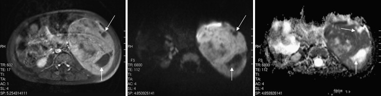
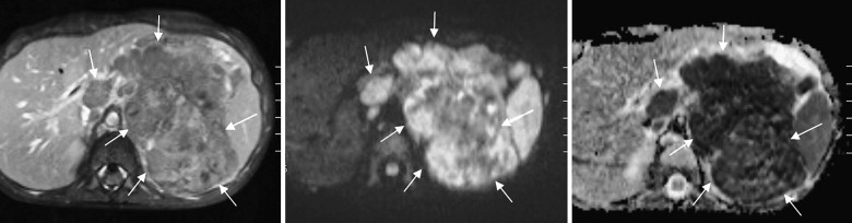
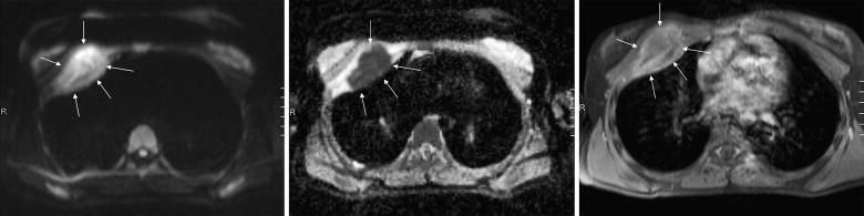
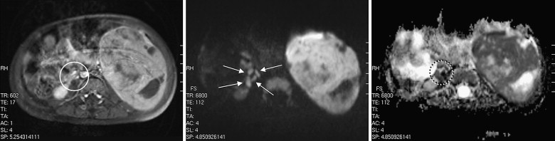
Twenty-nine pediatric and adolescent patients (17 males, 12 females, age, 2 months–20 years, mean age: 8.9 years) with clinically suspected malignant tumors were examined with use of a 1.5-T MR scanner with open bore design without sedation or general anesthesia. DWI images were acquired with a single-shot echo planar imaging (EPI) sequence in free breathing with b-values of 0, 500, and 1000 mm/s 2 . Images were assessed by two readers in consensus. Artifacts in DWI were graded as not relevant, acceptable, or nondiagnostic. DWI/apparent diffusion coefficient maps were correlated with T1-weighted post-contrast images, and the detectability and correct delineation of the tumors were graded using a three grade scale.
Results
Free-breathing DWI was successfully performed in all patients. In 27 patients, no relevant artifacts were observed; acceptable artifacts were seen in two patients. In all patients, malignancies were detected both on DWI and T1-weighted gadolinium images. Detection and delineation of tumors were possible in all cases with both sequences; T1-weighted gadolinium imaging was superior to DWI in only three patients. Additionally, small diffusion restricted lymph nodes were detected in three patients.
Conclusion
DWI is reliable for the accurate detection and delineation of malignant pediatric and adolescent tumors.
Diffusion weighted imaging (DWI) is an established imaging technique in magnetic resonance imaging (MRI) and widely used in imaging of the brain and central nervous system (CNS) pathologies, e.g., for early detection of ischemic and malignant changes. DWI applications outside the brain and CNS are becoming increasingly popular because rapid technologic developments of hardware and software, e.g., dedicated multichannel coils, faster and more powerful gradients, and parallel imaging techniques enable good image quality with high contrast-to-noise ratios (CNR) enabling acquisition of additional, functional MRI data within a reasonable scan time even during free breathing. A rapid increase in published data in the last 5 years has been recognized, reporting experiences with DWI in the chest , breast , abdomen , urogenital , and musculoskeletal system . Although these data are encouraging, published data about clinical studies using DWI in pediatric patients outside the brain is rare . More data about the usefulness of DWI regarding detection and delineation of pathology is needed to implement DWI into routine MR protocols.
The purpose of this study was to assess the value of DWI MR in pediatric and adolescent tumor imaging regarding detection and delineation of malignant tumors of the CNS, chest, abdomen, and musculoskeletal system.
Materials and methods
Get Radiology Tree app to read full this article<
Table 1
Tumor Entities Found on Histopathologic Specimen Work-up
Histopathologic Diagnosis n Ewing Sarcoma 4 Neuroblastoma 4 Ovarian teratoma 2 B-NHL 2 Medulloblastoma 2 Nephroblastoma 2 Rhabdomyosarcoma 2 ATRT 1 Burkitt-like lymphoma 1 Ependymoma 1 GBM 1 Hodgkin lymphoma 1 Optico-thalamic glioma 1 Pilocytic astrocytoma 1 PNET 1 Pontine astrocytoma 1 Renal sarcoma 1 T-cell lymphoma 1 Total 29
ATRT, atypical teratoid/rhabdoid tumor; GBM, glioblastoma multiforme; PNET, primitive neuroectodermal tumor.
Get Radiology Tree app to read full this article<
Get Radiology Tree app to read full this article<
Get Radiology Tree app to read full this article<
Get Radiology Tree app to read full this article<
Get Radiology Tree app to read full this article<
Get Radiology Tree app to read full this article<
Get Radiology Tree app to read full this article<
Get Radiology Tree app to read full this article<
Results
Get Radiology Tree app to read full this article<
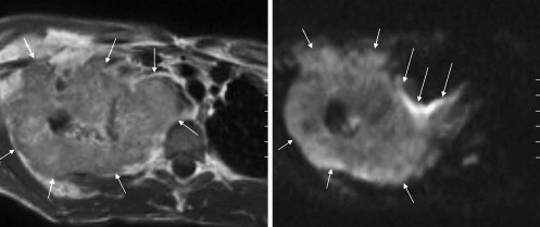
Table 2
Distribution and Quality of Artifacts on Diffusion Weighted Imaging
Artifact (rating) n No artifact (0) 27 Acceptable artifact (1) 2 Nondiagnostic image (2) 0
Get Radiology Tree app to read full this article<
Qualitative Assessment
Tumor Detection
Get Radiology Tree app to read full this article<
Get Radiology Tree app to read full this article<
Tumor Delineation
Get Radiology Tree app to read full this article<
Table 3
Comparison of Tumor Delineation, T1-weighted Post-Gadolinium versus Diffusion Weighted Imaging (DWI)
Tumor Delineation n T1-weighted superior 3 T1-weighted and DWI equal 26 DWI superior 0
Get Radiology Tree app to read full this article<
Qualitative Assessment
Get Radiology Tree app to read full this article<
Additional Findings
Get Radiology Tree app to read full this article<
Get Radiology Tree app to read full this article<
Discussion
Get Radiology Tree app to read full this article<
Get Radiology Tree app to read full this article<
Get Radiology Tree app to read full this article<
Get Radiology Tree app to read full this article<
Get Radiology Tree app to read full this article<
Limitations
Get Radiology Tree app to read full this article<
Conclusion
Get Radiology Tree app to read full this article<
Get Radiology Tree app to read full this article<
References
1. Baysal T., Bulut T., Gökirmak M., et. al.: Diffusion-weighted MR imaging of pleural fluid: differentiation of transudative versus exudative pleural effusions. Eur Radiol 2004; 14: pp. 890-896.
2. Woodhams R., Matsunaga K., Kan S., et. al.: ADC mapping of benign and malignant breast tumors. Magn Reson Med Sci 2005; 4: pp. 35-42.
3. Nasu K., Kuroki Y., Nawano S., et. al.: Hepatic metastases: diffusion weighted Sensitivity-encoding versus SPIO-enhanced MR imaging. Radiology 2006; 239: pp. 122-130.
4. Ichikawa T., Ertürk M.S., Motosugi U., et. al.: High-b value diffusion-weighted MRI for detecting pancreatic adenocarcinoma: preliminary results. AJR 2007; 188: pp. 409-414.
5. Ichikawa T., Ertürk M.S., Motosugi U., et. al.: High-b value diffusion-weighted MRI in colorectal cancer. AJR 2006; 187: pp. 181-184.
6. Thoeny H.C., De Keyzer F., Oyen R.H., et. al.: Diffusion-weighted MR imaging of kidneys in healthy volunteers and patients with parenchymal diseases: initial experience. Radiology 2005; 235: pp. 911-917.
7. Cova M., Squilacci E., Stacul F., et. al.: Diffusion-weighted MRI in the evaluation of renal lesions: preliminary results. Br J Radiol 2004; 77: pp. 851-857.
8. Sato C., Naganawa S., Nakamura T., et. al.: Differentiation of non cancerous tissue and cancer lesions by apparent diffusion coefficient values in transition and peripheral zones of the prostate. J Magn Reson Imaging 2005; 21: pp. 258-262.
9. Baur A., Stäbler A., Brüning R., et. al.: Diffusion-weighted MR imaging of bone marrow: differentiation of benign versus pathologic compression fractures. Radiology 1998; 207: pp. 349-356.
10. Raya J.G., Dietrich O., Reiser M.F., et. al.: Techniques for diffusion-weighted imaging of bone marrow. Eur J Radiol 2005; 55: pp. 64-73.
11. Abanoz R., Hakyemez B., Parlak M.: Diffusion-weighted imaging of acute vertebral compression: differential diagnosis of benign versus malignant pathologic fractures. Tani Girisim Radyol 2003; 9: pp. 176-183.
12. Uhl M., Altehoefer C., Kontny U., et. al.: MRI-diffusion imaging of neuroblastomas: first results and correlation to histology. Eur Radiol 2002; 12: pp. 2335-2338.
13. Uhl M., Saueressig U., Koehler G., et. al.: Evaluation of tumor necrosis during chemotherapy with diffusion-weighted MR imaging: preliminary results in osteosarcomas. Pediatr Radiol 2006; 36: pp. 1306-1311.
14. Humphries P.D., Sebire N.J., Siegel M.J., et. al.: Tumors in pediatric patients at diffusion-weighted MR imaging: apparent diffusion coefficient and tumor cellularity. Radiology 2007; 245: pp. 848-854.
15. Hayashida Y., Hirai T., Morishita S., et. al.: Diffusion-weighted imaging of metastatic brain tumors: comparison with histologic type and tumor cellularity. Am J Neuroradiol 2006; 27: pp. 1419-1425.
16. Chang S.C., Lai P.H., Chen W.L., et. al.: Diffusion-weighted MRI features of brain abscess and cystic or necrotic brain tumors: comparison with conventional MRI. Clin Imaging 2002; 26: pp. 227-236.
17. Cowper S.E., Robin H.S., Steinberg S.M., et. al.: Scleromyxedema-like cutaneous diseases in renal-dialysis patients. Lancet 2000; 356: pp. 1000-1001.
18. Auron A., Shao L., Warady B.A., et. al.: Nephrogenic fibrosing dermopathy in children. Pediatr Nephrol 2006; 21: pp. 1307-1311.
19. Jain S.M., Wesson S., Hassanein A., et. al.: Nephrogenic fibrosing dermopathy in pediatric patients. Pediatr Nephrol 2004; 19: pp. 467-470.
20. Jan F., Segal J.M., Dyer J., et. al.: Nephrogenic fibrosing dermopathy: two pediatric cases. J Pediatr 2003; 143: pp. 678-681.
21. Cowper S.E.: Nephrogenic fibrosing dermopathy: the first 6 years. Curr Opin Rheumatol 2003; 15: pp. 785-790.
22. Mendoza F.A., Artlett C.M., Sandorfi N., et. al.: Description of 12 cases of nephrogenic fibrosing dermopathy and review of the literature. Semin Arthritis Rheum 2006; 35: pp. 238-249.
23. Penfield J.G.: Nephrogenic systemic fibrosis and the use of gadolinium based contrast agents. Pediatr Nephrol 2008 Jun 10; [Epub ahead of print]