
Highlights
DBT use has continued to grow over the course of 2011–2016.
DBT use is now equally likely at academic centers versus in private practices.
Synthetic 2D has yet to replace conventional 2D for DBT interpretation.
Majority of DBT users do not currently perform DBT-guided procedures.
DBT remains a limited resource (40% 1 unit only), likely impacting usage.
Get Radiology Tree app to read full this article<
Rationale and Objectives
Get Radiology Tree app to read full this article<
Materials and Methods
Get Radiology Tree app to read full this article<
Results
Get Radiology Tree app to read full this article<
Conclusions
Get Radiology Tree app to read full this article<
Introduction
Get Radiology Tree app to read full this article<
Get Radiology Tree app to read full this article<
Materials and Methods
Get Radiology Tree app to read full this article<
Survey Methods
Get Radiology Tree app to read full this article<
Demographics—Geographic Location and Practice Type
Get Radiology Tree app to read full this article<
DBT Adoption—Current Use and Plan for Future Use
Get Radiology Tree app to read full this article<
DBT Utilization—User Characteristics, Equipment, and Payment
Get Radiology Tree app to read full this article<
Clinical Indications
Get Radiology Tree app to read full this article<
Interpretive Parameters—2D versus synthetic 2D (s2D)
Get Radiology Tree app to read full this article<
DBT-Guided Procedures
Get Radiology Tree app to read full this article<
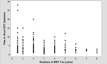
Performance Evaluation
Get Radiology Tree app to read full this article<
Radiologist Satisfaction
Get Radiology Tree app to read full this article<
Statistical Analysis
Get Radiology Tree app to read full this article<
Results
Demographics—Geographic Location and Practice Type
Get Radiology Tree app to read full this article<
TABLE 1
Demographics—DBT Utilization by Geography and by Practice Type
Parameters Number of All Respondents ‡ Number Using DBT (%) 95% CI (%)By geography 1092 ‡ 749 (68.6) 65.7–71.3 United States 732 577 (78.8) 75.7–81.7 Northeast 187 153 (81.8) 75.6–86.9 Midwest 166 134 (80.7) 74.0–86.4 West 118 88 (74.6) 65.8–82.1 South 223 172 (77.1) 71.2–82.5 Alaska \* 3 2 (66.7) N/A Hawaii \* 1 1 (100) N/A Unspecified 34 27 (79.4) 62.1–91.0 International 360 172 (47.8) 42.5–53.1By practice type 1099 ‡ 749 (68.2) 65.4–70.9 Academic 216 169 (78.2) 72.5–83.6 Private 596 423 (71.0) 67.3–74.6 Combined 198 118 (59.6) 52.7–66.5 Others † 89 39 (43.8) 33.3–54.8
95% CI, 95% confidence interval; DBT, digital breast tomosynthesis; N/A, does not apply.
Get Radiology Tree app to read full this article<
Get Radiology Tree app to read full this article<
Get Radiology Tree app to read full this article<
Get Radiology Tree app to read full this article<
DBT Adoption—Current Use and Plan for Future Use
Get Radiology Tree app to read full this article<
DBT Utilization—User Characteristics, Equipment, and Payment
Get Radiology Tree app to read full this article<
TABLE 2
Characteristics of DBT Users by Practice Type, Experience, Units, and Vendors
Total Number of DBT Users Number (%) 95% CI (%) Practice type_n_ = 749 † Academic 169 (22.6) 19.6–25.6 Private 423 (56.5) 52.9–60.0 Combined 118 (15.8) 13.2–18.5 Others \* 39 (5.2) 3.7–7.0 Years of DBT use_n_ = 722 † <1 176 (24.4) 21.2–27.7 1–3 435 (60.2) 56.6–63.8 >3 111 (15.4) 12.8–18.1 DBT units available_n_ = 728 † Only 1 290 (39.8) 36.3–43.4 >1 438 (60.2) 56.6–63.7 DBT brands in use ‡ n = 732 † Hologic 615 (84.0) 81.2–86.6 General Electric 104 (14.2) 11.7–16.9 Siemens 48 (6.6) 4.9–8.5 Fuji 13 (1.8) 0.9–3.0
95% CI, 95% confidence interval; DBT, digital breast tomosynthesis.
Get Radiology Tree app to read full this article<
Get Radiology Tree app to read full this article<
Get Radiology Tree app to read full this article<
Get Radiology Tree app to read full this article<
Get Radiology Tree app to read full this article<
Clinical Indications
Get Radiology Tree app to read full this article<
Interpretive Parameters—2D vs s2D
Get Radiology Tree app to read full this article<
TABLE 3
Images Included in DBT Interpretation Reported by Radiologists ( n = 702)
Combinations Used Number (%) 95% CI (%) 2D + 3D 286 (40.7) 37.0–44.4 2D + 3D + s2D 246 (35) 31.5–38.6 3D + s2D 170 (24.2) 21.0–27.5
95% CI, 95% confidence interval; 2D, conventional 2D mammogram; 3D, tomosynthesis image stack; DBT, digital breast tomosynthesis; s2D, synthetic 2D images derived from 3D source projection images.
Get Radiology Tree app to read full this article<
DBT-Guided Procedures
Get Radiology Tree app to read full this article<
TABLE 4
DBT Uptake and Pattern of Use Snapshots Over Time ( n = Number)
Parameters 2014 Hardesty Survey 2016 Current Survey All responders ( n ) 670 (30% use DBT) 1156 (65% use DBT) DBT users ( n ) \* 200 749 Academics ( n ) 73 (37%) 169 (23%) Private practice ( n ) 104 (52%) 423 (57%) Use 3D/2D/s2D ( n ) N/A 246 (35% † ) Use 3D/s2D ( n ) N/A 170 (24% † ) DBT procedures ( n ) N/A 231 (34% ‡ ) Demographic United States United States/International
2D, conventional 2D mammogram; 3D, tomosynthesis image stack; DBT, digital breast tomosynthesis; N/A, does not apply; s2D, synthetic 2D images derived from 3D source projection images.
Values with both the symbols † and ‡ have denominators that vary and reflect the total number of complete responses pertaining to each variable.
Get Radiology Tree app to read full this article<
Get Radiology Tree app to read full this article<
Get Radiology Tree app to read full this article<
Get Radiology Tree app to read full this article<
Performance Evaluation
Get Radiology Tree app to read full this article<
Get Radiology Tree app to read full this article<
Radiologist Satisfaction
Get Radiology Tree app to read full this article<
Discussion
Get Radiology Tree app to read full this article<
Get Radiology Tree app to read full this article<
Get Radiology Tree app to read full this article<
Get Radiology Tree app to read full this article<
Get Radiology Tree app to read full this article<
Get Radiology Tree app to read full this article<
Get Radiology Tree app to read full this article<
Get Radiology Tree app to read full this article<
Get Radiology Tree app to read full this article<
Get Radiology Tree app to read full this article<
Get Radiology Tree app to read full this article<
Get Radiology Tree app to read full this article<
Get Radiology Tree app to read full this article<
Get Radiology Tree app to read full this article<
Get Radiology Tree app to read full this article<
References
1. Andersson I., Ikeda D.M., Zackrisson S., et. al.: Breast tomosynthesis and digital mammography: a comparison of breast cancer visibility and BIRADS classification in a population of cancers with subtle mammographic findings. Eur Radiol 2008; 18: pp. 2817-2825.
2. Bernardi D., Caumo F., Macaskill P., et. al.: Effect of integrating 3D-mammography (digital breast tomosynthesis) with 2D-mammography on radiologists’ true-positive and false-positive detection in a population breast screening trial. Eur J Cancer 2014; 50: pp. 1232-1238.
3. Friedewald S.M., Rafferty E.A., Rose S.L., et. al.: Breast cancer screening using tomosynthesis in combination with digital mammography. JAMA 2014; 311: pp. 2499-2507.
4. Greenberg J.S., Javitt M.C., Katzen J., et. al.: Clinical performance metrics of 3D digital breast tomosynthesis compared with 2D digital mammography for breast cancer screening in community practice. AJR Am J Roentgenol 2014; 203: pp. 687-693.
5. Houssami N., Macaskill P., Bernardi D., et. al.: Breast screening using 2D-mammography or integrating digital breast tomosynthesis (3D-mammography) for single-reading or double-reading—evidence to guide future screening strategies. Eur J Cancer 2014; 50: pp. 1799-1807.
6. Kopans D.B.: Digital breast tomosynthesis from concept to clinical care. AJR Am J Roentgenol 2014; 202: pp. 299-308.
7. Statement on Breast Tomosynthesis. American College of Radiology; Available at https://www.acr.org/Advocacy/Economics-Health-Policy/Managed-Care-and-Private-Payer/Common-Coverage-Issues-with-Private-Payors/Digital-Breast-Tomosynthesis Accessed January 27, 2017
8. Skaane P., Bandos A.I., Gullien R., et. al.: Comparison of digital mammography alone and digital mammography plus tomosynthesis in a population-based screening program. Radiology 2013; 267: pp. 47-56.
9. Skaane P., Bandos A.I., Gullien R., et. al.: Prospective trial comparing full-field digital mammography (FFDM) versus combined FFDM and tomosynthesis in a population-based screening programme using independent double reading with arbitration. Eur Radiol 2013; 23: pp. 2061-2071.
10. United States Census Bureau : Geographic Terms and Concepts—Census Divisions and Census Regions. Available at http://www.census.gov/geo/reference/gtc/gtc_census_divreg.html Accessed April 3, 2016
11. Breast Series: Hot Topic Sessions. 101st Scientific Assembly and Annual Meeting. Radiological Society of North America. Chicago, IL. 12; Available at http://www.rsna.org/Past_Meetings.aspx Accessed June 18, 2016
12. American Roentgen Ray Society Annual Meeting. Los Angeles, CA. April 17–22; Available at http://www.arrs.org/uploadedFiles/ARRS/Education/Meetings/AM16/ProgramBook2016.PDF?hkey=a01e51ae-091e-4f58-9bb4-98817f35ee4d Accessed June 18, 2016
13. European Congress of Radiology. Vienna, Austria. Mar 2–6; Available at http://www.myesr.org/cms/website.php?id=/en/ecr_2016.htm Accessed June 18, 2016
14. American College of Radiology Society for Breast Imaging Annual Meeting. Austin, TX. Apr 7–10; Available at https://www.eventscribe.com/2016/SBI/aaSearchByPresentation.asp?BCFO=P Accessed June 18, 2016
15. Hardesty L.A., Kreidler S.M., Glueck D.H.: Digital breast tomosynthesis utilization in the United States: a survey of physician members of the Society of Breast Imaging. J Am Coll Radiol 2014; 11: pp. 594-599.
16. Svahn T.M., Macaskill P., Houssami N.: Radiologists’ interpretive efficiency and variability in true- and false-positive detection when screen-reading with tomosynthesis (3D-mammography) relative to standard mammography in population screening. Breast 2015; 24: pp. 687-693.
17. Skaane P., Bandos A.I., Eben E.B., et. al.: Two-view digital breast tomosynthesis screening with synthetically reconstructed projection images: comparison with digital breast tomosynthesis with full-field digital mammographic images. Radiology 2014; 271: pp. 655-663.
18. Zuley M.L., Guo B., Catullo V.J., et. al.: Comparison of two-dimensional synthesized mammograms versus original digital mammograms alone and in combination with tomosynthesis images. Radiology 2014; 271: pp. 664-671.
19. Freer P.E., Niell B., Rafferty E.A.: Preoperative tomosynthesis-guided needle localization of mammographically and sonographically occult breast lesions. Radiology 2015; 275: pp. 377-383.
20. Schrading S., Distelmaier M., Dirrichs T., et. al.: Digital breast tomosynthesis-guided vacuum-assisted breast biopsy: initial experiences and comparison with prone stereotactic vacuum-assisted biopsy. Radiology 2015; 274: pp. 654-662.
21. McDonald E.S., Oustimov A., Weinstein S.P., et. al.: Effectiveness of digital breast tomosynthesis compared with digital mammography: outcomes analysis from 3 years of breast cancer screening. JAMA Oncol 2016; 2: pp. 737-743.
22. Ciatto S., Houssami N., Bernardi D., et. al.: Integration of 3D digital mammography with tomosynthesis for population breast-cancer screening (STORM): a prospective comparison study. Lancet Oncol 2013; 14: pp. 583-589.
23. Conant E.F., Beaber E.F., Sprague B.L., et. al.: Breast cancer screening using tomosynthesis in combination with digital mammography compared to digital mammography alone: a cohort study within the PROSPR consortium. Breast Cancer Res Treat 2016; 156: pp. 109-116.
24. Skaane P.: Breast cancer screening with digital breast tomosynthesis. Breast Cancer 2016; 24: pp. 32-41.
25. Rafferty E.A., Durand M.A., Conant E.F., et. al.: Breast cancer screening using tomosynthesis and digital mammography in dense and nondense breasts. JAMA 2016; 315: pp. 1784-1786.
26. Gilbert F.J., Tucker L., Gillan M.G., et. al.: The TOMMY trial: a comparison of TOMosynthesis with digital MammographY in the UK NHS Breast Screening Programme–a multicentre retrospective reading study comparing the diagnostic performance of digital breast tomosynthesis and digital mammography with digital mammography alone. Health Technol Assess 2015; 19: pp. i-xxv. 1-136
27. Margolies L., Cohen A., Sonnenblick E., et. al.: Digital breast tomosynthesis changes management in patients seen at a tertiary care breast center. ISRN Radiol 2014; 2014: 658929
28. McCarthy A.M., Kontos D., Synnestvedt M., et. al.: Screening outcomes following implementation of digital breast tomosynthesis in a general-population screening program. J Natl Cancer Inst 2014; 106:
29. Dang P.A., Freer P.E., Humphrey K.L., et. al.: Addition of tomosynthesis to conventional digital mammography: effect on image interpretation time of screening examinations. Radiology 2014; 270: pp. 49-56.
30. Hardy K.: DBT Reimbursement. Radiology Today. Vol.16 No. 8 P. 12; Available at http://www.radiologytoday.net/archive/rt0815p12.shtml Accessed January 27, 2017
31. Eisenberg R.L., Bankier A.A., Boiselle P.M.: Compliance with Fleischner Society guidelines for management of small lung nodules: a survey of 834 radiologists. Radiology 2010; 255: pp. 218-224.
32. Callahan M.J., Servaes S., Lee E.Y., et. al.: Practice patterns for the use of iodinated i.v. contrast media for pediatric CT studies: a survey of the Society for Pediatric Radiology. AJR Am J Roentgenol 2014; 202: pp. 872-879.
33. Dym R.J., Burns J., Taragin B.H.: Appropriateness of imaging studies ordered by emergency medicine residents: results of an online survey. AJR Am J Roentgenol 2013; 201: pp. W619-W625.