
In the era of medical cost containment, radiologists must continually maintain their actual and perceived value to patients, payers, and referring providers. Exploitation of current and future digital technologies may be the key to defining and promoting radiology’s “brand” and assure our continued relevance in providing predictive, preventive, personalized, and participatory medicine. The Association of University of Radiologists Radiology Research Alliance Digitization of Medicine Task Force was formed to explore the opportunities and challenges of the digitization of medicine that are relevant to radiologists, which include the reporting paradigm, computational biology, and imaging informatics. In addition to discussing these opportunities and challenges, we consider how change occurs in medicine, and how change may be effected in medical imaging community. This review article is a summary of the research of the task force and hopefully can be used as a stimulus for further discussions and development of action plans by radiology leaders.
In the face of Medicare cost containment, all medical subspecialties must strive to eliminate waste and improve both the effectiveness and efficiency of patient care. Radiology has the added challenge of combating commoditization by finding ways to add value . Radiology must continually maintain its actual and perceived value to patients, payers, and referring providers. In the business world, this is considered “branding”; maintaining brand value is a constant responsibility of the organization . Exploitation of current and future digital technologies may be the key to defining and promoting radiology’s “brand” .
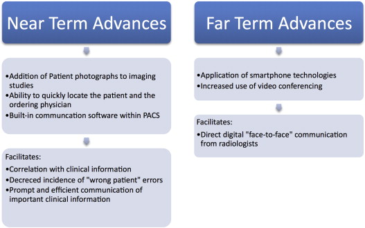
The three core services of radiology—scheduling, imaging, and reporting—can all be commoditized. Even image interpretation could be affected by computer-aided detection software. But before searching for new avenues of value added, a business struggling with commoditization should start with determining how existing products and services can be redefined to better meet its customers’ needs . Specific strategies include standardization, bundling, and customization—these strategies address the manner in which the same services are delivered, without actually changing the substance of those services. Standardization of reporting has certainly been an important topic in radiology and is discussed later; standardization of scheduling and imaging should also be important goals. One example of bundling radiology services would be to enable a referring clinician to click “schedule the recommended follow-up study” on the same page as the report where the recommendation was made, therein bundling the services of reporting and scheduling. An example of customizing radiology services would be to have a computerized order entry system that allows the provider to check the box “measure tumor volume” or “automatically fax report to my office.” In the non–health care setting, consumers have come to expect these standardized customer conveniences from online retailers, and the same expectations should apply to medical care. Failure to incorporate existing digital technologies into current radiology practice only enables the competition (within or outside radiology) to be the first to redefine how commoditized services can be delivered better. One aspect of radiology that cannot be commoditized is personal consultations (for patients and referring physicians) . How can digital technologies be used to make radiologists more effective consultants? Picture archiving and communication systems (PACS) may have made it easier for radiologists to isolate themselves, but digital technologies applied in the right way can also improve interpersonal interaction.
In many ways, efficiency in radiology has been driven by PACS, and this efficiency has become the envy of other clinical services. Radiology’s success to date has largely relied on building a wall around radiologists so they could focus on interpretation, report generation, and turnaround times. We envision that PACS would continue to provide radiologists with efficiencies in the future; however, the walls would have windows allowing radiologists to communicate with referring clinicians and patients. “Skype”-like (Microsoft Inc, Redmond, WA) technologies can easily be incorporated with PACS workstations to permit such online communications. Patient digital photographs and video-clips can be integrated with medical imaging examinations and serve as additional sources of clinical information that can enhance the interpretation of imaging studies.
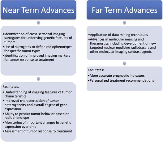
Referring clinicians in the future will face more data than any one physician can cope with (clinical, genetic, laboratory, radiologic, etc). Genetic screening will hopefully identify patients at risk for future disease, and those patients will need a lifelong monitoring plan and possibly even prophylactic therapy. If disease is later detected, primary treatment and follow-up will be necessary. Who will keep track of the massive amount of data that will be generated in the life of this patient? Who will analyze past and present data when acute situations arise, warranting prompt diagnosis? The existence of different medical specialties speaks to the fact that one human physician cannot know everything. A single physician cannot be expected to decipher the massive amount of data that modern tests are yielding, and this flood of information will only increase. Within medical specialties, online knowledge databases have been developed to assist in point-of-care clinical specialist decision-making. More data are yet to come, and these fragmented online databases will be inadequate.
There is clearly a need for a centralized information management system, and why not have a single medical specialty to serve as the shepherds for the flow of data, providing patient management and diagnostic decision support? Radiology is arguably in the best position (from an informatics standpoint) to take on this potential role. If radiology does not vie for a greater clinical presence, taking the opportunity to rebrand itself in this medical informatics role (either in part or whole), another specialty will. But how can the field of radiology effect these current, let alone future, changes? Radiology cannot expect to get reimbursed for new services unless those services can be shown to improve patient outcome and/or reduce cost. So, who is going to take the plunge in making the necessary digital technology investments and do the requisite research? Redefining the role of the radiologist in current and future practice will require the concerted effort and buy-in of multiple institutions. Rather than asking, “What can a radiologist do?” we will have more creative freedom by instead simply asking, “What needs to be done?” with all the patient information that is coming our way.
Get Radiology Tree app to read full this article<
Radiology report
Get Radiology Tree app to read full this article<
Get Radiology Tree app to read full this article<
Get Radiology Tree app to read full this article<
Get Radiology Tree app to read full this article<
Get Radiology Tree app to read full this article<
Radiology Lexicon
Get Radiology Tree app to read full this article<
Get Radiology Tree app to read full this article<
Get Radiology Tree app to read full this article<
Get Radiology Tree app to read full this article<
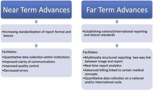
Reporting’s Future
Get Radiology Tree app to read full this article<
Get Radiology Tree app to read full this article<
Get Radiology Tree app to read full this article<
Get Radiology Tree app to read full this article<
Get Radiology Tree app to read full this article<
Get Radiology Tree app to read full this article<
Get Radiology Tree app to read full this article<
Get Radiology Tree app to read full this article<
Near-term Challenges and Opportunities in Radiology Reporting
Get Radiology Tree app to read full this article<
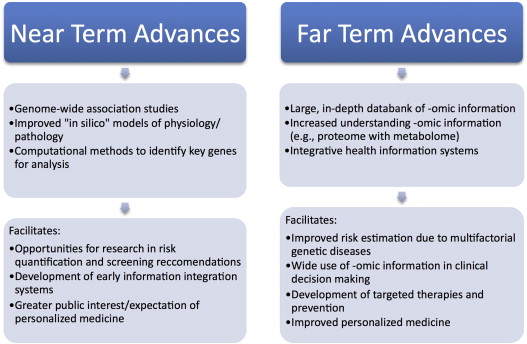
Computational biology
Get Radiology Tree app to read full this article<
Get Radiology Tree app to read full this article<
Get Radiology Tree app to read full this article<
Get Radiology Tree app to read full this article<
Get Radiology Tree app to read full this article<
Near-term Challenges and Opportunities in Computational Biology
Get Radiology Tree app to read full this article<
Get Radiology Tree app to read full this article<
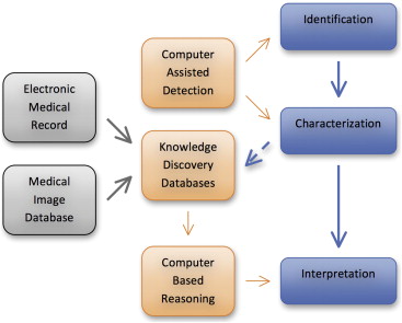
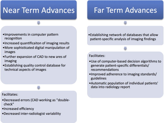
Imaging informatics
Get Radiology Tree app to read full this article<
Get Radiology Tree app to read full this article<
Get Radiology Tree app to read full this article<
Get Radiology Tree app to read full this article<
CAD
Get Radiology Tree app to read full this article<
Get Radiology Tree app to read full this article<
Get Radiology Tree app to read full this article<
Get Radiology Tree app to read full this article<
Get Radiology Tree app to read full this article<
Communication Technologies
Get Radiology Tree app to read full this article<
Get Radiology Tree app to read full this article<
Get Radiology Tree app to read full this article<
Get Radiology Tree app to read full this article<
Get Radiology Tree app to read full this article<
Linking Imaging Phenotype to Patient/Disease Genotype
Get Radiology Tree app to read full this article<
Get Radiology Tree app to read full this article<
Get Radiology Tree app to read full this article<
Get Radiology Tree app to read full this article<
Get Radiology Tree app to read full this article<
Get Radiology Tree app to read full this article<
Get Radiology Tree app to read full this article<
Near-term Challenges and Opportunities in Imaging Informatics
Get Radiology Tree app to read full this article<
How changes occur in the practice of medicine
Get Radiology Tree app to read full this article<
Get Radiology Tree app to read full this article<
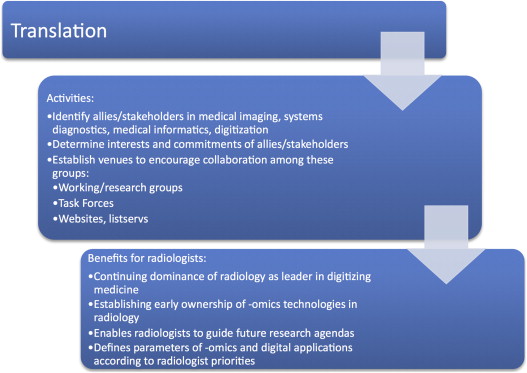
Translation
Get Radiology Tree app to read full this article<
Get Radiology Tree app to read full this article<
Get Radiology Tree app to read full this article<
Get Radiology Tree app to read full this article<
Get Radiology Tree app to read full this article<
Get Radiology Tree app to read full this article<
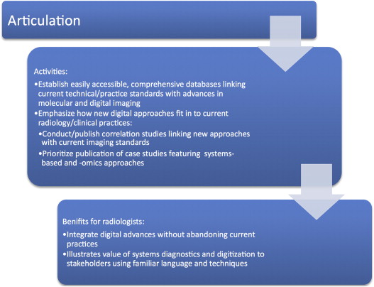
Articulation
Get Radiology Tree app to read full this article<
Get Radiology Tree app to read full this article<
Get Radiology Tree app to read full this article<
Get Radiology Tree app to read full this article<
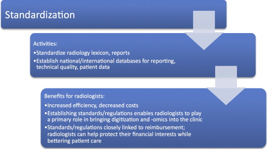
Standardization
Get Radiology Tree app to read full this article<
Get Radiology Tree app to read full this article<
Get Radiology Tree app to read full this article<
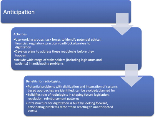
Anticipation
Get Radiology Tree app to read full this article<
Get Radiology Tree app to read full this article<
Get Radiology Tree app to read full this article<
Get Radiology Tree app to read full this article<
Competition for the Role of Medical Informatician
Get Radiology Tree app to read full this article<
Get Radiology Tree app to read full this article<
Get Radiology Tree app to read full this article<
Get Radiology Tree app to read full this article<
Get Radiology Tree app to read full this article<
Near-term Challenges and Opportunities in Effecting Change
Get Radiology Tree app to read full this article<
Conclusion
Get Radiology Tree app to read full this article<
References
1. Lin E.: Radiology 2011: the big picture. AJR Am J Roentgenol 2011; 196: pp. 136-139.
2. Longeteig K.: Competitive edge: the art and science of branding. Radiol Manage 2010; 32: pp. 44-47.
3. Rubin D.L.: Informatics in radiology—measuring and improving quality in radiology: meeting the challenge with informatics. Radiographics 2011; 31: pp. 1511-1527.
4. Kashani K.: Fighting commoditization: Strategies for creating novel customer values.2006.Institute for Management Development
5. Burnside E.S., Sickles E.A., Bassett L.W., et. al.: The ACR BI-RADS experience: learning from history. J Am Coll Radiol 2009; 6: pp. 851-860.
6. Hansell D.M., Bankier A.A., MacMahon H., et. al.: Fleischner Society: glossary of terms for thoracic imaging. Radiology 2008; 246: pp. 697-722.
7. Kahn C.E., Langlotz C.P., Burnside E.S., et. al.: Toward best practices in radiology reporting. Radiology 2009; 252: pp. 852-856.
8. Langlotz C.P.: RadLex: a new method for indexing online educational materials. Radiographics 2006; 26: pp. 1595-1597.
9. Shore M.W., Rubin D.L., Kahn C.E.: Integration of imaging signs into RadLex. J Digit Imaging 2012; 25: pp. 50-55.
10. Krestin G.P.: Commoditization in radiology: threat or opportunity?. Radiology 2010; 256: pp. 338-342.
11. Reiner B.I., Knight N., Siegel E.L.: Radiology reporting, past, present, and future: the radiologist’s perspective. J Am Coll Radiol 2007; 4: pp. 313-319.
12. Langlotz C.P.: Automatic structuring of radiology reports: harbinger of a second information revolution in radiology. Radiology 2002; 224: pp. 5-7.
13. Pool F., Goergen S.: Quality of the written radiology report: a review of the literature. J Am Coll Radiol 2010; 7: pp. 634-643.
14. Hawkins CM, Hall S, Hardin J, et al. Creation and implementation of department-wide standardized reports: an analysis of the impact on error rate in radiology reports. Society for Imaging Informatics in Medicine Annual Meeting Scientific Sessions, 2012. Available at: http://www.siim2012.org/abstract_SSD_Hawkins.shtml . Accessed January 23, 2013.
15. Marcovici P., Blume-Marcovici A.: Intuition versus rational thinking: psychological challenges in radiology and a potential solution. J Am Coll Radiol 2012; 10: pp. 25-29.
16. Schwartz L.H., Panicek D.M., Berk A.R., et. al.: Improving communication of diagnostic radiology findings through structured reporting. Radiology 2011; 260: pp. 174-181.
17. Naik S.S., Hanbidge A., Wilson S.R.: Radiology reports: examining radiologist and clinician preferences regarding style and content. AJR Am J Roentgenol 2001; 176: pp. 591-598.
18. Bosmans J.M., Weyler J.J., De Schepper A.M., et. al.: The radiology report as seen by radiologists and referring clinicians: results of the COVER and ROVER surveys. Radiology 2011; 259: pp. 184-195.
19. Sistrom C.L., Honeyman-Buck J.: Free text versus structured format: information transfer efficiency of radiology reports. AJR Am J Roentgenol 2005; 185: pp. 804-812.
20. Grieve F.M., Plumb A.A., Khan S.H.: Radiology reporting: a general practitioner’s perspective. Br J Radiol 2010; 83: pp. 17-22.
21. Plumb A.A., Grieve F.M., Khan S.H.: Survey of hospital clinicians’ preferences regarding the format of radiology reports. Clin Radiol 2009; 64: pp. 386-395.
22. Tennant G.: Six Sigma: SPC and TQM in manufacturing and services.2001.Gower Publishing Ltd
23. RSNA Radiology Reporting Initiative. Available at: http://www.rsna.org/Reporting_Initiative.aspx . Accessed January 23, 2013.
24. Lexicon (definition). Available at: http://dictionary.reference.com/browse/lexicon . Accessed January 23, 2013.
25. http://www.acr.org/Quality-Safety/Resources/BIRADS
26. http://www.acr.org/~/media/ACR/Documents/PDF/QualitySafety/Resources/LIRADS/lirads%20v20131.pdf
27. The Knowledge Graph, Google. Available at: http://googleblog.blogspot.com/2012/05/introducing-knowledge-graph-things-not.html . Accessed January 23, 2013.
28. RSNA RadLex. Available at: http://rsna.org/RadLex.aspx . Accessed January 23, 2013.
29. Martino A. Sketching a new reality: What will the radiology report of the future look like? In: ACR News, 2012. Available at: http://www.acr.org/News-Publications/News/News-Articles/2012/ACR-Bulletin/201203-Rad-Report-of-Future . Accessed January 23, 2013.
30. Aberle D.R., Adams A.M., Berg C.D., et. al.: Reduced lung-cancer mortality with low-dose computed tomographic screening. N Engl J Med 2011; 365: pp. 395-409.
31. Kitano H.: Computational systems biology. Nature 2002; 420: pp. 206-210.
32. Tawhai M.H., Bates J.H.: Multi-scale lung modeling. J Appl Physiol 2011; 110: pp. 1466-1472.
33. Yu T., Lloyd C.M., Nickerson D.P., et. al.: The Physiome Model Repository 2. Bioinformatics 2011; 27: pp. 743-744.
34. Oltvai Z.N., Barabasi A.L.: Systems biology. Life’s complexity pyramid. Science 2002; 298: pp. 763-764.
35. Barabasi A.L., Oltvai Z.N.: Network biology: understanding the cell’s functional organization. Nat Rev Genet 2004; 5: pp. 101-113.
36. Enzmann D.R.: Exploring the cell’s network with molecular imaging. J Magn Reson Imaging 2006; 24: pp. 257-266.
37. Han J.D.: Understanding biological functions through molecular networks. Cell Res 2008; 18: pp. 224-237.
38. Kitano H.: Systems biology: a brief overview. Science 2002; 295: pp. 1662-1664.
39. Buchman T.G.: The community of the self. Nature 2002; 420: pp. 246-251.
40. Noble D.: Claude Bernard, the first systems biologist, and the future of physiology. Exp Physiol 2008; 93: pp. 16-26.
41. Collins F.S., McKusick V.A.: Implications of the Human Genome Project for medical science. Jama 2001; 285: pp. 540-544.
42. Collins F.S., Green E.D., Guttmacher A.E., et. al.: A vision for the future of genomics research. Nature 2003; 422: pp. 835-847.
43. Bader J.S., Chant J.: Systems biology. When proteomes collide. Science 2006; 311: pp. 187-188.
44. Pieroni E., de la Fuente van Bentem S., Mancosu G., et. al.: Protein networking: insights into global functional organization of proteomes. Proteomics 2008; 8: pp. 799-816.
45. Pennisi E.: How will big pictures emerge from a sea of biological data?. Science 2005; 309: pp. 94.
46. Pennisi E.: Systems biology. Tracing life’s circuitry. Science 2003; 302: pp. 1646-1649.
47. Guttmacher A.E., Collins F.S.: Genomic medicine–a primer. N Engl J Med 2002; 347: pp. 1512-1520.
48. Guttmacher A.E., Collins F.S., Carmona R.H.: The family history: more important than ever. N Engl J Med 2004; 351: pp. 2333-2336.
49. Vogelstein B., Kinzler K.W.: Cancer genes and the pathways they control. Nat Med 2004; 10: pp. 789-799.
50. Eifert C., Powers R.S.: From cancer genomes to oncogenic drivers, tumour dependencies and therapeutic targets. Nat Rev Cancer 2012; 12: pp. 572-578.
51. Kaiser J.: Cancer genetics. A detailed genetic portrait of the deadliest human cancers. Science 2008; 321: pp. 1280-1281.
52. Koonin E.V., Wolf Y.I., Karev G.P.: The structure of the protein universe and genome evolution. Nature 2002; 420: pp. 218-223.
53. Moreau Y., Tranchevent L.C.: Computational tools for prioritizing candidate genes: boosting disease gene discovery. Nat Rev Genet 2012; 13: pp. 523-536.
54. Komili S., Silver P.A.: Coupling and coordination in gene expression processes: a systems biology view. Nat Rev Genet 2008; 9: pp. 38-48.
55. Bell J.: Predicting disease using genomics. Nature 2004; 429: pp. 453-456.
56. Enzmann D.R.: Radiology’s value chain. Radiology 2012; 263: pp. 243-252.
57. Feero W.G., Guttmacher A.E., Collins F.S.: Genomic medicine–an updated primer. N Engl J Med 2010; 362: pp. 2001-2011.
58. Hunter D.J., Kraft P., Jacobs K.B., et. al.: A genome-wide association study identifies alleles in FGFR2 associated with risk of sporadic postmenopausal breast cancer. Nat Genet 2007; 39: pp. 870-874.
59. Manolio T.A., Collins F.S.: Genes, environment, health, and disease: facing up to complexity. Hum Hered 2007; 63: pp. 63-66.
60. Katsuragawa S., Doi K.: Computer-aided diagnosis in chest radiography. Comput Med Imaging Graph 2007; 31: pp. 212-223.
61. De Boo D.W., Prokop M., Uffmann M., et. al.: Computer-aided detection (CAD) of lung nodules and small tumours on chest radiographs. Eur J Radiol 2009; 72: pp. 218-225.
62. Diederich S., Das M.: Solitary pulmonary nodule: detection and management. Cancer Imaging 2006; 6: pp. S42-S46.
63. Noble M., Bruening W., Uhl S., et. al.: Computer-aided detection mammography for breast cancer screening: systematic review and meta-analysis. Arch Gynecol Obstet 2009; 279: pp. 881-890.
64. Dehmeshki J., Halligan S., Taylor S.A., et. al.: Computer assisted detection software for CT colonography: effect of sphericity filter on performance characteristics for patients with and without fecal tagging. Eur Radiol 2007; 17: pp. 662-668.
65. Wittenberg R., Berger F.H., Peters J.F., et. al.: Acute pulmonary embolism: effect of a computer-assisted detection prototype on diagnosis–an observer study. Radiology 2012; 262: pp. 305-313.
66. Bagci U., Bray M., Caban J., et. al.: Computer-assisted detection of infectious lung diseases: a review. Comput Med Imaging Graph 2012; 36: pp. 72-84.
67. Bagci U., Yao J., Wu A., et. al.: Automatic detection and quantification of tree-in-bud (TIB) opacities from CT scans. IEEE Trans Biomed Eng 2012; 59: pp. 1620-1632.
68. Reiner B.: Uncovering and improving upon the inherent deficiencies of radiology reporting through data mining. J Digit Imaging 2010; 23: pp. 109-118.
69. Kahn C.E.: Artificial intelligence in radiology: decision support systems. Radiographics 1994; 14: pp. 849-861.
70. Ramamurthy S., Bhatti P., Arepalli C.D., et. al.: Integrating patient digital photographs with medical imaging examinations. J Digit Imaging 2013; Feb. 14 [Epub ahead of print]
71. Tridandapani S., Ramamurthy S., Galgano S.J., et. al.: Increasing rate of detection of wrong-patient radiographs: use of photographs obtained at time of radiography. AJR Am J Roentgenol 2013; 200: pp. W345-W352.
72. Diehn M., Nardini C., Wang D.S., et. al.: Identification of noninvasive imaging surrogates for brain tumor gene-expression modules. Proc Natl Acad Sci U S A 2008; 105: pp. 5213-5218.
73. Segal E., Sirlin C.B., Ooi C., et. al.: Decoding global gene expression programs in liver cancer by noninvasive imaging. Nat Biotechnol 2007; 25: pp. 675-680.
74. Fujimura J.H.: The molecular biological bandwagon in cancer-research: where social worlds meet. Soc Probl 1988; 35: pp. 261-283.
75. Latour B.: Science in action: how to follow scientists and engineers through society.1987.Harvard University PressCambridge, MA
76. Latour B.: Give me a laboratory and I will raise the world.Biagioli M.The science studies reader.1983.RoutledgeNew York, NY:pp. 258-275.
77. Reiner B., Siegel E., McKay P.: Adoption of alternative financing strategies to increase the diffusion of picture archiving and communication systems into the radiology marketplace. J Digit Imaging 2000; 13: pp. 108-113.
78. Huang H.K.: PACS and imaging informatics: basic principles and applications.2010.John Wiley & SonsHoboken, NJ
79. Harvey A.: From lab to lifestyle: translating genomics into healthcare practices. New Genet Soc 2011; 30: pp. 309-327.
80. Hedgecoe A., Martin P.: Genomics, STS and the making of sociotechnical futures.Hackett E. et. al.The handbook of science and technology studies.2008.MIT PressCambridge, MA:pp. 817-840.
81. Shostak S.: The emergence of toxicogenomics: a case study of molecularization. Social Studies of Science 2005; 35: pp. 367-403.
82. Bauman R.A., Lodwick G.S., Taveras J.M.: The digital computer in medical imaging: a critical review. Radiology 1984; 153: pp. 73-75.
83. Kundel H.L., Revesz G., Stauffer H.M.: Evaluation of a television image processing system. Invest Radiol 1968; 3: pp. 44-50.
84. Goodman L.R., Foley W.D., Wilson C.R., et. al.: Digital and conventional chest images: observer performance with Film Digital Radiography System. Radiology 1986; 158: pp. 27-33.
85. Wilson A.J., Hodge J.C.: Digitized radiographs in skeletal trauma: a performance comparison between a digital workstation and the original film images. Radiology 1995; 196: pp. 565-568.
86. Conoley P.M.: Productivity of radiologists in 1997: estimates based on analysis of resource-based relative value units. AJR Am J Roentgenol 2000; 175: pp. 591-595.
87. Siegel E., Reiner B.: Work flow redesign: the key to success when using PACS. AJR Am J Roentgenol 2002; 178: pp. 563-566.
88. Reiner B.I., Siegel E.L., Hooper F.J., et. al.: Radiologists’ productivity in the interpretation of CT scans: a comparison of PACS with conventional film. AJR Am J Roentgenol 2001; 176: pp. 861-864.
89. Timmermans S., Berg M.: Standardization in action: achieving local universality through medical protocols. Soc Stud Sci 1997; 27: pp. 273-305.
90. Kahn C.E., Carrino J.A., Flynn M.J., et. al.: DICOM and radiology: past, present, and future. J Am Coll Radiol 2007; 4: pp. 652-657.
91. Hillman B.J., Goldsmith J.C.: The Sorcerer’s apprentice: how medical imaging is changing health care.2011.Oxford University PressNew York, NY
92. Andriole K.P., Morin R.L., Arenson R.L., et. al.: Addressing the coming radiology crisis: the Society for Computer Applications in Radiology transforming the radiological interpretation process (TRIP) initiative. J Digit Imaging 2004; 17: pp. 235-243.
93. Faratian D.: Systems pathology. Breast Cancer Res 2010; 12: pp. S4.
94. Lisanti M.P., Tanowitz H.B.: Translational discoveries, personalized medicine, and living biobanks of the future. Am J Pathol 2012; 180: pp. 1334-1336.
95. Cordon-Cardo C., Kotsianti A., Verbel D.A., et. al.: Improved prediction of prostate cancer recurrence through systems pathology. J Clin Invest 2007; 117: pp. 1876-1883.
96. Donovan M.J., Costa J., Cordon-Cardo C.: Systems pathology: a paradigm shift in the practice of diagnostic and predictive pathology. Cancer 2009; 115: pp. 3078-3084.
97. Faratian D., Clyde R.G., Crawford J.W., et. al.: Systems pathology–taking molecular pathology into a new dimension. Nat Rev Clin Oncol 2009; 6: pp. 455-464.
98. Tonellato P.J., Crawford J.M., Boguski M.S., et. al.: A national agenda for the future of pathology in personalized medicine: report of the proceedings of a meeting at the Banbury Conference Center on genome-era pathology, precision diagnostics, and preemptive care: a stakeholder summit. Am J Clin Pathol 2011; 135: pp. 668-672.