
Rationale and Objectives
As liquid crystal displays (LCDs) age, the whitepoint shifts toward a yellow hue, changing the appearance of the displayed images. This study examined whether this shift in whitepoint influences observer performance and visual search efficiency of radiologists interpreting clinical images.
Materials and Methods
Six radiologists viewed 50 digital radiography chest images (half with a solitary pulmonary nodule, half without) on three LCDs that had their whitepoint adjusted to simulate monitor age: new, 1 year old, and 2.5 years old. Presence or absence of nodules was reported along with reader confidence. Results were analyzed using receiver operating characteristic techniques. Visual search was measured on a subset of 15 images using eye position recording techniques.
Results
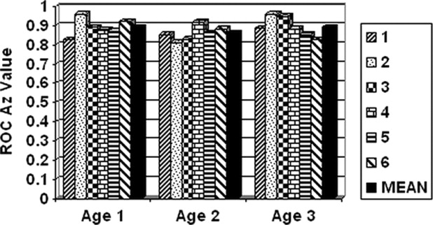
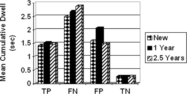
There was no statistically significant difference in receiver operating characteristic performance as a function of monitor age (F = 0.4901, P = .6187). In terms of total viewing time, there were not statistically significant differences between the three monitors (F = 0.056, P = .9452). The dwell times associated with each decision type (true and false, positive and negative) did not differ significantly as a function of monitor age for any decision.
Conclusion
At least up to 2.5 years of age, the shift in whitepoint toward the yellow range does not significantly impact diagnostic accuracy or visual search efficiency of radiologists.
As digital display monitors age, there are visible changes in their appearance ( ). The useful lifetime of digital displays varies, but because they are often used continuously in radiology their lifetime is shorter than the typical desktop monitor, having to be replaced every few years. With liquid crystal displays (LCDs), failure (i.e., below the maximum luminance point at which the manufacturer recommends typical use) of the backlight is the most common point of failure with age, although drifts in luminance and whitepoint (the color of light emitted by a source) can occur long before actual failure ( ). Most LCDs use a CCFL (cold cathode fluorescent lamp) as the white-light source. White light, of course, is a mixture of wavelengths of various colors that are perceived as colorless, and the whitepoint is defined as a point in a color space. An example is an x,y coordinate in CIE 1931 color space (Commission Internationale de l’Eclairage, or the International Commission on Illumination) ( ). As a digital display ages, this whitepoint tends to drift towards a yellow hue because the blue fluorescent substance in the CCFL degrades more quickly than the other colors, altering the appearance of even monochrome displays and thus the images portrayed on them.
Although guidelines for medical-grade monitor quality assessment and quality control (QA/QC) note that display characteristics such as luminance can drift with age and recommend regular calibration ( ), there have been no studies to date that have evaluated the impact of monitor aging on diagnostic performance. In a previous study ( ), we demonstrated that color monitors negatively impact diagnostic accuracy and visual search performance of radiologists compared to monochrome monitors, but that does not address the question of whether changes in the whitepoint of monochrome displays toward yellow will affect performance as well. The present study was designed to address the question of whether shifts in display whitepoint toward yellow impact observer performance.
Materials and methods
Displays
Get Radiology Tree app to read full this article<
Get Radiology Tree app to read full this article<
Observers and Reading Protocol
Get Radiology Tree app to read full this article<
Get Radiology Tree app to read full this article<
Get Radiology Tree app to read full this article<
Visual Search
Get Radiology Tree app to read full this article<
Results
Diagnostic Performance
Get Radiology Tree app to read full this article<
Get Radiology Tree app to read full this article<
Visual Search Performance
Get Radiology Tree app to read full this article<
Get Radiology Tree app to read full this article<
Get Radiology Tree app to read full this article<
Discussion
Get Radiology Tree app to read full this article<
Get Radiology Tree app to read full this article<
Get Radiology Tree app to read full this article<
Get Radiology Tree app to read full this article<
References
1. Roehrig H.: The monochrome cathode ray tube display and its performance.Kim Y.Horii S.C.Handbook of Medical Imaging.2000.SPIE PressBellingham, WA:pp. 155-220.
2. Behlen F.M., Hemminger B.M., Horii S.C.: Displays.Kim Y.Horii S.C.Handbook of Medical Imaging.2000.SPIE PressBellingham, WA:pp. 403-440.
3. International Commission on Illumination. http://www.cie.co.at/cie/ . Accessed October 31, 2006.
4. Samei E.: AAPM/RSNA physics tutorial for residents: Technological and psychophysical considerations for digital mammographic displays. RadioGraphics 2005; 25: pp. 491-501.
5. Samei E., Badano A., Chakraborty D., et. al.: Assessment of display performance for medical imaging systems: Executive summary of AAPM TG18 report. Med Phys 2005; 32: pp. 1205-1225.
6. Krupinski E.A., Roehrig H.: Pulmonary nodule detection and visual search: P45 and P104 monochrome versus color monitor displays. Acad Radiol 2002; 9: pp. 638-645.
7. NEC Xlight displays. http://www.necdisplay.com/medical/radiology/index.htm . Accessed January 8, 2007.
8. Miyamoto T., Hayashida K., Matsui S., Mori K.: High resolution gray scale medical displays: Technical developments and actual products. NEC Technical J 2006; 1: pp. 70-73.
9. Dorfman D.D., Berbaum K.S., Metz C.E.: Receiver operating characteristic rating analysis: Generalization to the population of readers and patients with the jackknife method. Invest Radiol 1992; 27: pp. 723-731.
10. MRMC sample size estimates. http://perception.radiology.uiowa.edu/SampleSize/KrupinskiGonzalesGonzalesetal2000/tabid/191/Default.aspx . Accessed November 29, 2006.
11. Krupinski E.A., Berger W., Dallas W., Roehrig H.: Pulmonary nodule detection: What features attract attention?. Proc SPIE Med Imag 2004; 5372: pp. 122-127.
12. Krupinski E.A., Lund P.J.: Differences in time to interpretation for evaluation of bone radiographs with monitor and film viewing. Acad Radiol 1997; 4: pp. 177-182.
13. Krupinski E.A., Weinstein R.S., Rozek L.S.: Experience-related differences in diagnoses from medical images displayed on monitors. Telemed J 1996; 2: pp. 101-108.
14. Krupinski E.A.: Visual scanning patterns of radiologists searching mammograms. Acad Radiol 1996; 3: 317–144.
15. Hu C.H., Kundel H.L., Nodine C.F., Krupinski E.A., Toto L.C.: Searching for bone fractures: A comparison with pulmonary nodule search. Acad Radiol 1994; 1: pp. 25-32.
16. Krupinski E.A., Roehrig H., Furukawa T.: Influence of film and monitor display luminance on observer performance and visual search. Acad Radiol 1999; 6: pp. 411-418.
17. Krupinski E.A., Roehrig H.: The influence of a perceptually linearized display on observer performance and visual search. Acad Radiol 2000; 7: pp. 8-13.
18. Krupinski E.A., Roehrig H.: Pulmonary nodule detection and visual search: P45 and P104 monochrome versus color monitor displays. Acad Radiol 2002; 9: pp. 638-645.
19. Nodine C.F., Kundel H.L., Toto L.C., Krupinski E.A.: Recording and analyzing eye-position data using a microcomputer workstation. Behav Res Meth Instrum Comput 1992; 24: pp. 475-485.